. 2022 Jul 28:13:917232.
doi: 10.3389/fimmu.2022.917232. eCollection 2022.

Alveolar macrophages in early stage COPD show functional deviations with properties of impaired immune activation

Affiliations

Alveolar macrophages in early stage COPD show functional deviations with properties of impaired immune activation

Kevin Baßler et al. Front Immunol. .

Abstract

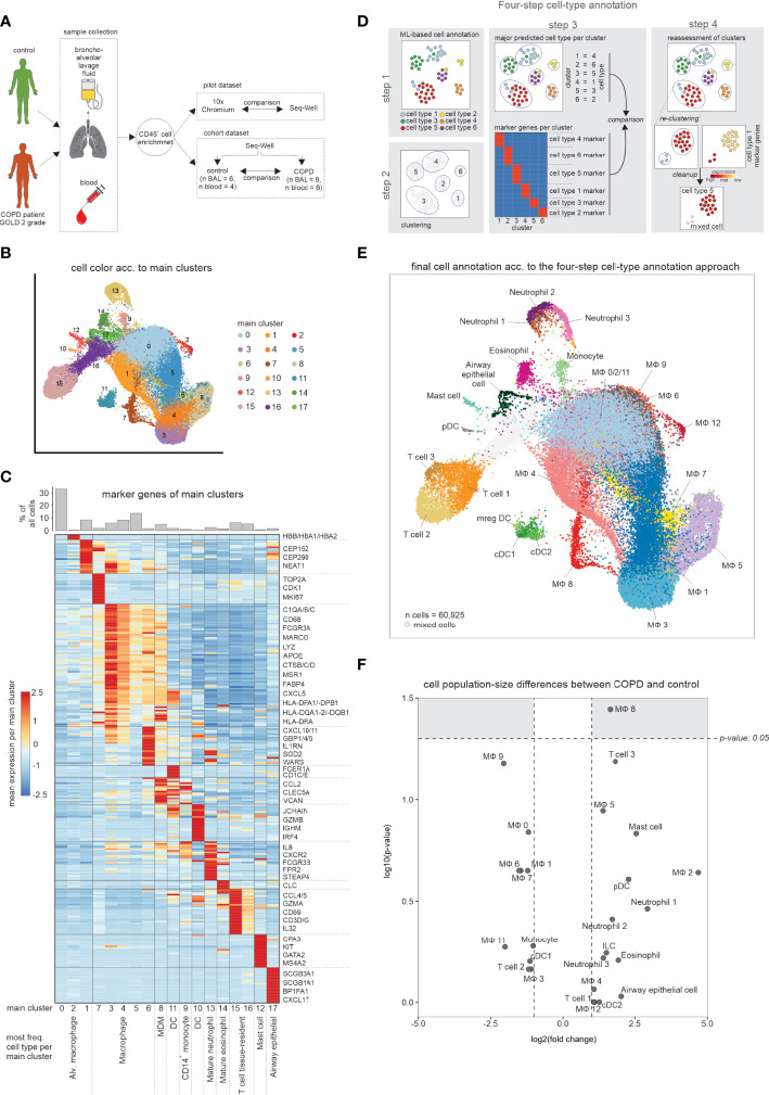
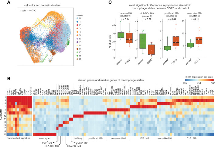
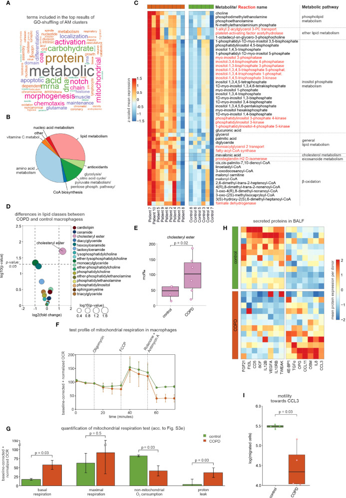
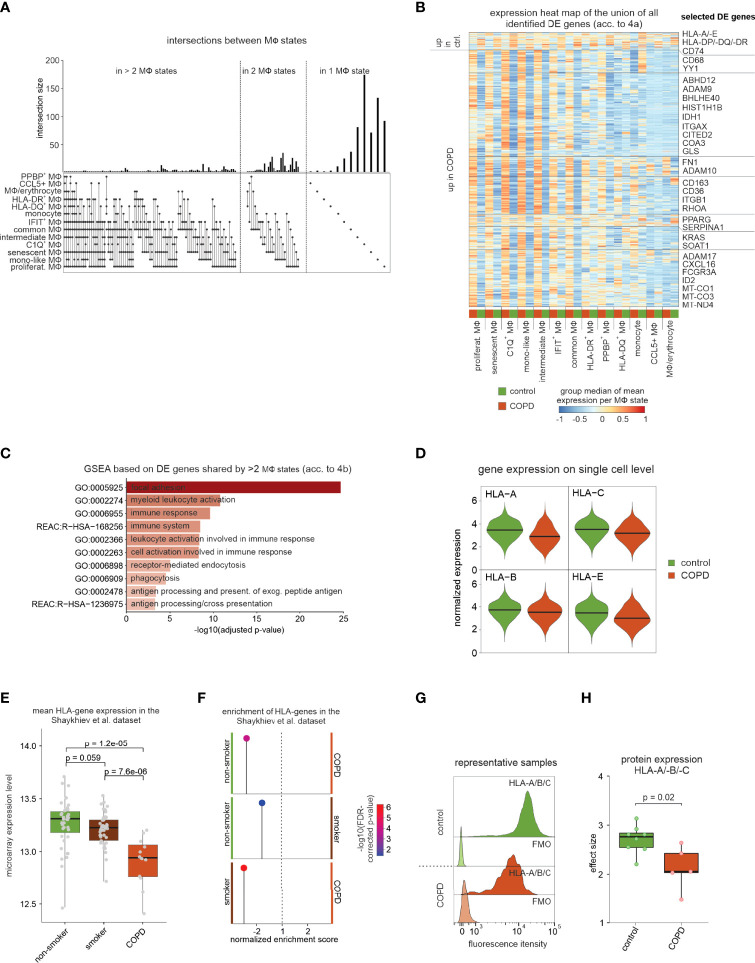
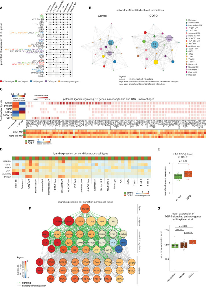
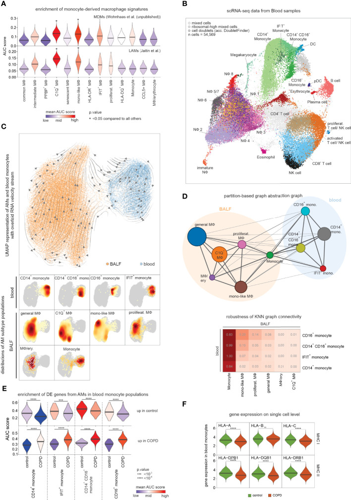
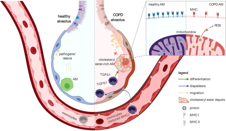
Despite its high prevalence, the cellular and molecular mechanisms of chronic obstructive pulmonary disease (COPD) are far from being understood. Here, we determine disease-related changes in cellular and molecular compositions within the alveolar space and peripheral blood of a cohort of COPD patients and controls. Myeloid cells were the largest cellular compartment in the alveolar space with invading monocytes and proliferating macrophages elevated in COPD. Modeling cell-to-cell communication, signaling pathway usage, and transcription factor binding predicts TGF-β1 to be a major upstream regulator of transcriptional changes in alveolar macrophages of COPD patients. Functionally, macrophages in COPD showed reduced antigen presentation capacity, accumulation of cholesteryl ester, reduced cellular chemotaxis, and mitochondrial dysfunction, reminiscent of impaired immune activation.

Keywords: TGF-β1; blood; bronchoalveolar lavage; chronic obstructive pulmonary disease; impaired immune activation; macrophage; monocyte.


Conflict of interest statement

The handling editor [AH] declared a shared affiliation with the author(s) [AW, NY] at the time of review. BR, FB, MK and HD were employed by CommaSoft. CW and PB were employed by Boehringer Ingelheim. The remaining authors declare that the research was conducted in the absence of any commercial or financial relationships that could be constructed as a potential conflict of interest.

Figures

Figure 1
Figure 1
scRNA-seq data of BALF samples obtained from COPD patients and healthy controls. (A) Schematic workflow of the present study. Bronchoalveolar lavage fluid (BALF) and peripheral blood was obtained from control donors and COPD patients (GOLD stage 2). After enrichment for immune cells (CD45+ cells), single-cell RNA-seq was performed. (B) UMAP representation of integrated BALF data obtained from all COPD patients and control donors. Coloring and numbering according to identified main clusters. (C) Heatmap of the calculated marker genes per main cluster with a bar chart representation of the relative cell type proportions at the top. The marker gene expression per cluster is represented as a z-transformed value (across all clusters). Rows of the heatmap are clustered hierarchically. At the bottom of the plot, the main cell type is displayed, which is contained in the respective main cluster. (D) Schematic workflow of the four-step annotation approach, including machine learning-based cell type annotation, clustering, assignment and subsequent confirmation of a cluster to a cell type according to the machine learning-based cell type annotation, and identification of ‘contaminating’ cells (referred to as ‘mixed cells’). (E) Final cell type annotation of integrated BALF data according to the four-step annotation approach. (F) Volcano plot visualization of log2 fold changes and negative log10 p-values (Wilcoxon rank sum test) of changes in cell type occurrence in BALF of samples obtained from COPD patients and controls. BALF, bronchoalveolar lavage fluid; alv., alveolar; MDM, monocyte-derived macrophage; DC, dendritic cell; n, number; MФ, macrophage.
Figure 2
Figure 2
Exploration of the macrophage and monocyte cell types and states in human BALF. (A) UMAP representation and clustering of cells annotated as monocytes or macrophages by the four-step annotation approach (according to Figure 1). (B) Heatmap of marker genes per macrophage/monocyte cluster (referred to as ‘macrophage states’; according to Figure 2A). The marker gene expression per macrophage state is represented as a z-transformed value across all macrophage states. On the left side of the heatmap, conserved macrophage markers are depicted. Columns and rows of the heatmap are sorted by hierarchical clustering. (C) Box visualization plot (with marked median values) of most significant differences in population sizes within the identified macrophage states between COPD and control (error bars indicating the standard deviation; statistics based on Wilcoxon rank sum test). n, number; MФ, macrophage.
Figure 3
Figure 3
Modeling of the metabolic landscape and alterations in macrophages. (A) Word cloud of the most common words in the top predicted terms of the GO-shuffling approach across all macrophage clusters. (B) Compass results of the modeled metabolic landscape in macrophages. The pie chart summarizes and categorizes the predicted metabolites and pathways that are significantly different between COPD and control. (C) Heatmap showing the metabolites and pathways that were predicted by Compass as altered in COPD and that were associated with lipid metabolism. Metabolites are shown in black and reactions in red. Columns and rows of the heat map are sorted by hierarchical clustering. (D) Volcano plot visualization of log2 fold changes and negative log10 p-values (Wilcoxon rank sum test) of lipid class levels between COPD and control macrophages obtained by lipidomics analysis. (E) Box plot with marked median values of cholesteryl ester proportions with the representation of individual donors. (F) Evaluation of mitochondrial function via the time-dependent course of the oxygen consumption rate (OCR) in macrophages using baseline-corrected values. Error bars indicate the standard deviations (control n = 2, COPD n = 3). Dashed arrows represent the injection of various compounds (shown at the top of the plot) used to assess different aspects of mitochondrial function (according to Figure S3 E). (G) Bar plots showing quantifications of different aspects of mitochondrial function inferred from the OCR measurement in Figure 3F (according to Figure S3 E; error bars indicating the standard deviation; statistics based on t-test). (H) Heatmap representation of proteins detected in BALF with a p-value < 0.1 according to the Wilcoxon rank sum test between COPD patients and control donors (control n = 11, COPD n = 12). The mean protein expression (identified by Olink Proteomics) per donor is represented as a z-transformed value (across all donors). Columns of the heatmap are sorted by hierarchical clustering. (I) Quantification of the migratory capability of macrophages towards CCL3 displayed in a box plot with marked median values and the representation of individual donors (control n = 4, COPD n = 4; error bars indicating the standard deviation; statistics based on t-test). BALF, bronchoalveolar lavage fluid; OCR, oxygen consumption rate.
Figure 4
Figure 4
DE gene analysis of identified macrophage states. (A) UpSet plot of calculated DE genes across macrophage states. DE genes found in the same states are binned and the size of the bins is represented as a bar chart. At the bottom, dots indicate which macrophage states contained and shared these DE genes. (B) Heat map representation of the union of all DE genes found in the macrophage states. Depicted is the group median (group = COPD or control) of the z-transformed mean expression data per donor and macrophage state across all macrophage states, and the names of some selected DE genes are shown on the right side of the plot. Columns and rows of the heat map are sorted by hierarchical clustering. (C) Selected functional gene sets from GSEA based on DE genes that reach the defined significance cutoffs for more than two macrophage states (acc. to Figure 4B). (D) Violin plot with marked median of HLA-A/-B/-C and -E expression in all macrophages based on scRNA-seq data. The plot shows the expression across the donors, whereby the donors were downsampled to the same number of cells, followed by downsampling to the same number of cells between COPD and control. The plot displays cells with an expression > 0. (E) Box plots (with marked median values) showing the mean expression per sample of HLA genes expressed in macrophages (error bars indicating the standard deviation; statistics are based on the Wilcoxon rank sum test). The data are obtained from Shaykhiev et al. (17). (F) Pin plot representing the enrichments in the samples of Shaykhiev et al. of HLA genes expressed in macrophages. (G) Fluorescence intensity histograms showing representative samples of flow cytometric analysis of HLA-A/-B/-C expression on the cell surface of isolated macrophages (FMO = fluorescence minus one). (H) Box plots with marked median of the calculated effect sizes of HLA-A/-B/-C expression in COPD and control with the representation of individual donors (control n = 8, COPD n = 5; error bars indicating the standard deviation; statistics based on Wilcoxon rank sum test). MФ, macrophage; mono, monocyte; DE, differentially expressed; GSEA, gene set enrichment analysis; FDR, false discovery rate; FMO, fluorescence minus one.
Figure 5
Figure 5
Modeling the cell-to-cell interactions of BALF cells. (A) UpSet plot of predicted transcriptional regulators of DE genes. Dots indicate which clusters contain and share predicted transcriptional regulators. The names of selected regulators are shown on the right side of the plot with the font color indicating the association with NOTCH, WNT, TGF-β1, TNF or circadian rhythm signaling. (B) Network representation of predicted cell-to-cell interactions derived from CellPhoneDB. The names of the two most interconnected cell types are displayed (edge: identified cell-to-cell interaction; edge width: proportional to number of interactions between two cell types; node size: proportional to number of overall interactions). (C) Results of NicheNet analysis, in which the heatmap in the top left corner displays the z-normalized ligand activity scores (based on area under the precision recall curve (AUPR)) of the top 3 ligands for either the DE genes from C1Q+ macrophages or monocyte-like macrophages, respectively. On the right the top 250 interaction scores of the ligands’ target genes are colored by their interaction score. The heatmap at the bottom represents the mean expression (z-transformed by gene across all macrophage states; according to Figure S5 D) of the ligands’ target genes in C1Q+ macrophages or monocyte-like macrophages from control and COPD. (D) The mean expression of the top 6 ligands in all identified BALF cell types for either COPD or control patients (z-transformed by gene) is displayed. (E) Box plot with marked median of the measured protein expression (by Olink Proteomics) in BALF of LAP TGF-β1 in COPD and control with representation of individual donors (control n = 11, COPD n = 12; error bars indicating the standard deviation; statistics based on Wilcoxon rank sum test). (F) Representation of inferred ligand-to-target signaling path for TGF-β1 derived from the NicheNet analysis. The nodes representing the genes are colored by the expression fold change between COPD and control patients. (G) Box plots (with marked median values) showing the mean expression per sample of TGF-β-signaling genes (error bars indicating the standard deviation; statistics are based on the Wilcoxon rank sum test). The underlying data are obtained from Shaykhiev et al. (17). signal., signaling; TF, transcription factor; MФ, macrophage; regulat., regulatory; mono, monocyte; DC, dendritic cell; transcript., transcriptional.
Figure 6
Figure 6
Assessing the relationship between blood monocytes and BALF macrophages. (A) Violin plots (with marked median values) displaying enrichment of human orthologues of murine monocyte-derived macrophage signature genes across macrophage states in COPD and control based on Area Under the Curve (AUC). (B) Integrated scRNA-seq data of blood immune cells annotated according to the four-step annotation approach (according to Figure 1B). (C) UMAP of embedded macrophages/monocytes from BALF and blood monocytes. Inferred main average vector flow is indicated by velocity streamlines that are projected as vectors. Locations of the main cell types (acc. to the combined labels from Figure S6A) in the UMAP are indicated by the heat maps at the bottom. (D) PAGA graph derived from embedded BALF and blood data (according to Figure 6C). The weight of an edge, which reflects a statistical measure of connectivity, is represented as the edge width. The table below summarizes the results of the PAGA connectivity calculation, where a value of 1 indicates a strong connection and 0 indicates a weak connection between two cell types. (E) Violin plots (with marked median values) displaying enrichment of macrophage-related DE genes (according to Figures 4B, C) in blood monocytes based on AUC. (F) Violin plots with marked median of the expression of HLA genes, in blood monocytes based on scRNA-seq data. The plots show the expression across the donors, whereby the donors were downsampled to the same number of cells, followed by downsampling to the same number of cells between COPD and control. The plots display cells with an expression > 0. BALF, bronchoalveolar lavage fluid; mono, monocyte; MФ, macrophage; NФ, neutrophil; proliferat., proliferating; MDM, monocyte-derived macrophage; LAM, lipid-associated macrophage; ns, means not significant.
Figure 7
Figure 7
Schematic representation of the key findings of the present study In healthy lungs, alveolar macrophages survey the alveoli and remove pathogens and debris to enable proper gas exchange. In the alveoli of COPD patients, the alveolar macrophages accumulate cholesteryl esters. In addition, blood monocytes invade the alveoli and differentiate into alveolar macrophages. The transcriptome of COPD alveolar macrophages indicate TGF-β1-associated cell signaling especially in the early stages of monocyte-to-macrophage differentiation. The alveolar macrophages in COPD show a reduced ability to migrate towards chemokine. Furthermore, they express fewer MHC molecules; especially MHC class I. Together with the reduced phagocytosis of alveolar macrophages in COPD, the ability of these cells for immune surveillance is severely limited during the disease. In addition, their mitochondria are leaking (e.g. to protons) and therefore produce high amounts of reactive oxygen species. Taken together, the guardians of normal lung function (alveolar macrophages) are severely altered in COPD, preventing them from fulfilling their important physiological functions properly. Furthermore, the observation of reduced MHC expression in blood monocytes indicates that the manifestation of COPD has a strong systemic component.

References

    1. GBD 2017 Causes of Death Collaborators . Global, regional, and national age-sex-specific mortality for 282 causes of death in 195 countries and territories, 1980-2017: A systematic analysis for the global burden of disease study 2017. Lancet (2018) 392:1736–88. doi: 10.1016/S0140-6736(18)32203-7 - DOI - PMC - PubMed
    1. Celli BR, Wedzicha JA. Update on clinical aspects of chronic obstructive pulmonary disease. N Engl J Med (2019) 381:1257–66. doi: 10.1056/NEJMra1900500 - DOI - PubMed
    1. Barnes PJ, Burney PGJ, Silverman EK, Celli BR, Vestbo J, Wedzicha JA, et al. Chronic obstructive pulmonary disease. Nat Rev Dis Primers (2015) 1:15076. doi: 10.1038/nrdp.2015.76 - DOI - PubMed
    1. Barnes PJ. Alveolar macrophages as orchestrators of COPD. COPD (2004) 1:59–70. doi: 10.1081/COPD-120028701 - DOI - PubMed
    1. Zheng GXY, Terry JM, Belgrader P, Ryvkin P, Bent ZW, Wilson R, et al. Massively parallel digital transcriptional profiling of single cells. Nat Commun (2017) 8:14049. doi: 10.1038/ncomms14049 - DOI - PMC - PubMed

Publication types