. 2022 Aug 8:2022:5392966.
doi: 10.1155/2022/5392966. eCollection 2022.

Chronic Cerebral Hypoperfusion Aggravates Parkinson's Disease Dementia-Like Symptoms and Pathology in 6-OHDA-Lesioned Rat through Interfering with Sphingolipid Metabolism

Affiliations

Chronic Cerebral Hypoperfusion Aggravates Parkinson's Disease Dementia-Like Symptoms and Pathology in 6-OHDA-Lesioned Rat through Interfering with Sphingolipid Metabolism

Yaohua Fan et al. Oxid Med Cell Longev. .

Abstract

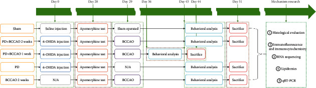
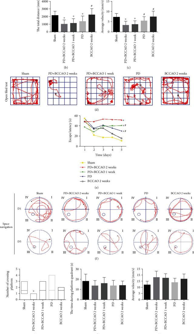
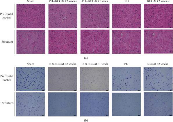
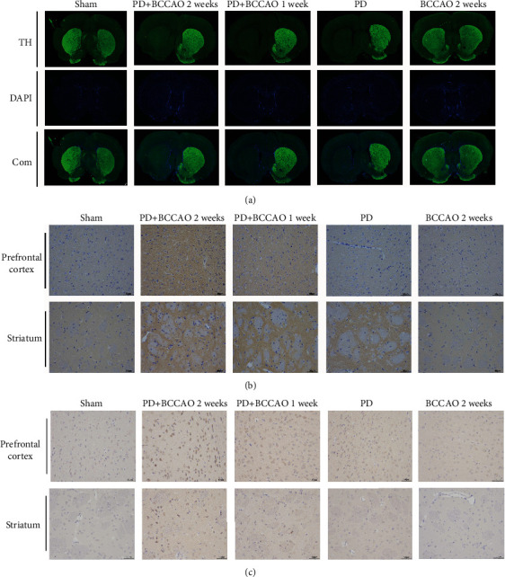
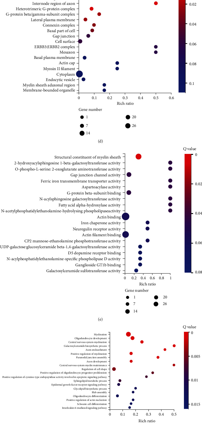
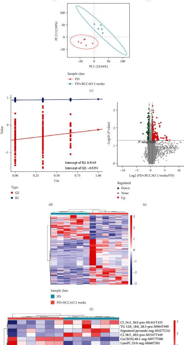
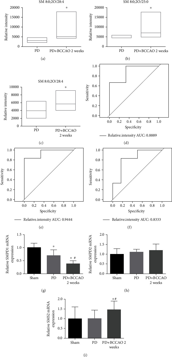
Chronic cerebral hypoperfusion (CCH) is a cardinal risk factor for Parkinson's disease dementia (PDD), but this potential causality lacks mechanistic evidence. We selected bilateral common carotid artery occlusion (BCCAO) to simulate chronic cerebral hypoperfusion in the rat model of PD induced by typical neurotoxin 6-hydroxy dopamine (6-OHDA). Four weeks after unilateral injection of 6-OHDA into the medial forebrain bundle, rats underwent BCCAO. Male Sprague-Dawley rats were divided into five groups of ten, including sham, PD+BCCAO 2 weeks, PD+BCCAO 1 week, PD, and BCCAO 2 weeks. Then, open field test (OFT) and Morris water maze test (MWM) were used to assess the PDD-like symptoms in rats. Also, the pathological manifestations and mechanisms of BCCAO impairing cognitive functions have been explored via hematoxylin-eosin staining, Nissl staining, immunohistochemistry, immunofluorescence, RNA sequencing analysis, lipidomics, and quantitative real-time polymerase chain reaction. In this study, we found that CCH could aggravate PDD-like cognitive symptoms (i.e., learning memory and spatial cognition) and PDD-like pathology (higher expression of α-Syn and Aβ in prefrontal cortex and striatum). Moreover, a potential relationship between differentially expressed mRNAs and lipid metabolism was revealed by RNA sequencing analysis. Lipidomics showed that CCH could affect the intensity of 5 lipids, including sphingomyelin (SM 9:0;2O/26:2; SM 8:1;2O/25:0; and SM 8:0;2O/28:4), cardiolipin, lysophosphatidylcholine, cholesteryl ester, and triacylglycerol. Interestingly, the KEGG pathway analysis of both RNA sequencing analysis and lipidomics suggested that CCH leaded to learning impairment by affecting sphingolipid metabolism. Finally, we found that CCH disrupts the sphingolipid metabolism by affecting the mRNA expression of SMPD1 and SMS2, leading to the accumulation of sphingomyelin in the prefrontal cortex. In summary, CCH, an independent exacerbating reason for impairment in learning and memory within the pathopoiesis of PD, aggravates Parkinson's disease dementia-like symptoms and pathology in 6-OHDA-lesioned rat through interfering with sphingolipid metabolism.


Conflict of interest statement

The authors declare that they have no competing interests.

Figures

Figure 1
Figure 1
The timeline of the experiment. Abbreviations: PD: Parkinson's disease; BCCAO: bilateral common carotid artery occlusion; 6-OHDA: 6-hydroxy dopamine; N/A: no operation; RNA: ribonucleic acid; qRT-PCR: quantitative real-time polymerase chain reaction.
Figure 2
Figure 2
Behavioral performance in apomorphine-induced rotation tests, OFT, and MWM (n = 10). (a) The number of rotations of each group in apomorphine-induced rotation tests. (b) The total distance of each group in OFT test. (c) Average velocity of each group in OFT test. (d) Representative movement trajectories of rats in the OFT. (e) Escape latency of each group in space navigation of MWM. (f) Representative movement trajectories of rats on the first and fifth days of space navigation. (g) Number of crossing platform of each group in space exploration of MWM. (h) The time during platform quadrant of each group in space exploration of MWM. (i) Average speed of each group in space exploration of MWM. (j) Representative movement trajectories of rats in space exploration of MWM. P < 0.05 compared with the sham group and #P < 0.05 compared to the BCCAO 2 weeks group.
Figure 3
Figure 3
Hematoxylin and eosin (H&E) staining and Nissl staining sections from prefrontal cortex and striatum (n = 3). (a) Representative images of HE staining in prefrontal cortex and striatum of each group. In PD+BCCAO 2 weeks group, the interstitial space of the striatum is relatively loose, while the boundary of the striatal interstitial space is unclear, and many vacuoles can be seen throughout the striatum. The prefrontal cortex in PD+BCCAO 2 weeks group showed that a large number of neurons in the prefrontal cortex were shrunken, the staining of the cells was deepened, the boundary between the nucleus and cytoplasm was unclear, and the cells were loosely arranged. (b) Representative images of Nissl staining in prefrontal cortex and striatum of each group. Dark blue represents Nissl bodies. Nissl bodies are large and numerous, indicating that nerve cells have a strong function of synthesizing proteins; on the contrary, when nerve cells are damaged, the number of Nissl bodies will decrease or even disappear. The numbers of Nissl-stained neurons in prefrontal cortex and striatum were significantly decreased in the PD+BCCAO 2 weeks group. Scale bars represent 50 μm.
Figure 4
Figure 4
CCH aggravated PDD-like pathology in prefrontal cortex and striatum of 6-OHDA-lesioned rat (n = 3). (a) Representative images of TH expression in prefrontal cortex and striatum of each group. TH (green) and DAPI nuclear stain (blue). CCH could not aggravate the loss of TH-positive cells in striatum, compared to the PD group. (b) Representative images of α-Syn expression in prefrontal cortex and striatum of each group. α-Syn (brown) and cell nucleus (blue). CCH combined with 6-OHDA treatment could increase the expression of α-Syn in prefrontal cortex. (c) Representative images of Aβ expression in prefrontal cortex and striatum of each group. Aβ (brown) and cell nucleus (blue). The expression of Aβ in striatum and prefrontal cortex of PD+BCCAO 2 weeks was higher than that of other groups. Scale bars represent 50 μm.
Figure 5
Figure 5
CCH modulated mRNA expression in prefrontal cortex of 6-OHDA-lesioned rat (n = 3). (a) Volcano map of differentially expressed mRNAs characterized by RNA sequencing. Compared with the PD group, green represents decrease in the PD+BCCAO 2 weeks group, and red represents increase. (b) Correlation heatmap of differentially expressed mRNAs. The color represents the expression level; the higher the expression level, the redder the color, otherwise the bluer. (c) PPI of differentially expressed mRNAs. (d) Enrichment of GO biological process of differentially expressed mRNAs. (e) Enrichment of GO cellular component of differentially expressed mRNAs. (f) Enrichment of GO molecular function of differentially expressed mRNAs. (g) Enrichment of KEGG pathways of differentially expressed mRNAs.
Figure 6
Figure 6
CCH aggravates PDD-like symptoms and pathology in 6-OHDA-lesioned rat through interfering with sphingolipid metabolism (n = 6). (a) Metabolic ions identified in positive and negative ion mode into six major categories of lipids. (b) PCA analysis of differentially metabolic ions. The horizontal axis is the first principal component, and the vertical axis is the second principal component. (c) PLS-DA analysis of differentially metabolic ions. (d) Response ranking test plot for the PLS-DA analytical model. (e) Volcano map of differentially metabolic ions. (f) Correlation heatmap of differentially metabolic ions. (g) Correlation heatmap of differentially secondary metabolites. (h) Enrichment of KEGG pathways of differentially metabolic ions.
Figure 7
Figure 7
CCH disrupts the sphingolipid metabolism by affecting the mRNA expression of SMPD1 and SMS2. (a–c) Relative intensity analysis of metabolic ions. pos-M716T311 (SM 9:0;2O/26:2), pos-M690T309 (SM 8:1;2O/25:0), and pos-M726T325 (SM 8:0;2O/28:4) are higher in the PD+BCCAO 2 weeks group compared to the PD group. (d) Receiver operating characteristic curve analysis showed the AUC of pos-M726T325 (SM 8:0;2O/28:4) was 0.8889. (e) Receiver operating characteristic curve analysis showed the AUC of pos-M690T309 (SM 8:1;2O/25:0) was 0.9444. (e) Receiver operating characteristic curve analysis showed the AUC of pos-M716T311 (SM 9:0;2O/26:2) was 0.8333. (g–i) Relative mRNA expression of each group. The relative expression of SMPD1 was significantly decreased in the prefrontal cortex of PD+BCCAO 2 weeks group, but SMS2 was significantly elevated, while SMPD2 did not change. P < 0.05 compared with the sham group and #P < 0.05 compared to BCCAO 2 weeks group.

Similar articles

Cited by

References

    1. Emre M., Ford P. J., Bilgiç B., Uç E. Y. Cognitive impairment and dementia in Parkinson's disease: practical issues and management. Movement Disorders . 2014;29(5):663–672. doi: 10.1002/mds.25870. - DOI - PubMed
    1. Gratwicke J., Jahanshahi M., Foltynie T. Parkinson's disease dementia: a neural networks perspective. Brain . 2015;138(6):1454–1476. doi: 10.1093/brain/awv104. - DOI - PMC - PubMed
    1. Fan Y., Han J., Zhao L., et al. Experimental models of cognitive impairment for use in Parkinson's disease research: the distance between reality and ideal. Frontiers in Aging Neuroscience . 2021;13, article 745438 doi: 10.3389/fnagi.2021.745438. - DOI - PMC - PubMed
    1. Han J., Fan Y., Wu P., et al. Parkinson's disease dementia: synergistic effects of alpha-synuclein, tau, beta-amyloid, and iron. Frontiers in Aging Neuroscience . 2021;13, article 743754 doi: 10.3389/fnagi.2021.743754. - DOI - PMC - PubMed
    1. Huang X., Wang C., Chen L., Zhang T., Leung K. L., Wong G. Human amyloid beta and α-synuclein co-expression in neurons impair behavior and recapitulate features for Lewy body dementia in Caenorhabditis elegans. Biochimica et Biophysica Acta - Molecular Basis of Disease . 2021;1867(10, article 166203) doi: 10.1016/j.bbadis.2021.166203. - DOI - PubMed