

Antimicrobial peptides: Defending the mucosal epithelial barrier
- PMID: 35979535
- PMCID: PMC9376388
- DOI: 10.3389/froh.2022.958480
Antimicrobial peptides: Defending the mucosal epithelial barrier
Abstract
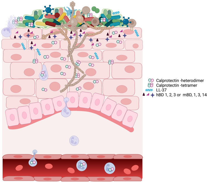
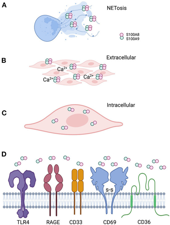
The recent epidemic caused by aerosolized SARS-CoV-2 virus illustrates the importance and vulnerability of the mucosal epithelial barrier against infection. Antimicrobial proteins and peptides (AMPs) are key to the epithelial barrier, providing immunity against microbes. In primitive life forms, AMPs protect the integument and the gut against pathogenic microbes. AMPs have also evolved in humans and other mammals to enhance newer, complex innate and adaptive immunity to favor the persistence of commensals over pathogenic microbes. The canonical AMPs are helictical peptides that form lethal pores in microbial membranes. In higher life forms, this type of AMP is exemplified by the defensin family of AMPs. In epithelial tissues, defensins, and calprotectin (complex of S100A8 and S100A9) have evolved to work cooperatively. The mechanisms of action differ. Unlike defensins, calprotectin sequesters essential trace metals from microbes, which inhibits growth. This review focuses on defensins and calprotectin as AMPs that appear to work cooperatively to fortify the epithelial barrier against infection. The antimicrobial spectrum is broad with overlap between the two AMPs. In mice, experimental models highlight the contribution of both AMPs to candidiasis as a fungal infection and periodontitis resulting from bacterial dysbiosis. These AMPs appear to contribute to innate immunity in humans, protecting the commensal microflora and restricting the emergence of pathobionts and pathogens. A striking example in human innate immunity is that elevated serum calprotectin protects against neonatal sepsis. Calprotectin is also remarkable because of functional differences when localized in epithelial and neutrophil cytoplasm or released into the extracellular environment. In the cytoplasm, calprotectin appears to protect against invasive pathogens. Extracellularly, calprotectin can engage pathogen-recognition receptors to activate innate immune and proinflammatory mechanisms. In inflamed epithelial and other tissue spaces, calprotectin, DNA, and histones are released from degranulated neutrophils to form insoluble antimicrobial barriers termed neutrophil extracellular traps. Hence, calprotectin and other AMPs use several strategies to provide microbial control and stimulate innate immunity.
Keywords: LL-37; antimicrobial peptides/proteins; calprotectin; defensins; disease; epithelium; health.
Copyright © 2022 Johnstone and Herzberg.
Conflict of interest statement
The authors declare that the research was conducted in the absence of any commercial or financial relationships that could be construed as a potential conflict of interest.
Figures
Similar articles
-
Understanding the Dynamics of Human Defensin Antimicrobial Peptides: Pathogen Resistance and Commensal Induction.Appl Biochem Biotechnol. 2024 Oct;196(10):6993-7024. doi: 10.1007/s12010-024-04893-8. Epub 2024 Mar 13. Appl Biochem Biotechnol. 2024. PMID: 38478321 Review.
-
Antimicrobial peptides of the vaginal innate immunity and their role in the fight against sexually transmitted diseases.New Microbes New Infect. 2019 Dec 10;34:100627. doi: 10.1016/j.nmni.2019.100627. eCollection 2020 Mar. New Microbes New Infect. 2019. PMID: 31993204 Free PMC article. Review.
-
Regulation of antimicrobial peptide gene expression by nutrients and by-products of microbial metabolism.Eur J Nutr. 2012 Dec;51(8):899-907. doi: 10.1007/s00394-012-0415-4. Epub 2012 Jul 15. Eur J Nutr. 2012. PMID: 22797470 Free PMC article. Review.
-
Antimicrobial peptides: the ancient arm of the human immune system.Virulence. 2010 Sep-Oct;1(5):440-64. doi: 10.4161/viru.1.5.12983. Virulence. 2010. PMID: 21178486 Review.
-
Shosaikoto increases calprotectin expression in human oral epithelial cells.J Periodontal Res. 2010 Feb;45(1):79-86. doi: 10.1111/j.1600-0765.2009.01203.x. Epub 2009 Jul 8. J Periodontal Res. 2010. PMID: 19602113
Cited by
-
An Overview of the Potentialities of Antimicrobial Peptides Derived from Natural Sources.Antibiotics (Basel). 2022 Oct 26;11(11):1483. doi: 10.3390/antibiotics11111483. Antibiotics (Basel). 2022. PMID: 36358138 Free PMC article. Review.
-
A fish-specific antimicrobial peptide MsPiscidin2 inactivates MSRV and confers protection in largemouth bass.Front Immunol. 2025 Jun 23;16:1629256. doi: 10.3389/fimmu.2025.1629256. eCollection 2025. Front Immunol. 2025. PMID: 40625750 Free PMC article.
-
Probiotic intervention alters immune gene expression and tumor characteristics in experimental breast cancer.Mol Biol Rep. 2025 Aug 8;52(1):809. doi: 10.1007/s11033-025-10873-w. Mol Biol Rep. 2025. PMID: 40779195
-
Multiple mechanisms enable broad-spectrum activity of the Pelargonium sidoides root extract EPs 7630 against acute respiratory tract infections.Front Pharmacol. 2024 Oct 14;15:1455870. doi: 10.3389/fphar.2024.1455870. eCollection 2024. Front Pharmacol. 2024. PMID: 39469622 Free PMC article. Review.
-
Intestinal Barrier Dysfunction and Gut Microbiota in Non-Alcoholic Fatty Liver Disease: Assessment, Mechanisms, and Therapeutic Considerations.Biology (Basel). 2024 Apr 6;13(4):243. doi: 10.3390/biology13040243. Biology (Basel). 2024. PMID: 38666855 Free PMC article. Review.
References
Publication types
Grants and funding
LinkOut - more resources
Full Text Sources
Miscellaneous