

Surface CD3-negative monomorphic epitheliotropic intestinal T-cell lymphoma
- PMID: 35979577
- PMCID: PMC9635036
- DOI: 10.3960/jslrt.22005
Surface CD3-negative monomorphic epitheliotropic intestinal T-cell lymphoma
Abstract
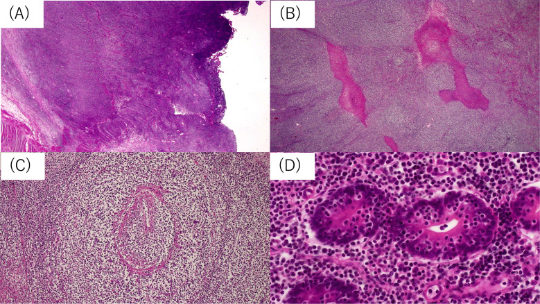
Intestinal T/NK-cell lymphomas include enteropathy-associated T-cell lymphoma (EATL), monomorphic epitheliotropic intestinal T-cell lymphoma (MEITL), indolent T-cell lymphoproliferative disorders of the GI tract (ITCLPD), extranodal NK/T-cell lymphoma, nasal type (ENKTL), and intestinal T-cell lymphoma NOS (ITCL-NOS). Here we describe a case of surface CD3-negative MEITL. A 63-year-old Japanese female had a tumor located in the conglomerated ileum, which formed multiple mass lesions. The resected tissue showed a diffuse infiltration of monomorphic medium-sized lymphocytes with epitheliotropism. Flowcytometry using a fresh specimen of the tumor revealed positivity for CD7, CD8, CD38, and CD56, but not surface CD3. On immunohistochemistry, the tumor showed positivity for cytoplasmic CD3, CD8, CD56, TIA-1, Granzyme B, and perforin. EBER with in situ hybridization was negative. Moreover, H3K36me3, which is negative in MEITL with SETD2-mutation, was positive. This is an important case of MEITL due to its oncogenesis.
Keywords: flowcytometry; intestinal T/NK-cell lymphoma; monomorphic epitheliotropic intestinal T-cell lymphoma; surface CD3.
Conflict of interest statement
CONFLICT OF INTEREST
The authors have no potential conflicts of interest to declare with respect to the research, authorship, and/or publication of this article.
Figures


References
-
- Chan JKC, Fukayama M. Haematolymphoid tumours of digestive system: Introduction. In : The WHO Classification of Tumours Editorial Board (eds) : Digestive System Tumours. Lyon (France), International Agency for Research on Cancer. 2019; pp. 376-377.
-
- Olszewska-Szopa M, Wróbel T. Gastrointestinal non-Hodgkin lymphomas. Adv Clin Exp Med. 2019; 28: 1119-1124. - PubMed
-
- Jaffe ES, Bhagat G, Chott A, et al. Intestinal T-cell lymphoma. In : The WHO Classification of Tumours Editorial Board (eds) : Digestive System Tumours. Lyon (France), International Agency for Research on Cancer. 2019; pp. 372-380.
-
- Chan JKC, Chan ACL, Cheuk W, et al. Type II enteropathy-associated T-cell lymphoma: a distinct aggressive lymphoma with frequent γδ T-cell receptor expression. Am J Surg Pathol. 2011; 35: 1557-1569. - PubMed
Publication types
MeSH terms
Substances
LinkOut - more resources
Full Text Sources
Research Materials
Miscellaneous