. 2022 Nov;55(11):1225-1240.
doi: 10.1111/iej.13814. Epub 2022 Aug 25.

MicroRNA-22 suppresses NLRP3/CASP1 inflammasome pathway-mediated proinflammatory cytokine production by targeting the HIF-1α and NLRP3 in human dental pulp fibroblasts

Affiliations

MicroRNA-22 suppresses NLRP3/CASP1 inflammasome pathway-mediated proinflammatory cytokine production by targeting the HIF-1α and NLRP3 in human dental pulp fibroblasts

Wenkai Jiang et al. Int Endod J. 2022 Nov.

Abstract

Aim: To investigate the synergetic regulatory effect of miR-22 on HIF-1α and NLRP3, subsequently regulating the production of the NLRP3/CASP1 inflammasome pathway-mediated proinflammatory cytokines IL-1β and IL-18 in human dental pulp fibroblasts (HDPFs) during the progression of pulpitis.

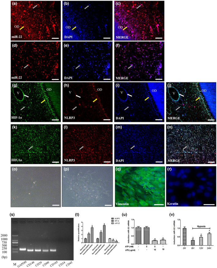
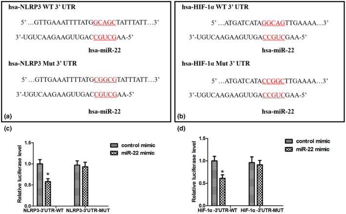
Methodology: Fluorescence in situ hybridization (FISH) and immunofluorescence (IF) were performed to determine the localization of miR-22-3p, NLRP3 and HIF-1α in human dental pulp tissues (HDPTs). The miR-22 mimics and inhibitor or plasmid of NLRP3 or HIF-1α were used to upregulate or downregulate miR-22 or NLRP3 or HIF-1α in HDPFs, respectively. Computational prediction via TargetScan 5.1 and a luciferase reporter assay were conducted to confirm target association. The mRNA and protein expression of HIF-1α, NLRP3, caspase-1, IL-1β and IL-18 were determined by qRT-PCR and western blotting, respectively. The release of IL-1β and IL-18 was analysed by ELISA. The significance of the differences between the experimental and control groups was determined by one-way analysis of variance, p < .05 indicated statistical significance.

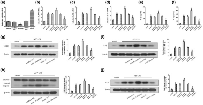
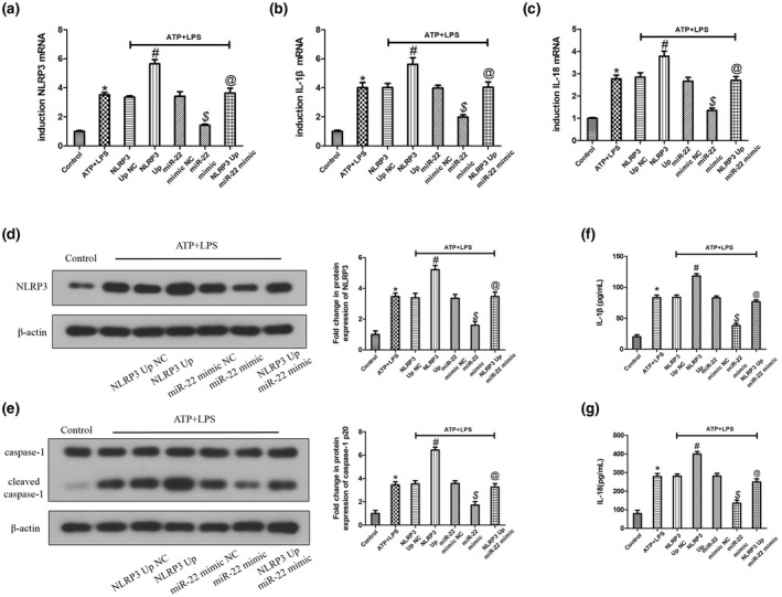
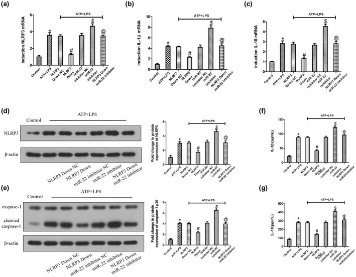
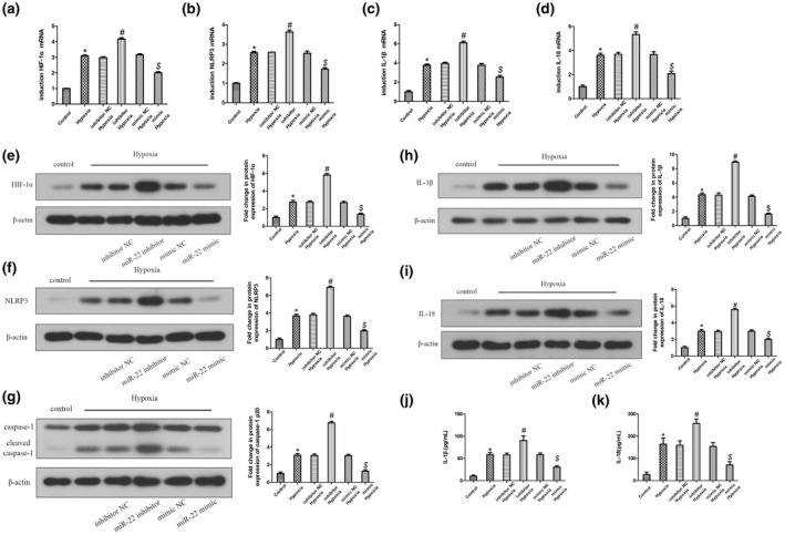
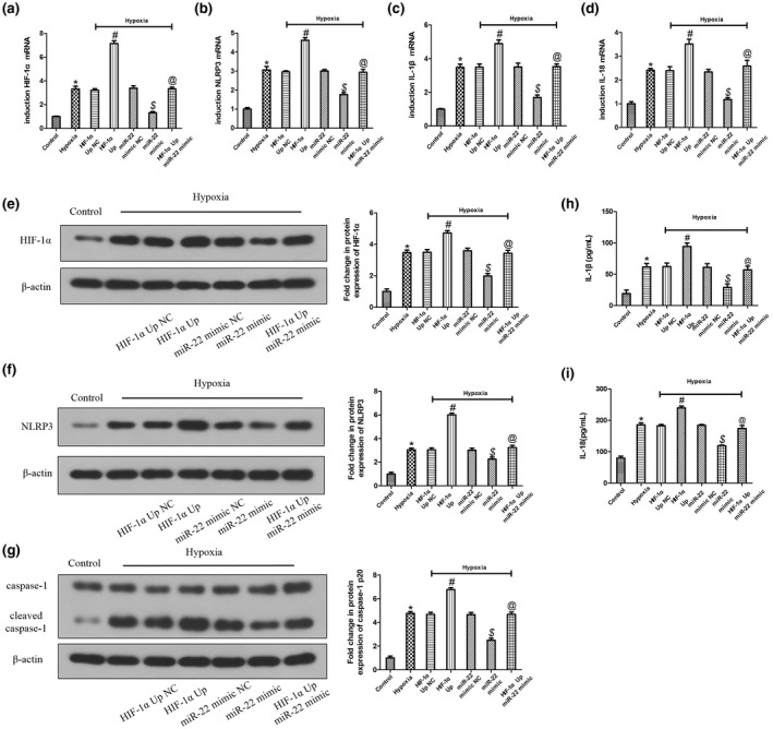
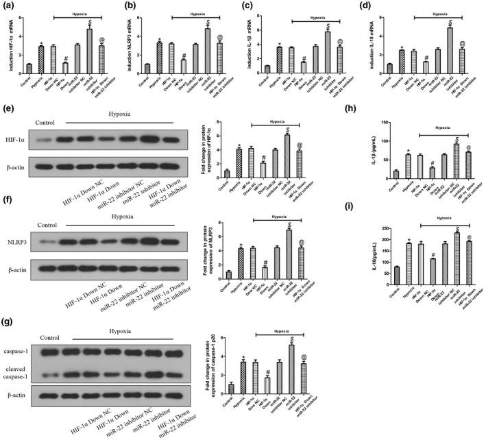
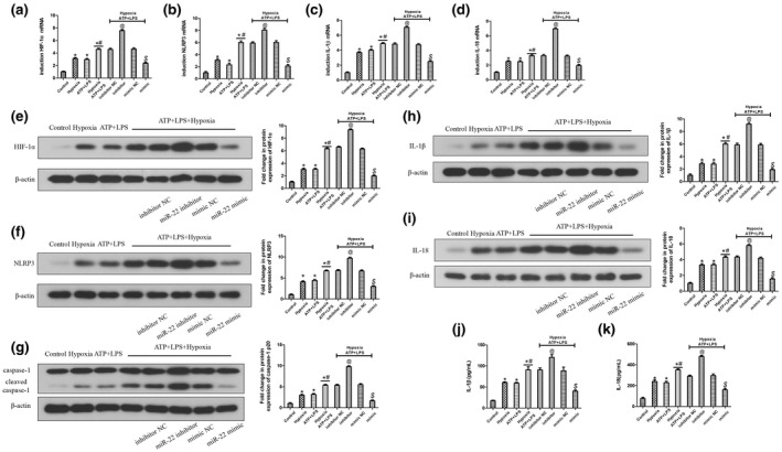
Results: A decrease in miR-22 and an increase in HIF-1α and NLRP3 in HDPTs occurred during the transformation of reversible pulpitis into irreversible pulpitis compared with that in the healthy pulp tissues (p < .05). In the normal HDPTs, miR-22-3p was extensively expressed in dental pulp cells. HIF-1α and NLRP3 were mainly expressed in the odontoblasts and vascular endothelial cells. Whereas in the inflamed HDPTs, the odontoblast layers were disrupted. HDPFs were positive for miR-22-3p, HIF-1α and NLRP3. Computational prediction via TargetScan 5.1 and luciferase reporter assays confirmed that both NLRP3 and HIF-1α were direct targets of miR-22 in HDPFs. The miR-22 inhibitor further promoted the activation of NLRP3/CASP1 inflammasome pathway induced by ATP plus LPS and hypoxia (p < .05). In contrast, the miR-22 mimic significantly inhibited the NLRP3/CASP1 inflammasome pathway activation induced by ATP plus LPS and hypoxia (p < .05).

Conclusion: MiR-22, as a synergetic negative regulator, is involved in controlling the secretion of proinflammatory cytokines mediated by the NLRP3/CASP1 inflammasome pathway by targeting NLRP3 and HIF-1α. These results provide a novel function and mechanism of miR-22-HIF-1α-NLRP3 signalling in the control of proinflammatory cytokine secretion, thus indicating a potential therapeutic strategy for future endodontic treatment.

Keywords: HIF-1α; IL-1β; NLRP3; human dental pulp fibroblasts; miR-22; pulpitis.


Conflict of interest statement

The authors declare that there is no conflict of interest regarding the publication of this paper.

Figures

FIGURE 1
FIGURE 1
PRILE 2021 flowchart (Nagendrababu et al., 2021). For further details visit: http://pride‐endodonticguidelines.org/prile
FIGURE 2
FIGURE 2
The expression of miR‐22, NLRP3 and HIF‐1α in human dental pulp tissues (HDPTs) and isolation and characterization of human dental pulp fibroblasts (HDPFs). Two groups were stained for miR‐22 (red staining) (a–f), HIF‐1α (green staining) and NLRP3 (red staining) (g–n): the normal dental pulp tissues and the irreversible pulpitis tissues. Nulei were stained with DAPI (blue staining). Odontoblast (yellow arrows), vascular endothelial cells (white arrows), fibroblasts (grey arrows). The morphological observation of primary cultured HDPFs at day 1 (o) and at day 7 (p). The characterization of HDPFs by immunocytochemical staining and RT‐PCR; positive immunostaining for vimentin (q); negative immunostaining for keratin (r); positive for the markers CD146, CD29, CD90, CD105; and negative for the markers CD34 and CD45 (s). Bar: 50 μm. The mRNA expression of miR‐22, NLRP3 and HIF‐1α was analysed by qRT‐PCR (t–v). *p < .05 compared with the normal dental pulp tissues or the control group.
FIGURE 3
FIGURE 3
miR‐22 directly targets NLRP3 and HIF‐1α. The targeting site in the 3′UTR of NLRP3 and the corresponding mutant sequence (a). The targeting site in the 3′UTR of HIF‐1α and the corresponding mutant sequence (b). Relative luciferase activity of cells after cotransfection with wild‐type (WT) or mutant (MUT) NLRP3 3′UTR luciferase reporter vector and the miR‐22 mimic or control mimic (c). Relative luciferase activity of cells after cotransfection with wild‐type (WT) or mutant (MUT) HIF‐1α 3′UTR luciferase reporter vector and the miR‐22 mimic or control mimic (d). *p < .05 when compared with the miR‐con group.
FIGURE 4
FIGURE 4
miR‐22 is a negative regulator of ATP plus LPS‐induced NLRP3/CASP1 inflammasome pathway activation in human dental pulp fibroblasts (HDPFs). The mRNA expression of miR‐22, NLRP3, IL‐1β and IL‐18 was analysed by qRT‐PCR (a–d). The protein expression of NLRP3, caspase‐1, IL‐1β, IL‐18 and β‐actin was analysed by western blotting, and the relative band intensities were determined by densitometry (g–j). The release of IL‐1β and IL‐18 was analysed by ELISA (e, f). *p < .05 compared with the control group. # p < .05 compared with the ATP plus LPS‐induced group and inhibitor NC ATP plus LPS‐induced group. $ p < .05 when compared with the ATP plus LPS‐induced group and mimic NC ATP plus LPS‐induced group.
FIGURE 5
FIGURE 5
Overexpression of miR‐22 prohibited the activation of NLRP3/CASP1 inflammasome pathway induced by ATP plus LPS, which was alleviated by upregulating NLRP3 in human dental pulp fibroblasts (HDPFs). The mRNA expression of NLRP3, IL‐1β and IL‐18 was analysed by qRT‐PCR (a–c). The protein expression of NLRP3, caspase‐1 and β‐actin was analysed by western blotting, and the relative band intensities were determined by densitometry (d, e). The release of IL‐1β and IL‐18 was analysed by ELISA (f, g). *p < .05 compared with the control group. # p < .05 compared with the ATP plus LPS‐induced group and NLRP3 Up NC group. $ p < .05 compared with the ATP plus LPS‐induced group and miR‐22 mimic NC group. @ p < .05 compared with the NLRP3 Up group and miR‐22 mimic group.
FIGURE 6
FIGURE 6
Knockdown of miR‐22 stimulated the activation of NLRP3/CASP1 inflammasome pathway induced by ATP plus LPS, which was attenuated by downregulating NLRP3 in human dental pulp fibroblasts (HDPFs). The mRNA expression of NLRP3, IL‐1β and IL‐18 was analysed by qRT‐PCR (a–c). The protein expression of NLRP3, caspase‐1 and β‐actin was analysed by western blotting, and the relative band intensities were determined by densitometry (d, e). The release of IL‐1β and IL‐18 was analysed by ELISA (f, g). *p < .05 compared with the control group. # p < .05 compared with the ATP plus LPS‐induced group and NLRP3 Down NC group. $ p < .05 compared with the ATP plus LPS‐induced group and miR‐22 inhibitor NC group. @ p < .05 compared with the NLRP3 Down group and miR‐22 inhibitor group.
FIGURE 7
FIGURE 7
miR‐22 is a negative regulator of hypoxia‐induced NLRP3/CASP1 inflammasome pathway activation in human dental pulp fibroblasts (HDPFs). The mRNA expression of HIF‐1α, NLRP3, IL‐1β and IL‐18 was analysed by qRT‐PCR (a–d). The protein expression of HIF‐1α, NLRP3, caspase‐1, IL‐1β, IL‐18 and β‐actin was analysed by western blotting, and the relative band intensities were determined by densitometry (e–i). The release of IL‐1β and IL‐18 was analysed by ELISA (j, k). *p < .05 compared with the control group. # p < .05 when compared with the hypoxia‐induced group and inhibitor NC hypoxia‐induced group. $ p < .05 compared with the hypoxia‐induced group and mimic NC hypoxia‐induced group.
FIGURE 8
FIGURE 8
Overexpression of miR‐22 prohibited the activation of NLRP3/CASP1 inflammasome pathway induced by hypoxia, which was alleviated by upregulating HIF‐1α in human dental pulp fibroblasts (HDPFs). The mRNA expression of HIF‐1α, NLRP3, IL‐1β and IL‐18 was analysed by qRT‐PCR (a–d). The protein expression of HIF‐1α, NLRP3, caspase‐1 and β‐actin was analysed by western blotting, and the relative band intensities were determined by densitometry (e–g). The release of IL‐1β and IL‐18 was analysed by ELISA (h, i). *p < .05 compared with the control group. # p < .05 compared with the hypoxia‐induced group and HIF‐1α Up NC group. $ p < .05 compared with the hypoxia‐induced group and miR‐22 mimic NC group. @ p < .05 compared with the HIF‐1α Up group and miR‐22 mimic group.
FIGURE 9
FIGURE 9
Knockdown of miR‐22 stimulated the activation of NLRP3/CASP1 inflammasome pathway induced by hypoxia, which was attenuated by downregulaing HIF‐1α in human dental pulp fibroblasts (HDPFs). The mRNA expression of HIF‐1α, NLRP3, IL‐1β and IL‐18 was analysed by qRT‐PCR (a–d). The protein expression of HIF‐1α, NLRP3, caspase‐1 and β‐actin was analysed by western blotting, and the relative band intensities were determined by densitometry (e–g). The release of IL‐1β and IL‐18 was analysed by ELISA (h, i). *p < .05 compared with the control group. # p < .05 compared with the hypoxia‐induced group and HIF‐1α Down NC group. $ p < .05 compared with the hypoxia‐induced group and miR‐22 inhibitor NC group. @ p < .05 compared with the HIF‐1α Down group and miR‐22 inhibitor group.
FIGURE 10
FIGURE 10
miR‐22 negatively regulates hypoxia and ATP plus LPS‐induced NLRP3/CASP1 inflammasome pathway activation in human dental pulp fibroblasts (HDPFs). The mRNA expression of HIF‐1α, NLRP3, IL‐1β and IL‐18 was analysed by qRT‐PCR (a–d). The protein expression of HIF‐1α, NLRP3, caspase‐1, IL‐1β, IL‐18 and β‐actin was analysed by western blotting, and the relative band intensities were determined by densitometry (e–i). The release of IL‐1β and IL‐18 was analysed by ELISA (j, k). *p < .05 compared with the control group. # p < .05 compared with the hypoxia‐induced group and ATP plus LPS‐induced group. @ p < .05 compared with the hypoxia and ATP plus LPS‐induced group and inhibitor NC hypoxia and ATP plus LPS‐induced group. $ p < .05 compared with the hypoxia and ATP plus LPS‐induced group and mimic NC hypoxia and ATP plus LPS‐induced group.

Similar articles

Cited by

References

    1. Acuna, S.M. , Floeter‐Winter, L.M. & Muxel, S.M. (2020) MicroRNAs: biological regulators in pathogen‐host interactions. Cell, 9, 113. - PMC - PubMed
    1. Chandan, K. , Gupta, M. & Sarwat, M. (2019) Role of host and pathogen‐derived MicroRNAs in immune regulation during infectious and inflammatory diseases. Frontiers in Immunology, 10, 3081. - PMC - PubMed
    1. Chang, M.C. , Lin, L.D. , Zwei‐Ching Chang, J. , Huang, C.F. , Chuang, F.H. , Lee, J.J. et al. (2012) Regulation of vascular cell adhesion molecule‐1 in dental pulp cells by interleukin‐1beta: the role of prostanoids. Journal of Endodontis, 38, 774–779. - PubMed
    1. Chang, M.C. , Tsai, Y.L. , Chang, H.H. , Lee, S.Y. , Lee, M.S. , Chang, C.W. et al. (2016) IL‐1beta‐induced MCP‐1 expression and secretion of human dental pulp cells is related to TAK1, MEK/ERK, and PI3K/Akt signaling pathways. Archives of Oral Biology, 61, 16–22. - PubMed
    1. Chen, Y. , Chen, J. , Wang, H. , Shi, J. , Wu, K. , Liu, S. et al. (2013) HCV‐induced miR‐21 contributes to evasion of host immune system by targeting MyD88 and IRAK1. PLoS Pathogens, 9, e1003248. - PMC - PubMed

MeSH terms