. 2022 Aug;12(8):e945.
doi: 10.1002/ctm2.945.

FAM135B sustains the reservoir of Tip60-ATM assembly to promote DNA damage response

Affiliations

FAM135B sustains the reservoir of Tip60-ATM assembly to promote DNA damage response

Kai Zhang et al. Clin Transl Med. 2022 Aug.

Abstract

Background: Recently, the mechanism by which cells adapt to intrinsic and extrinsic stresses has received considerable attention. Tat-interactive protein 60-kDa/ataxia-telangiectasia-mutated (TIP60/ATM) axis-mediated DNA damage response (DDR) is vital for maintaining genomic integrity.

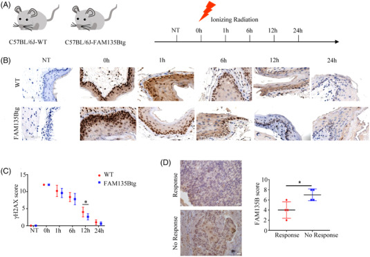
Methods: Protein levels were detected by western blot, protein colocalisation was examined by immunofluorescence (IF) and protein interactions were measured by co-immunoprecipitation, proximity ligation assay and GST pull-down assays. Flow cytometry, comet assay and IF assays were used to explore the biological functions of sequence similarity 135 family member B (FAM135B) in DDR. Xenograft tumour, FAM135B transgenic mouse models and immunohistochemistry were utilised to confirm in vitro observations.

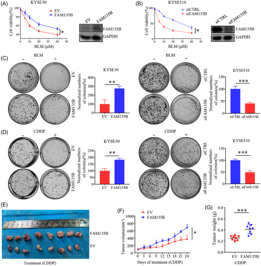
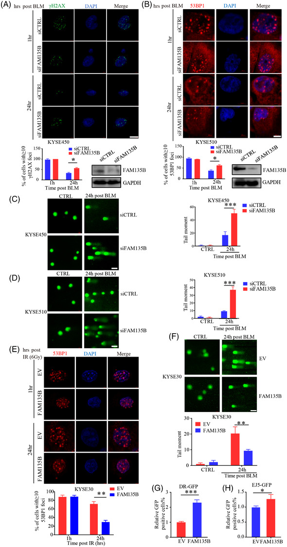
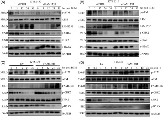
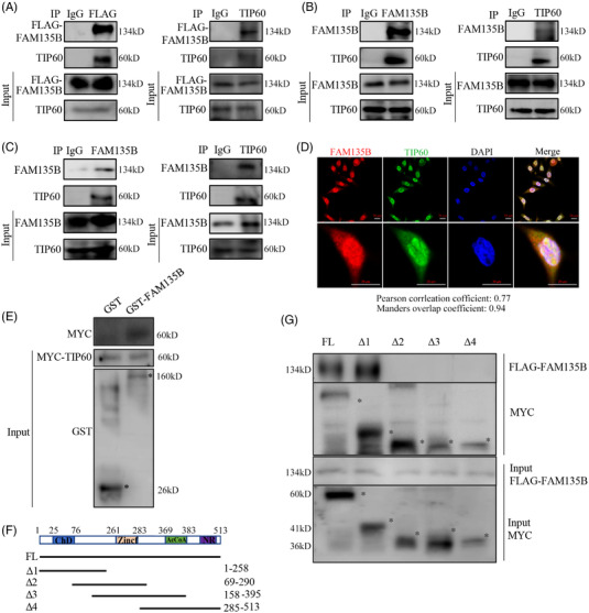
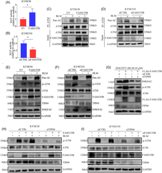
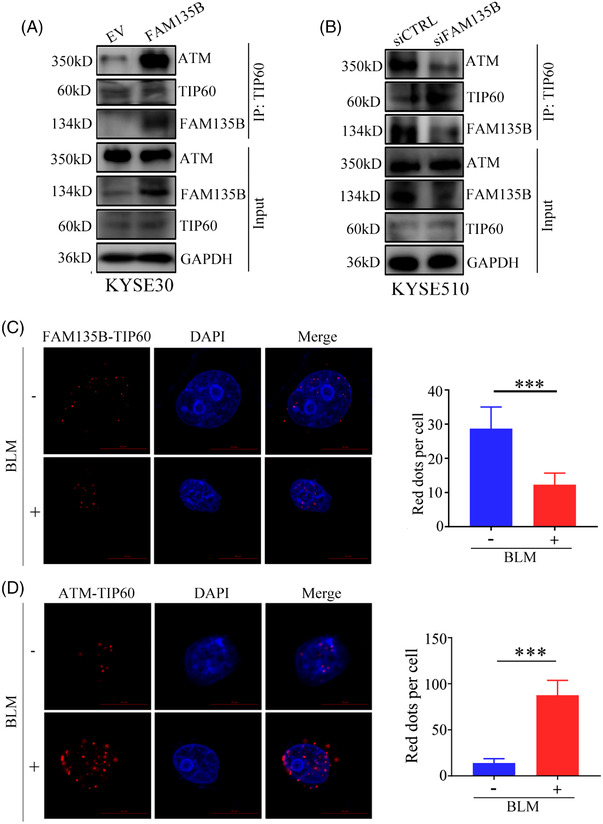
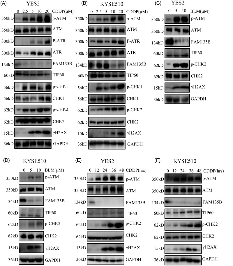
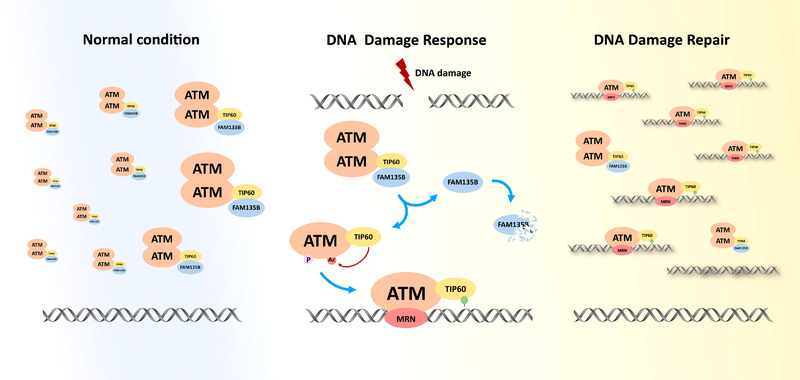
Results: We identified a novel DDR regulator FAM135B which could protect cancer cells from genotoxic stress in vitro and in vivo. The overexpression of FAM135B promoted the removal of γH2AX and 53BP1 foci, whereas the elimination of FAM135B attenuated these effects. Consistently, our findings revealed that FAM135B could promote homologous recombination and non-homologous end-joining repairs. Further study demonstrated that FAM135B physically bound to the chromodomain of TIP60 and improved its histone acetyltransferase activity. Moreover, FAM135B enhanced the interactions between TIP60 and ATM under resting conditions. Intriguingly, the protein levels of FAM135B dramatically decreased following DNA damage stress but gradually increased during the DNA repair period. Thus, we proposed a potential DDR mechanism where FAM135B sustains a reservoir of pre-existing TIP60-ATM assemblies under resting conditions. Once cancer cells suffer DNA damage, FAM135B is released from TIP60, and the functioning pre-assembled TIP60-ATM complex participates in DDR.

Conclusions: We characterised FAM135B as a novel DDR regulator and further elucidated the role of the TIP60-ATM axis in response to DNA damage, which suggests that targeting FAM135B in combination with radiation therapy or chemotherapy could be a potentially effective approach for cancer treatment.


Conflict of interest statement

The authors have declared that no competing interest exists.

Figures

FIGURE 1
FIGURE 1
FAM135B mediates tumour cells resistance to DNA damage in vitro and in vivo. (A and B) Cell growth curve examined by 3‐(4,5‐dimethylthiazol‐2‐yl)‐5‐(3‐carboxymethoxyphenyl)‐2‐(4‐sulfophenyl)‐2H‐tetrazolium (MTS) assay. KYSE30 cells (A) were transfected with FLAG‐FAM135B or an empty vector and KYSE510 cells (B) were transfected with FAM135B (siFAM135B) or control siRNA (siCTRL). After 48 h, a part of the cells was seeded in 96‐well plates and treated with different concentrations of bleomycin (BLM). Another part of the cells was used for western blotting to detect protein expression efficiency. The MTS assay was used to monitor cell proliferation rate. (C and D) Colony formation assays were performed on tumour cells transfected with an empty vector or FAM135B (in KYSE30 cells) and siFAM135B or siCTRL (in KYSE510 cells) and treated with different kinds of DNA damaging drugs, including Cisplatin (CDDP, 100 nM; C) or BLM (1 μM; D) for 24 h, then refreshed with fresh medium. After 7–10 days, cells were fixed and stained. Data are presented as mean ± SD. Images are the representative of from the collation of three independent experiments. Quantification of the number of normalised colonies was presented in a histogram. (E–G) A xenograft model was used for evaluating the effect of FAM135B on CDDP response. Xenograft tumours from KYSE30 cells transfected with an empty vector or FAM135B were treated with 5‐mg/kg CDDP when they were approximately 100 mm3. (E) The images of xenograft tumours dissected at the endpoint. (F) The xenograft tumour growth curve. (G) The xenograft tumour mass weight at the endpoint
FIGURE 2
FIGURE 2
FAM135B regulates the impact of DNA damage and accelerates DNA repair (A and B) Immunofluorescence analysis of the formation of γH2AX and 53BP1 foci. KYSE450 cells were transient transfected with FAM135B siRNA (siFAM135B) or control siRNA (siCTRL). After 48 h, a western blot was performed to evaluate knockdown efficiency. The KYSE450‐siCTRL or KYSE450‐siFAM135B (A) and KYSE510‐siCTRL or KYSE510‐siFAM135B cells (B) were treated with 10‐μM BLM for 12 h then released into fresh media without BLM to enable DNA damage response at different time points. Cells were fixed at 1 or 24 h after the removal of BLM and were immunostained with anti‐γH2AX (green) or anti‐53BP1 antibodies (red) with DAPI (blue). Top: representative images, bottom: Quantification data of γH2AX/53BP1 foci; more than 50 cells were measured. Scale bar = 10 μm. (C and D) Representative images and quantification of comet assay. KYSE450‐siCTRL or KYSE450‐siFAM135B (C) and KYSE510‐siCTRL or KYSE510‐siFAM135B cells (D) were treated with 10‐μM BLM for 12 h, then released into fresh median without BLM to enable DNA damage response for 24 h. Left: representative images, right: quantification data. Scale bar = 40 μm. (E) Immunofluorescence analysis of the formation of 53BP1 foci. KYSE30 cells were transfected with FLAG‐FAM135B or empty vectors for 48 h then subjected to 6 Gy of γ‐irradiation then permitted to DNA damage response at different time points. Top, the representative image of 53BP1 foci, scale bar = 10 μm. Bottom, the quantification data. (F) Representative images and quantification of comet assay. KYSE30 cells were transfected with FLAG‐FAM135B or an empty vector for 48 h and then were treated with 10‐μM BLM for 12 h, then released into fresh medium without BLM for permitting DNA damage response for 24 h. Top, representative images of the comet assay, right, quantification data. Scale bar = 40 μm. (G) Homologous recombination (HR) rate was monitored by pDRGFP reporter Assay KYSE30 cells co‐transfected with FLAG‐FAM135B or empty vectors with pDRGFP and ISceI‐GR‐RFP. After 72 h, cells were examined for GFP‐positive fluorescence cells by flow cytometry. Histogram showed relative GFP‐positive fluorescence compared to the control cells. (H) Non‐homologous end‐joining (NHEJ) DNA repair rate were monitored by pimEJ5GFP reporter assays. KYSE30 cells were co‐transfected with FLAG‐FAM135B or empty vectors with pimEJ5GFP and ISceI‐GR‐RFP. After 72 h, cells were examined for GFP‐positive fluorescence cells by flow cytometry. Histogram showed the relative GFP‐positive fluorescence compared to the control cells.
FIGURE 3
FIGURE 3
FAM135B affects ATM activity in DNA repair. (A and B) KYSE450 (A) and KYSE510 (B) cells were transfected with siFAM135B or siCTRL for 48 h and subsequently treated with 10‐μM BLM for 12 h, then cells were harvested at the indicated time point and subjected to western blots with indicated antibodies. (C and D) KYSE30 cells were transfected with FAM135B‐FLAG or empty vector (EV) for 48 h and subsequently treated with 10‐μM BLM for 12 h (C) or 6‐Gy IR (D), then cells were harvested at the indicated time points and subjected to western blot with indicated antibodies.
FIGURE 4
FIGURE 4
FAM135B physically interacts with TIP60. (A–C) Co‐immunoprecipitation (Co‐IP) assays identified the interaction between FAM135B and TIP60. Stably expressed FAM135B‐FLAG in KYSE30 cells was used for Co‐IP assays by anti‐FLAG (left panel) and anti‐TIP60 (right panel) antibodies (A) or anti‐FAM135B (left panel) and anti‐TIP60 (right panel) antibodies (B). Endogenous interactions between the TIP60 and FAM135B were confirmed by Co‐IP assays in KYSE510 cells using anti‐FAM135B and anti‐TIP60 antibodies (C). IgG was used as the negative control. (D) The immunofluorescence (IF) assay revealed the colocalisation of FAM135B and TIP60 in the nuclei of KYSE30‐FAM135B cells. Scale bar = 20 μm. (E) The GST pull‐down assay confirmed the interaction between FAM135B and TIP60. GST‐FAM135B fusion proteins were purified and incubated with HEK‐293T cell lysates that expressed MYC‐TIP60 and analysed by SDS–PAGE western blotting (* indicates correct band). Purified GST proteins served as negative controls. (F and G) FAM135B bound to the chromodomain of TIP60. Schematic depiction of serial truncated deletion of MYC‐TIP60 was constructed (F). HEK293T cells were co‐transfected with FAM135B‐FLAG and different MYC‐TIP60 fragments (full length [FL] and Δ1–4) for 48 h, and then cells were harvested and supplied for Co‐IP assay. * indicates correct band, aa, amino acids
FIGURE 5
FIGURE 5
FAM135B increases TIP60 histone acetyltransferase (HAT) activity and promotes TIP60 and ATM interactions. (A and B) FAM135B affects the HAT activity of TIP60. KYSE30 cells (A) were transfected with FLAG‐FAM135B or empty vector and MYC‐TIP60. KYSE510 cells (B) were transfected with FAM135B (siFAM135B) or control siRNA (siCTRL) and MYC‐TIP60. Nuclear proteins were extracted from the cells treated earlier, then the protein MYC‐Tip60 complex was purified using anti‐MYC magnetic beads, and the HAT activity of Tip60 was determined by incubating 50 g of MYC‐Tip60 complex using HAT assays. The data are presented as mean ± SD. (C) KYSE30 cells overexpressed FAM135B and related controls for 48 h and were then treated with or without 10‐μM BLM for 12 h. Cell lysates were subjected to immunoprecipitation (IP) analysis. (D) KYSE510 cells were transfected with siFAM135B and related controls for 48 h, then treated with or without 10‐μM BLM for 12 h. Cell lysates were subjected to IP analysis. Pan‐AC at an identical molecular weight as ATM indicated ATM acetylation. (E and F) KYSE30 cells with overexpressed FAM135B (E) and KYSE510 cells transfected with siFAM135B (F), and their related controls for 48 h were then treated with or without 10‐μM BLM for 12 h, then cells were harvested for western blotting. Pan‐AC at an identical molecular weight as ATM indicated ATM acetylation, and H4K9AC indicated TIP60 activity. (G) HEK‐293T cells transfected with FAM135B‐FLAG, TIP60 siRNA and their controls as indicated for 48 h were treated with 10‐μM BLM for 12 h, then cells were harvested for western blot analysis. (H) FAM135B's effects on the DNA damage response (DDR) are TIP60‐dependent, KYSE30 cells transfected with TIP60 siRNA, FLAG‐FAM135B and their controls as indicated for 48 h, then treated with 10‐μM BLM for 12 h, then cells were harvested for western blot analysis. (I) FAM135B's effects on the DDR are TIP60‐dependent, KYSE510 cells transfected with TIP60 siRNA, FAM135B siRNA and their controls for 48 h were treated with 10‐μM BLM for 12 h, then cells were harvested for western blot analysis. The GAPDH antibody was used to monitor equal loading in western blots. Data are represented as the mean ± SD from three independent experiments, *p < .05.
FIGURE 6
FIGURE 6
DNA damage can decrease FAM135B and TIP60 interaction. (A) The interaction between TIP60 and ATM after overexpression of FAM135B. KYSE30 cells overexpressed FAM135B or control vectors were subjected to co‐immunoprecipitation (Co‐IP) by anti‐TIP60 antibody and western blot was used to detect indicated proteins. (B) The interaction between TIP60 and ATM after the knock‐down of FAM135B. KYSE510 cells transfected with siFAM135B or control siRNA were subjected to Co‐IP by anti‐TIP60 antibody and western blot analysis was used to detect indicated proteins. (C) The fluorescence shows proximity ligation assay (PLA) pictures of the normal (FAM135B and TIP60) and DNA damage conditions (FAM135B and TIP60). (D) The fluorescence shows PLA pictures of the normal (TIP60 and ATM) and DNA damage conditions (TIP60 and ATM). The histograms on the right show the red spot per cell. ***p < .001
FIGURE 7
FIGURE 7
FAM135B dramatically downregulated upon genotoxic stress. (A–D), YES2 and KYSE510 were treated with different concentrations of CDDP (A and B) for 24 h and BLM (C and D) for 12 h and then subjected to western blot for detecting the indicated proteins. (E and F) YES2 (E) and KYSE510 (F) cells were treated with 10‐μM CDDP at various time points (0, 12, 24, 36, 48 h) and then subjected to western blot analysis for detecting the indicated proteins.
FIGURE 8
FIGURE 8
FAM135B facilitates DNA repair in vivo. (A) Schematic picture of mice IR assay. FAM135B transgenic (FAM135Btg) and wild‐type (WT) mice were used in this assay. In each group, one mouse was used as a negative control, five underwent 6‐Gy IR, and subsequently, mice were euthanised at the indicated time points and oesophagus tissues were taken for further immunohistochemistry (IHC) assay. (B) Representative IHC images of γpres for both FAM135Btg and WT mice, respectively. (C) Quantification of the γQuan IHC score. Five visual fields were analysed per sample. (D) Representative IHC images of FAM135B in patients with platinum‐based chemotherapy (left panel). Quantification of the FAM135B IHC score is shown in the right panel. *p < .05
FIGURE 9
FIGURE 9
Schematic diagram showing the model where FAM135B promotes the pre‐assemble of TIP60‐ATM under normal conditions and is released from TIP60 and degraded, thus enabling pre‐existing TIP60‐ATM assembly to be recruited to the DNA damage sites rapidly upon DNA damage.

Similar articles

Cited by

References

    1. Jackson SP, Bartek J. The DNA‐damage response in human biology and disease. Nature. 2009;461(7267):1071‐1078. - PMC - PubMed
    1. Yoshioka KI, Kusumoto‐Matsuo R, Matsuno Y, et al. Genomic instability and cancer risk associated with erroneous DNA repair. Int J Mol Sci. 2021;22(22):12254. - PMC - PubMed
    1. Ka NL, Na TY, Na H, et al. NR1D1 recruitment to sites of DNA damage inhibits repair and is associated with chemosensitivity of breast cancer. Cancer Res. 2017;77(9):2453‐2463. - PubMed
    1. Ciccia A, Elledge SJ. The DNA damage response: making it safe to play with knives. Mol Cell. 2010;40(2):179‐204. - PMC - PubMed
    1. Harper JW, Elledge SJ. The DNA damage response: ten years after. Mol Cell. 2007;28(5):739‐745. - PubMed

Publication types

Substances