. 2022 Dec;22(12):3111-3119.
doi: 10.1111/ajt.17174. Epub 2022 Sep 8.

Donor-derived acute myeloid leukemia in solid organ transplantation

Affiliations

Donor-derived acute myeloid leukemia in solid organ transplantation

Luigi Marchionni et al. Am J Transplant. 2022 Dec.

Abstract

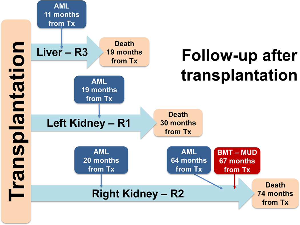
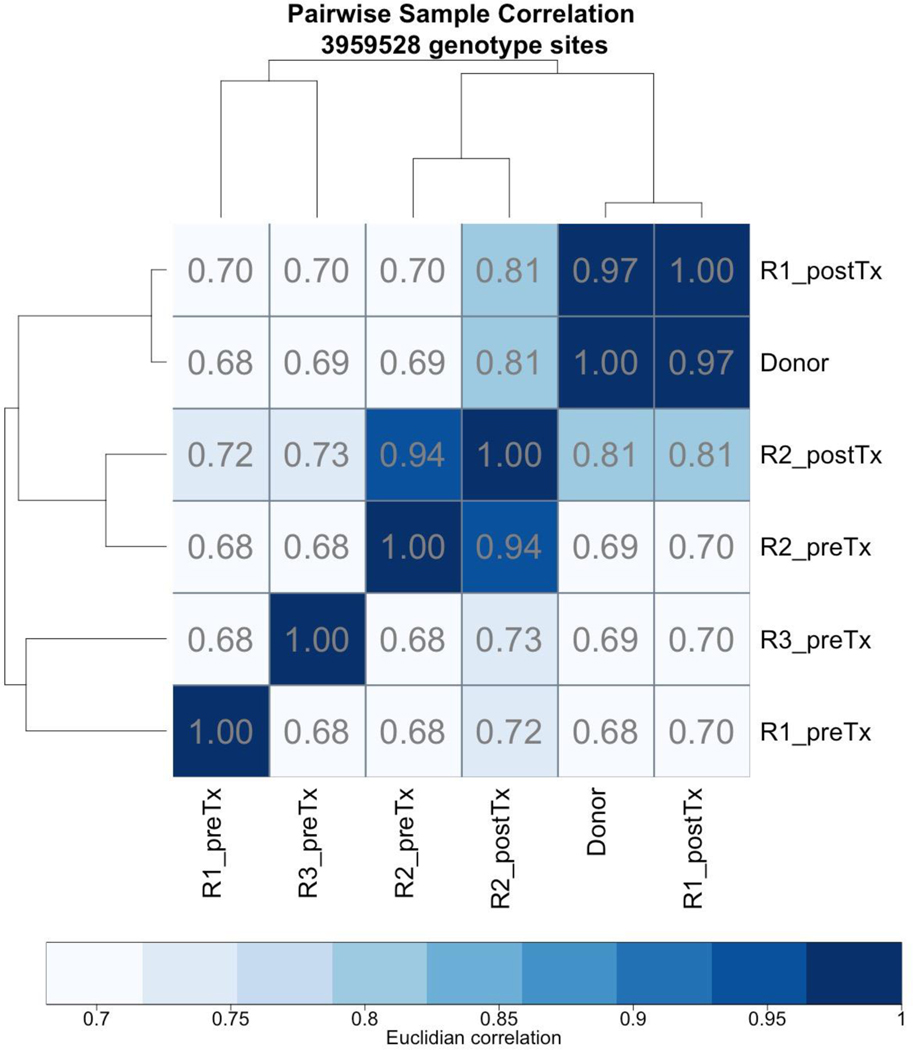
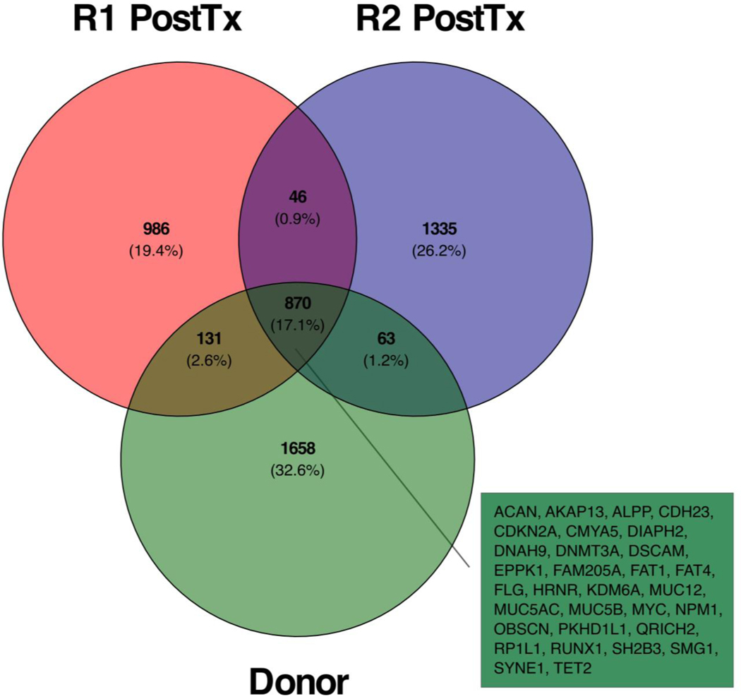
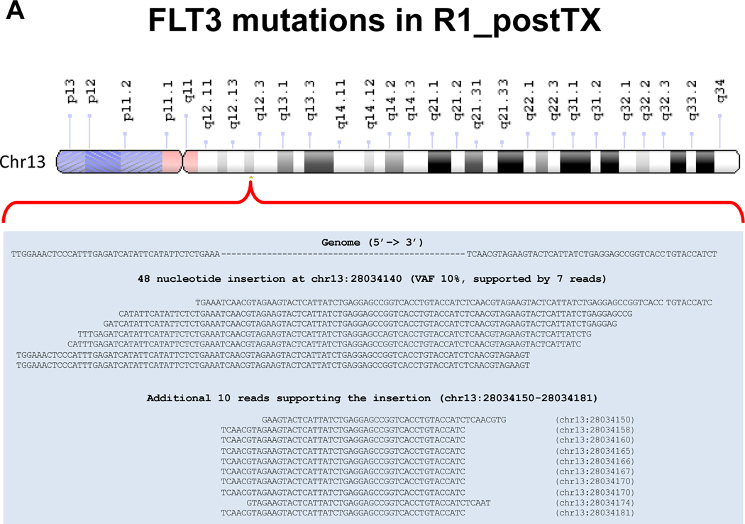
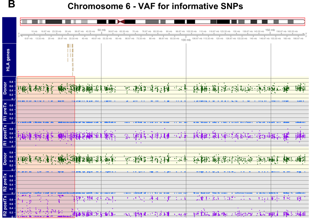
We report the transmission of acute myeloid leukemia (AML) undetected at donation from a deceased organ donor to two kidneys and one liver recipients. We reviewed the medical records, and performed molecular analyses and whole exome sequencing (WES) to ascertain AML donor origin and its molecular evolution. The liver recipient was diagnosed 11 months after transplantation and died from complications 2 months later. The two kidney recipients (R1 and R2) were diagnosed 19 and 20 months after transplantation and both received treatment for leukemia. R1 died of complications 11 months after diagnosis, while R2 went into complete remission for 44 months, before relapsing. R2 died 10 months later of complications from allogenic bone marrow transplantation. Microsatellite analysis demonstrated donor chimerism in circulating cells from both kidney recipients. Targeted molecular analyses and medical records revealed NPM1 mutation present in the donor and recipients, while FLT3 was mutated only in R1. These findings were confirmed by WES, which revealed additional founder and clonal mutations, and HLA genomic loss in R2. In conclusion, we report the first in-depth genomic analysis of AML transmission following solid organ transplantation, revealing distinct clonal evolution, and providing a potential molecular explanation for tumor escape.

Keywords: basic (laboratory) research science; complication: malignant; donors and donation; genetics; genomics; hematology/oncology; solid organ transplantation; translational research/science.


Conflict of interest statement

Disclosure

The authors declare that they do not have actual or potential conflict of interest in relation to this study.

Figures

Figure 1.
Figure 1.. Follow up timeline.
The clinical course of the three organ recipients is show. DNA for whole exome sequencing and other molecular analyses was obtained at AML diagnosis.
Figure 2.
Figure 2.. Genotype analysis in the donor and the kidney recipients.
Pair-wise correlation based on allele frequencies from ~4 million high-quality variants. Post-Tx samples are highly similar to the donor’s one (R ≅ 0.97 for R1 post-TX and R ≅ 0.81 for R2 post-TX). The correlation between samples before and after transplant reflects the amount of chimerism: while R2 post-Tx still show correlation with the sample obtained pre-TX, R1 is almost identical to the donor. Frequencies of shared reference alleles were used as input with Euclidian distance and the average clustering method.
Figure 3.
Figure 3.. Mutations detected in coding regions.
Venn diagrams showing the counts of all high-quality variants detected in coding regions with a predicted impact on the encoded protein. Common genes are highlighted in the colored box adjacent to the Venn diagram. Complete tables with gene locations and mutation descriptions are reported in the corresponding Supplementary Tables S4–S10.
Figure 4.
Figure 4.. Distinct molecular alterations associated with AML evolution in the kidney recipients.
Panel A. FLT3 mutation in R1 post-Tx. Results from the analysis of longer structural variants using Pindel. A 48 nucleotide insertion, supported by 7 reads out a total of 70, was identified at chr13:28034140 in R1 post-Tx. Additional 9 independent reads, supporting the same insertion, were identified in the vicinity (chr13:28034150–28034181). Panel B. Copy number loss of chromosome 6 in R2 post-Tx. The figure shows alternative allele frequency (VAF) for the donor and the two kidney recipients, before and after transplantation, for the alleles informative of chimerism and chromosomal loss. To this end, we first identified common SNPs that were homozygous (AA, BB) in a recipient before transplantation and heterozygous in the donor (AB), and then analyzed their frequency after transplantation. Since post-transplantation the samples are mixtures of recipient and donor DNA, the VAF in post-Tx samples is expected to be 0 or 1, if donor DNA is absent, and expected to approach 0.5, if donor DNA is present. Departures from this behavior are suggestive of chimerism and LOH. In the figure, informative VAFs (y-axis) are shown along genomic locations (x-axis) on chromosome 6, using different colors: green for the donor, blue for samples before transplantation, and purple for samples after transplantation. In the case of R1 post-TX, the VAF is centered around 0.5, confirming that virtually all DNA is derived from the donor. In the case of R2 post-TX, the informative VAF is either closer to 0 or 1 (centered around 0.15 or 0.85), suggesting that this sample is a mixture of donor and recipient DNA. Notably, for a large portion of the short arm of chromosome 6 (highlighted in the figure), the VAF distributions are suggestive of a sub-clonal copy number loss and chromothripsis. This region spans the HLA I and II loci.

Comment in

  • Donor-derived myeloid leukemia.
    Stratta RJ. Stratta RJ. Am J Transplant. 2023 Jul;23(7):1080-1081. doi: 10.1016/j.ajt.2023.03.007. Epub 2023 Mar 17. Am J Transplant. 2023. PMID: 36933832 No abstract available.

References

    1. Myron Kauffman H, McBride MA, Cherikh WS, Spain PC, Marks WH, Roza AM. Transplant tumor registry: donor related malignancies. Transplantation. 2002;74(3):358–362. doi: 10.1097/00007890-200208150-00011. - DOI - PubMed
    1. Desai R, Collett D, Watson CJ, Johnson P, Evans T, Neuberger J. Cancer Transmission From Organ Donors—Unavoidable But Low Risk. Transplantation. 2012;94(12):1200–1207. doi: 10.1097/TP.0b013e318272df41. - DOI - PubMed
    1. Ison MG, Nalesnik MA. An update on donor-derived disease transmission in organ transplantation. Am J Transplant. 2011;11(6):1123–1130. doi: 10.1111/j.1600-6143.2011.03493.x. - DOI - PubMed
    1. National Transplantation Center – Italian Ministry of Health. Linee Guida Per La Valutazione Di Idoneità Del Donatore E Protocolli Specifici. 2005:1–77. http://www.trapianti.salute.gov.it/imgs/C_17_normativa_24_allegato.pdf.
    1. Falini B, Brunetti L, Sportoletti P, Martelli MP. NPM1-mutated acute myeloid leukemia: from bench to bedside. Blood. 2020;136(15):1707–1721. doi: 10.1182/blood.2019004226. - DOI - PubMed

Publication types