Review
. 2022 Oct;311(1):205-223.
doi: 10.1111/imr.13125. Epub 2022 Aug 18.

At the crux of maternal immune activation: Viruses, microglia, microbes, and IL-17A

Affiliations
Review

At the crux of maternal immune activation: Viruses, microglia, microbes, and IL-17A

Ashley M Otero et al. Immunol Rev. 2022 Oct.

Abstract

Inflammation during prenatal development can be detrimental to neurodevelopmental processes, increasing the risk of neuropsychiatric disorders. Prenatal exposure to maternal viral infection during pregnancy is a leading environmental risk factor for manifestation of these disorders. Preclinical animal models of maternal immune activation (MIA), established to investigate this link, have revealed common immune and microbial signaling pathways that link mother and fetus and set the tone for prenatal neurodevelopment. In particular, maternal intestinal T helper 17 cells, educated by endogenous microbes, appear to be key drivers of effector IL-17A signals capable of reaching the fetal brain and causing neuropathologies. Fetal microglial cells are particularly sensitive to maternally derived inflammatory and microbial signals, and they shift their functional phenotype in response to MIA. Resulting cortical malformations and miswired interneuron circuits cause aberrant offspring behaviors that recapitulate core symptoms of human neurodevelopmental disorders. Still, the popular use of "sterile" immunostimulants to initiate MIA has limited translation to the clinic, as these stimulants fail to capture biologically relevant innate and adaptive inflammatory sequelae induced by live pathogen infection. Thus, there is a need for more translatable MIA models, with a focus on relevant pathogens like seasonal influenza viruses.

Keywords: TH17 cells; influenza virus; maternal immune activation; microbial signaling; microglia; neurodevelopment.


Conflict of interest statement

The authors declare no conflicts of interest.

Figures
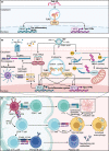
FIGURE 1
FIGURE 1
Influenza A virus initiates a more complex immune cascade than poly I:C. (A) Poly I:C activates the innate immune system by binding to cells that express endosomal toll‐like receptor 3 (TLR3). Recognition of poly I:C activates TIR‐domain‐containing adaptor‐inducing interferon‐β (TRIF), resulting in activation of interferon regulatory factor 3 (IRF3) and nuclear factor kappa B (NFκB). These transcriptional regulators mediate production of type I antiviral interferons (IFNs) and pro‐inflammatory cytokines. (B) Influenza A virus (IAV) initiates a more complex innate response in infected respiratory epithelial cells. IAV enters cells by binding of hemagglutinin (HA) glycoprotein to sialic acid residues. ssRNA from the virus itself, along with unidentified dsRNA from dying cells, activate TLR7 and TLR3, respectively. TLR7 signals through myeloid differentiation primary response 88 (Myd88), where it further activates IRF7 and NFκB. Unmodified 5′ triphosphate ends of ssRNA are recognized by retinoic acid‐inducible gene 1 (RIG‐I). Binding of ssRNA to RIG‐1 exposes the caspase activating and recruitment domain (CARD) to recruit polyubiquitinated (Ub) chains. Polyubiquitinated CARDs bind mitochondrial antiviral‐signaling proteins (MAVS), which ultimately activate IRF3 and NFκB pathways. NFκB produces pro‐IL‐18 and pro‐IL‐1β, which prime the nod‐like receptor family pyrin domain containing 3 (NLRP3) inflammasome. A second signal, such as ion flux, allows NLRP3 to form oligomers with adaptor protein ASC. ASC interacts with pro‐caspase 1 through CARD for cleavage of pro‐caspase 1 into active caspase 1. Cleaved caspase 1 then cleaves pro‐IL‐18 and pro‐IL‐1β into active IL‐18 and IL‐1β. Binding of viral Z‐RNA to Z‐DNA binding protein 1 (ZPB1) activates programmed cell death pathways and NFκB via receptor‐interacting protein kinases (RIPK) 1 and 3. The infected cell alerts surrounding immune cells to prime the (C) adaptive immune response. Respiratory dendritic cells (DCs) migrate to draining lymph nodes to present viral peptides via major histocompatibility complex (MHC) class I to naive CD8+ T cells, which differentiate into effector cytotoxic T cells (CTLs) that kill IAV‐infected cells. DCs also present viral peptides via MHC class II to naive CD4+ T cells. IFN‐γ and IL‐12 promote differentiation of T helper (TH) 1 cells. These effector TH1 cells produce IFN‐γ, which activates alveolar macrophages for direct lyses of virus. CD4+ T follicular helper (TFH) cells or TH1 cells activate primed B cells through antigen recognition and binding of CD40L/CD40. B cells differentiate into plasma cells that produce antibodies against HA glycoprotein. dsRNA, double‐stranded RNA; NA, neuraminidase; ssRNA, single‐stranded RNA; TCR, T‐cell receptor
FIGURE 2
FIGURE 2
Intestinal TH17 cells in poly I:C and influenza models. (A) RORγt+ TH17 cells are constitutively expressed in the intestine and rely on commensal segmented filamentous bacteria (SFB) for homeostatic regulation. Upon intraperitoneal administration of poly I:C, intestinal dendritic cells (DC) detect poly I:C directly through TLR3, initiating production of IL‐1β, IL‐6, and IL‐23. These secreted cytokines stimulate pre‐existing resident TH17 cells to produce IL‐17A. This innate immune signaling pattern is initiated quickly, within 4‐8 h, resulting in a measurable accumulation of IL‐17A in maternal circulation at 48 h. (B) During respiratory influenza A virus (IAV) infection, CCR9+CD4+ TH1 cells—stimulated in the lungs—are recruited to the uninfected intestine by chemokine CCL25, where they produce copious amounts of IFN‐γ. Subsequent disruption of endogenous gut microbes (ie, dysbiosis) stimulates intestinal epithelial cells to produce IL‐15. In conjunction with stimulation by intestinal antigen presenting cells (APC), cytokines IL‐15, IL‐6, IL‐23, and IL‐1β prompt naive CD4+ T cells to express transcription factor RORγt, promoting polarization towards TH17 lineage. Polarized effector TH17 cells then produce IL‐17A, which has been linked to intestinal injury during IAV infection. This adaptive immune response takes up to 5 d, resulting in measurable increases in intestinal TH17 cells within 6‐7 d after IAV infection. d, days; h, hours; RORγt, retinoic acid receptor‐related orphan receptor γt; TLR, toll‐like receptor. Images were adapted from Cua and Tato (2010), and the description of the common mucosal immune response to IAV was informed by Wang et al (2014).
FIGURE 3
FIGURE 3
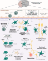
Embryonic microglia as central players in maternal immune activation neuropathologies. Maternally derived immune and microbial signals direct neurodevelopmental trajectories during embryonic development. These signals are believed to cross the immature blood‐brain barrier and enter the brain parenchyma. Once there, they are thought to bind receptors on various CNS cell types, including microglia. A multitude of studies have revealed microglial‐directed alterations to various neurodevelopmental processes—including neural progenitor cell (NPC) proliferation and differentiation, radial glia scaffold extension, dopaminergic axonal outgrowth, and wiring of inhibitory interneuron circuits—resulting from abnormal microglial behaviors. Gestational insult leads to increased production of chemokines CCL3 and CCL4 by a subpopulation of microglia adjacent to NPCs, altering the proliferative capacity and maturation fate of these cells. Additionally, microglial upregulation of phagocytic markers CD68 and MHCII in response to maternally derived factors, including IL‐17A, may result in exaggerated engulfment of NPCs. Gestational insult may also lead to a more activated microglial phenotype through upregulation of CD45 and downregulation of purinergic receptor P2RY12. Neuroprotective TAM receptor signaling on microglia allows them to sense disruptions in projecting radial glia, resulting in increased degeneration and phagocytosis of radial glia cells following insult. MIA has also been shown to reduce microglial proliferation, measured in part by reduced Ki‐67 expression. This is driven by increased type I interferon (IFN) production originating in the yolk sac, which binds interferon alpha receptor 1 (IFNAR1) on microglia. Impaired chemotactic ability of microglia following MIA may also be related to IL‐17A‐specific inhibition of microglial migratory capacity, which alters their localization in the developing brain. IL‐17A has also been shown to direct microglial expression of G Protein‐Coupled Receptor 56 (GPR56), which, in conjunction with increased TNFα, appears to impair parvalbumin‐positive (PV+) interneuron generation. This impairment ultimately leads to miswiring of inhibitory interneuron circuits, triggering hyperinhibition in the neocortex. MIA‐induced inflammation in the embryonic brain is also propagated by increased production of CCL2 in cerebral spinal fluid (CSF), prompting macrophage recruitment and entry into the choroid plexus (ChP). Figure and legend creation was informed by data cited in the main text. TAM, family of receptor tyrosine kinases TYRO, AXL, and MERTK

Similar articles

Cited by

References

    1. Mednick SA. Adult schizophrenia following prenatal exposure to an influenza epidemic. Arch Gen Psychiatry. 1988;45(2):189. doi:10.1001/archpsyc.1988.01800260109013 - DOI - PubMed
    1. Al‐Haddad BJS, Jacobsson B, Chabra S, et al. Long‐term risk of neuropsychiatric disease after exposure to infection in utero. JAMA Psychiatry. 2019;76(6):594. doi:10.1001/jamapsychiatry.2019.0029 - DOI - PMC - PubMed
    1. Brown AS, Begg MD, Gravenstein S, et al. Serologic evidence of prenatal influenza in the Etiology of schizophrenia. Arch Gen Psychiatry. 2004;61(8):774. doi:10.1001/archpsyc.61.8.774 - DOI - PubMed
    1. Brown AS, Derkits EJ. Prenatal infection and schizophrenia: a review of epidemiologic and translational studies. Am J Psychiatry. 2010;167(3):261‐280. doi:10.1176/appi.ajp.2009.09030361 - DOI - PMC - PubMed
    1. Miller BJ, Culpepper N, Rapaport MH, Buckley P. Prenatal inflammation and neurodevelopment in schizophrenia: a review of human studies. Prog Neuro‐Psychopharmacol Biol Psychiatry. 2013;42:92‐100. doi:10.1016/j.pnpbp.2012.03.010 - DOI - PubMed