Review
. 2022 Aug;34(8):e13169.
doi: 10.1111/jne.13169. Epub 2022 Aug 18.

Aggressive corticotroph tumors and carcinomas

Affiliations
Review

Aggressive corticotroph tumors and carcinomas

Hélène Lasolle et al. J Neuroendocrinol. 2022 Aug.

Abstract

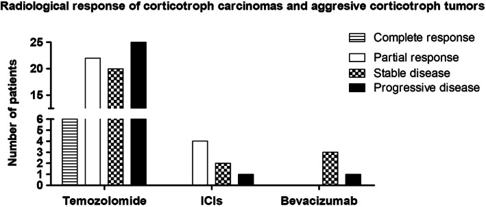
Pituitary tumors are generally benign, although in rare cases aggressive pituitary tumors (APTs) and carcinomas present important diagnostic and therapeutic challenges and are associated with a high mortality rate. Almost half of these APTs and carcinomas are corticotroph tumors, suggesting a specific prognosis. Clinical, pathological and molecular prognostic markers are limited and do not allow early management of these tumors. Temozolomide remains the first-line treatment once a diagnosis of aggressive pituitary tumor or carcinoma has been made. Novel alternative treatments exist, including immune checkpoint inhibitors, which can be used in the case of temozolomide treatment failure. The aim of this review is to present the clinical, pathological and molecular characteristics of aggressive corticotroph tumors and carcinomas, and to describe the results obtained with currently available treatments.

Keywords: Cushing's disease; aggressive pituitary tumor; corticotroph tumor; pituitary carcinoma; temozolomide.


Conflict of interest statement

The authors have no conflicts of interest to declare.

Figures

FIGURE 1
FIGURE 1
Histopathology of corticotroph carcinomas. (A) Corticotroph carcinomas are composed of basophilic cells. Nuclear atypia may be found but is not a reliable marker of malignancy. Elevated mitotic activity is a common finding but is not observed in all cases (circle: mitosis) (hematoxylin, phloxine, saffron staining, original magnification [OM] × 400). (B) Expression of ACTH in corticotroph carcinomas is variable and may be heterogeneous (ACTH immunohistochemistry, OM × 200). (C) Corticotroph carcinomas express the T‐PIT transcription factor as do all other tumors of the corticotroph lineage (T‐PIT immunohistochemistry, OM × 200). (D) In corticotroph carcinomas, Ki67 proliferation index is usually high, exceeding 3% (in this example, around 15%) (Ki67 immunohistochemistry, OM × 100). (E) Strong nuclear immunopositivity for P53 is suggestive of an aggressive behavior and is in some cases related to TP53 gene mutations (P53 immunohistochemistry, OM × 200). (F) In this example, MGMT nuclear expression is lost in most neoplastic cells (internal positive control: endothelial cells). For some authors, this may be predictive of sensitivity to temozolomide treatment (MGMT immunohistochemistry, OM × 200).
FIGURE 2
FIGURE 2
Histotypes of corticotroph aggressive tumors. (A) Silent corticotroph tumors are typically composed of regular cells with chromophobic or slightly basophilic cytoplasm (hematoxylin, phloxine, saffron [HPS] staining, original magnification [OM] × 200). (B) Silent corticotroph tumors express the T‐PIT transcription factor as do all other tumors of the corticotroph lineage (T‐PIT immunohistochemistry, OM × 100). (C) Some silent corticotroph tumors show immunoreactivity for GATA3, a transcription factor also expressed in gonadotroph tumors (GATA3 immunohistochemistry, OM × 100). (D) Crooke cell adenoma is composed of neoplastic cells harboring the ring‐like hyaline change typical of normal corticotroph cells of patients with hypercortisolism (HPS staining, OM × 200). (E) The ring‐like hyaline material corresponds to an accumulation of low‐molecular‐weight cytokeratins (cytokeratin 18 immunohistochemistry, OM × 200). (F) Expression of ACTH in Crooke cell adenomas is typically restricted to the paranuclear and peripheral regions of the cytoplasm (ACTH immunohistochemistry, OM × 200).
FIGURE 3
FIGURE 3
SSTR5 expression in aggressive corticotroph tumors and carcinomas (SSTR5 immunohistochemistry, Original magnification × 200). (A) Strong SSTR5 immunopositivity in a corticotroph carcinoma with Cushing's disease. (B) Absence of SSTR5 immunoexpression in a T‐PIT+/GATA3+ silent corticotroph tumor.
FIGURE 4
FIGURE 4
Radiological response of corticotroph carcinomas and aggressive corticotroph tumors treated with temozolomide, immune checkpoint inhibitors (ICIs), and bevacizumab. Derived from data from, , , , , , , ,

Similar articles

Cited by

References

    1. Pivonello R, De Leo M, Cozzolino A, Colao A. The treatment of Cushing's disease. Endocr Rev. 2015;36(4):385‐486. doi:10.1210/er.2013-1048 - DOI - PMC - PubMed
    1. Mete O, Lopes MB. Overview of the 2017 WHO classification of pituitary tumors. Endocr Pathol. 2017;28(3):228‐243. doi:10.1007/s12022-017-9498-z - DOI - PubMed
    1. Raverot G, Burman P, McCormack A, et al. European Society of Endocrinology Clinical Practice Guidelines for the management of aggressive pituitary tumours and carcinomas. Eur J Endocrinol. 2018;178(1):G1‐G24. doi:10.1530/EJE-17-0796 - DOI - PubMed
    1. Dekkers OM, Karavitaki N, Pereira AM. The epidemiology of aggressive pituitary tumors (and its challenges). Rev Endocr Metab Disord. 2020;21:209‐212. doi:10.1007/s11154-020-09556-7 - DOI - PMC - PubMed
    1. McCormack A, Dekkers OM, Petersenn S, et al. Treatment of aggressive pituitary tumours and carcinomas: results of a European Society of Endocrinology (ESE) survey 2016. Eur J Endocrinol. 2018;178(3):265‐276. doi:10.1530/EJE-17-0933 - DOI - PubMed