

[Effect of modified titanium loaded with endothelial progenitor cells-exosomes on osteogenic and angiogenic differentiations of adipose-derived stem cells]
- PMID: 35979798
- PMCID: PMC9379449
- DOI: 10.7507/1002-1892.202203132
[Effect of modified titanium loaded with endothelial progenitor cells-exosomes on osteogenic and angiogenic differentiations of adipose-derived stem cells]
Abstract
Objective: To investigate the effects of titanium modified by ultrasonic acid etching/anodic oxidation (UAT) loaded with endothelial progenitor cells-exosome (EPCs-exo) on proliferation and osteogenic and angiogenic differentiations of adipose-derived stem cells (ADSCs).
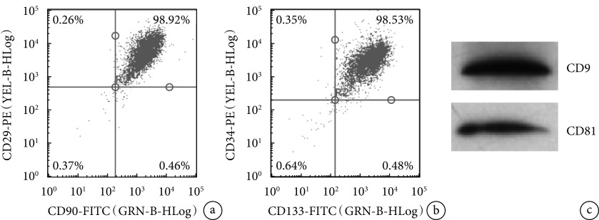
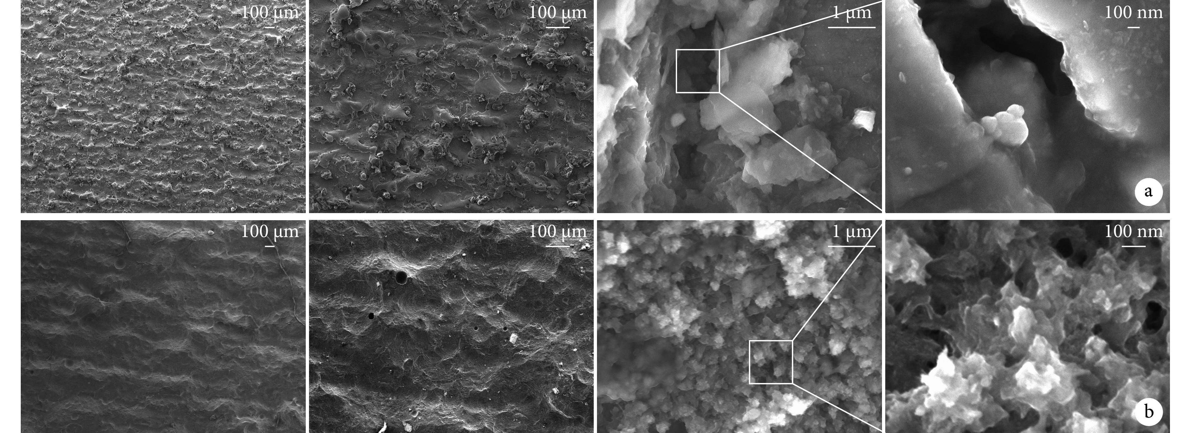
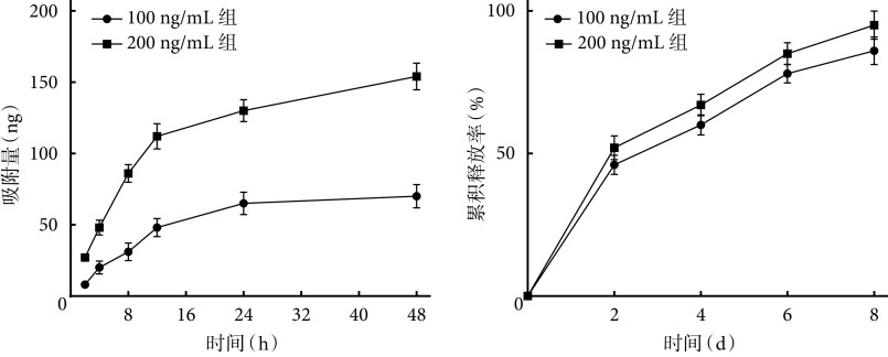
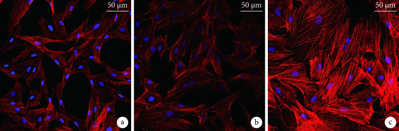
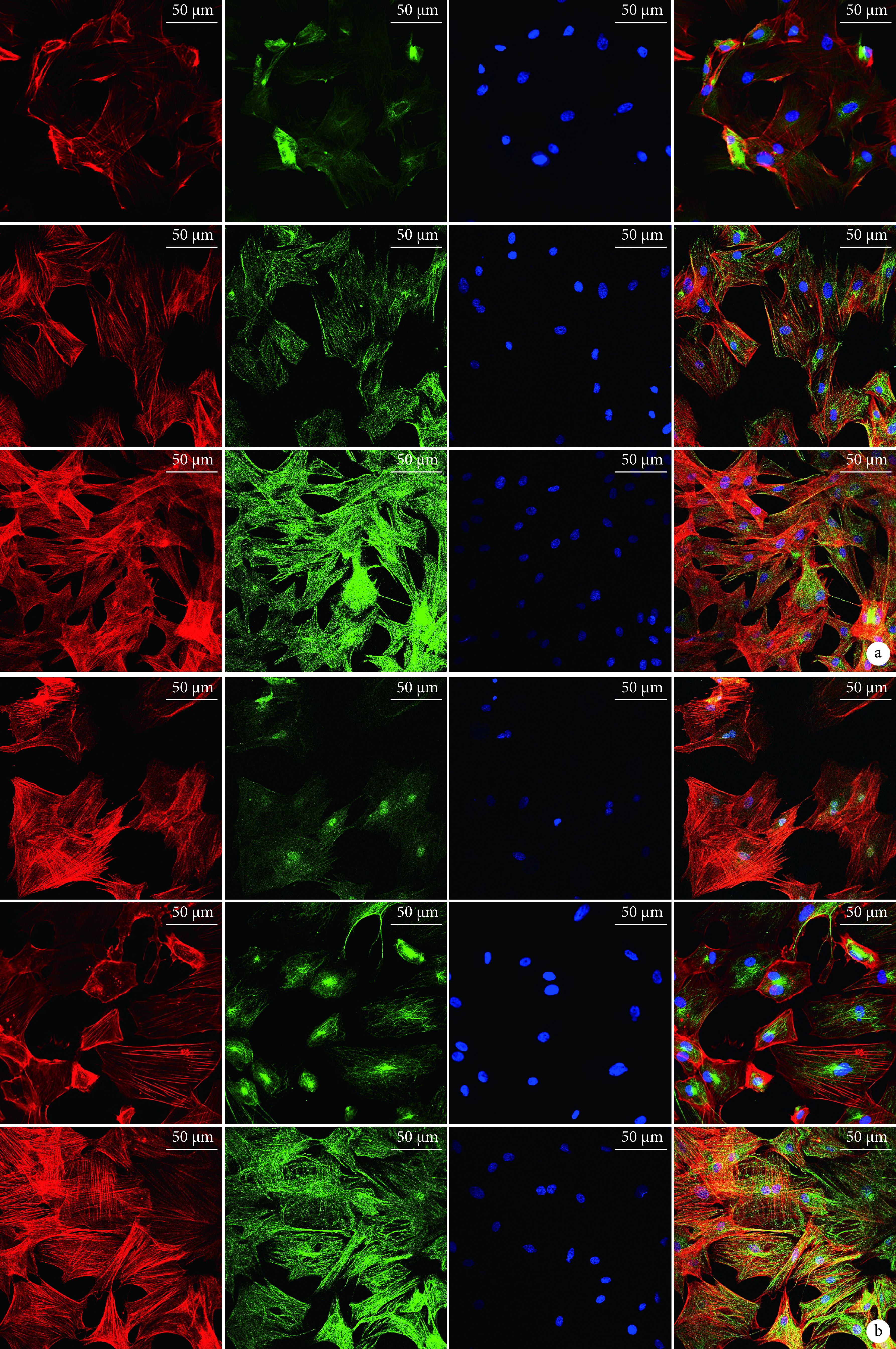
Methods: The adipose tissue and bone marrow of 10 Sprague Dawley rats were harvested. Then the ADSCs and EPCs were isolated and cultured by collagenase digestion method and density gradient centrifugation method, respectively, and identified by flow cytometry. Exo was extracted from the 3rd to 5th generation EPCs using extraction kit, and CD9 and CD81 were detected by Western blot for identification. The three-dimensional printed titanium was modified by ultrasonic acid etching and anodic oxidation to prepare the UAT. The surface characteristics of UAT before and after modification was observed by scanning electron microscopy; UAT was placed in EPCs-exo solutions of different concentrations (100, 200 ng/mL), and the in vitro absorption and release capacity of EPCs-exo was detected by BCA method. Then, UAT was placed in DMEM medium containing different concentrations of EPCs-exo (0, 100, 200 ng/mL), and co-cultured with the 3rd generation ADSCs to construct UAT-ADSCs-exo. Cell morphology by laser confocal microscopy, live/dead cell staining, and cell proliferation were observed to evaluate biocompatibility; alkaline phosphatase (ALP) staining and alizarin red staining, RT-PCR detection of osteogenesis-related genes [osteocalcin (OCN), RUNT-related transcription factor 2 (Runx2), ALP, collagen type 1 (COL-1)] and angiogenesis-related gene [vascular endothelial growth factor (VEGF)], immunofluorescence staining for osteogenesis (OCN)- and angiogenesis (VEGF)-related protein expression were detected to evaluate the effect on the osteogenic and angiogenic differentiation ability of ADSCs.
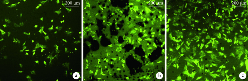
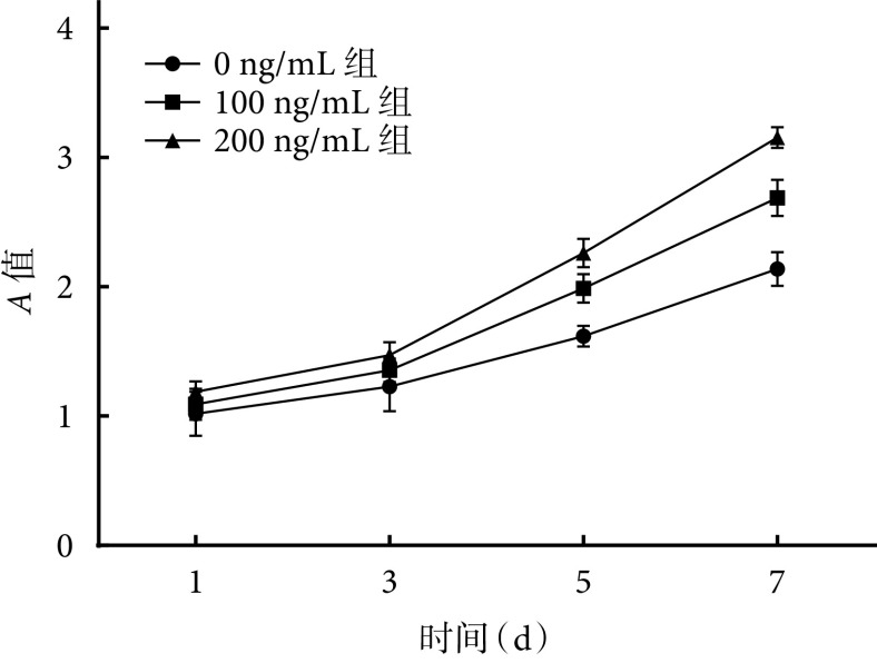
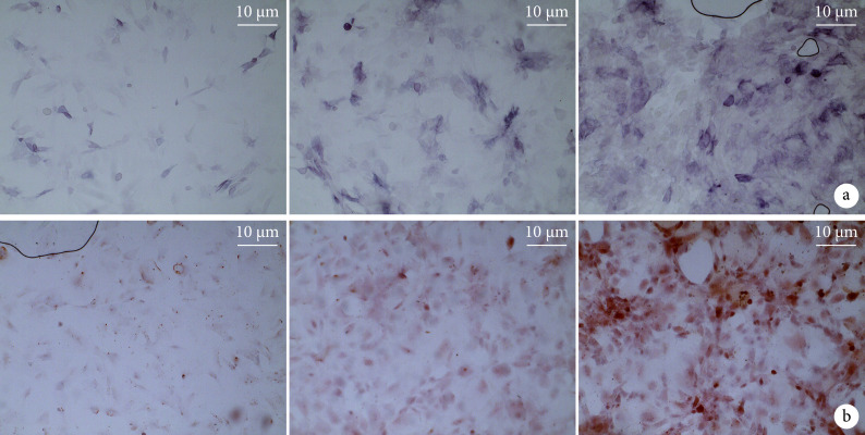
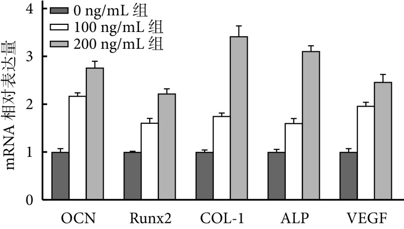
Results: Scanning electron microscopy showed that micro-nano multilevel composite structures were formed on the surface of UAT. About 77% EPCs-exo was absorbed by UAT within 48 hours, while EPCs-exo absorbed on the surface of UAT showed continuous and stable release within 8 days. The absorption and release amount of 200 ng/mL group were significantly higher than those of 100 ng/mL group ( P<0.05). Biocompatibility test showed that the cells in all concentration groups grew well after culture, and the 200 ng/mL group was better than the other groups, with fully spread cells and abundant pseudopodia, and the cell count and cell activity were significantly higher than those in the other groups ( P<0.05). Compared with the other groups, 200 ng/mL group showed enhanced ALP activity and mineralization ability, increased expressions of osteogenic and angiogenic genes (OCN, Runx2, COL-1, ALP, and VEGF), as well as increased expressions of OCN and VEGF proteins, with significant differences ( P<0.05).
Conclusion: EPCs-exo can effectively promote the adhesion, proliferation, and osteogenic and angiogenic differentiation of ADSCs on UAT surface, the effect is the most significant when the concentration is 200 ng/mL.
目的: 探讨经超声酸蚀/阳极氧化联合改性钛(titanium modified by ultrasonic acid etching/anodic oxidation,UAT)负载内皮祖细胞外泌体(endothelial progenitor cells-exosome,EPCs-exo)后,对脂肪来源干细胞(adipose-derived stem cells,ADSCs)生长以及成骨、成血管分化的影响。.
方法: 取10只SD大鼠脂肪组织及骨髓,分别采用胶原酶消化法和密度梯度离心法分离培养ADSCs及EPCs,并经流式细胞仪鉴定。取第3~5代EPCs采用试剂盒提取exo,并经Western blot检测CD9、CD81进行鉴定。采用超声酸蚀和阳极氧化方法对3D打印技术制备的钛片进行联合改性制备UAT,扫描电镜观察改性前后材料表面形貌;将UAT分别置于不同浓度(100、200 ng/mL)EPCs-exo溶液中,采用BCA法检测其体外吸附及释放EPCs-exo能力。然后,将UAT置于含不同浓度EPCs-exo(0、100、200 ng/mL)的DMEM培养液,并与第3代ADSCs复合培养,构建UAT-ADSCs-exo。通过激光共聚焦显微镜观察细胞形态、活/死细胞染色、细胞增殖检测,评价生物相容性;ALP染色及茜素红染色,RT-PCR检测成骨相关基因骨钙素(osteocalcin,OCN)、RUNT相关基因2(RUNT-related transcription factor 2,Runx2)、ALP、Ⅰ型胶原蛋白(collagen type 1,COL-1)和成血管相关基因VEGF,免疫荧光染色观测成骨(OCN)及成血管(VEGF)相关蛋白表达,评价负载EPCs-exo的UAT对ADSCs成骨、成血管分化能力的影响。.
结果: 扫描电镜观察示改性后UAT表面形成了微纳米多级复合结构;UAT 48 h内能吸附约77% EPCs-exo,而吸附在UAT表面的EPCs-exo在8 d内呈连续稳定释放,200 ng/mL组吸附及释放量明显优于100 ng/mL组,差异均有统计学意义( P<0.05)。生物相容性检测示,复合培养后各组细胞均生长良好,其中200 ng/mL组优于其他组,细胞完全铺展,有丰富伪足,活细胞数量及细胞活性均高于其他组( P<0.05)。ADSCs成骨和成血管能力检测示,与其余两组比较,200 ng/mL组ADSCs的ALP活性和矿化能力增强,成骨和成血管基因OCN、Runx2、COL-1、ALP 和VEGF表达均增高,OCN和VEGF蛋白亦增高,差异均有统计学意义( P<0.05)。.
结论: EPCs-exo能有效促进UAT表面ADSCs的黏附、增殖及成骨、成血管分化,其中浓度为200 ng/mL时效果最显著。.
Keywords: Modified titanium; adipose-derived stem cells; angiogenic differentiation; endothelial progenitor cells; exosome; osteogenic differentiation.
Conflict of interest statement
利益冲突 在课题研究和文章撰写过程中不存在利益冲突;经费支持没有影响文章观点和对研究数据客观结果的统计分析及其报道
Figures
Similar articles
-
[Effect of vascular endothelial growth factor 165-loaded porous poly (ε-caprolactone) scaffolds on the osteogenic differentiation of adipose-derived stem cells].Zhongguo Xiu Fu Chong Jian Wai Ke Za Zhi. 2018 Mar 15;32(3):270-275. doi: 10.7507/1002-1892.201710064. Zhongguo Xiu Fu Chong Jian Wai Ke Za Zhi. 2018. PMID: 29806274 Free PMC article. Chinese.
-
[Role of M2 Macrophage Exosomes in Osteogenic Differentiation of Mouse Bone Marrow Mesenchymal Stem Cells under High-Glucose and High-Insulin].Sichuan Da Xue Xue Bao Yi Xue Ban. 2022 Jan;53(1):63-70. doi: 10.12182/20220160207. Sichuan Da Xue Xue Bao Yi Xue Ban. 2022. PMID: 35048602 Free PMC article. Chinese.
-
Three-Dimensional Printed Titanium Scaffolds Enhance Osteogenic Differentiation and New Bone Formation by Cultured Adipose Tissue-Derived Stem Cells Through the IGF-1R/AKT/Mammalian Target of Rapamycin Complex 1 (mTORC1) Pathway.Med Sci Monit. 2019 Oct 27;25:8043-8054. doi: 10.12659/MSM.918517. Med Sci Monit. 2019. PMID: 31655847 Free PMC article.
-
Influence of the Anatomical Site on Adipose Tissue-Derived Stromal Cells' Biological Profile and Osteogenic Potential in Companion Animals.Vet Sci. 2023 Nov 24;10(12):673. doi: 10.3390/vetsci10120673. Vet Sci. 2023. PMID: 38133224 Free PMC article. Review.
-
Biomaterials combined with ADSCs for bone tissue engineering: current advances and applications.Regen Biomater. 2023 Sep 12;10:rbad083. doi: 10.1093/rb/rbad083. eCollection 2023. Regen Biomater. 2023. PMID: 37808955 Free PMC article. Review.
Cited by
-
Osseointegration-Related Exosomes for Surface Functionalization of Titanium Implants.Biomater Res. 2024 Dec 20;28:0124. doi: 10.34133/bmr.0124. eCollection 2024. Biomater Res. 2024. PMID: 39711824 Free PMC article. Review.
References
-
-
Ren B, Wan Y, Liu C, et al. Improved osseointegration of 3D printed Ti-6Al-4V implant with a hierarchical micro/nano surface topography: An in vitro and in vivo study. Mater Sci Eng C Mater Biol Appl, 2021, 118: 111505. doi: 10.1016/j.msec.2020.111505.
-
MeSH terms
Substances
LinkOut - more resources
Full Text Sources