. 2022 Sep 15;11(9):bio059436.
doi: 10.1242/bio.059436. Epub 2022 Sep 27.

Focal sources of FGF-10 promote the buckling morphogenesis of the embryonic airway epithelium

Affiliations

Focal sources of FGF-10 promote the buckling morphogenesis of the embryonic airway epithelium

Kara E Peak et al. Biol Open. .

Abstract

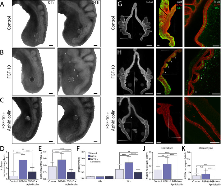
During airway branching morphogenesis, focal regions of FGF-10 expression in the pulmonary mesenchyme are thought to provide a local guidance cue, which promotes chemotactically the directional outgrowth of the airway epithelium. Here, however, we show that an ectopic source of FGF-10 induces epithelial buckling morphogenesis and the formation of multiple new supernumerary buds. FGF-10-induced budding can be modulated by altered epithelial tension and luminal fluid pressure. Increased tension suppresses the formation of ectopic branches, while a collapse of the embryonic airway promotes more expansive buckling and additional FGF-10-induced supernumerary buds. Our results indicate that a focal source of FGF-10 can promote epithelial buckling and suggest that the overall branching pattern cannot be explained entirely by the templated expression of FGF-10. Both FGF-10-mediated cell behaviors and exogenous mechanical forces must be integrated to properly shape the bronchial tree.

Keywords: Biomechanics; Branching morphogenesis; Chick embryo; Lung development; Mechanobiology.


Conflict of interest statement

Competing interests The authors declare no competing or financial interests.

Figures

Fig. 1.
Fig. 1.
Individual ectopic sources of FGF-10 elicit the formation of multiple supernumerary buds. (A) Schematic representation of the spatial pattern of FGF-10 expression in the embryonic mouse lung (top), as well as the implantation of ectopic sources of FGF-10 within the pulmonary mesenchyme of embryonic avian lungs (bottom). (B,C) Bright-field images of representative embryonic lung explants cultured ex vivo with either (B) PBS- or (C) FGF-10-loaded beads. White dashed lines indicate the location of the bead. White asterisks indicate supernumerary buds. Scale bars: 200 µm; inset scale bars: 100 µm. (D) Schematic representation of metrics used to quantify epithelial morphology. (E-H) Quantification of (E) the number of supernumerary buds, (F) the bud wavelength λ, (G) the epithelial contour ratio Λ=L/L0, and (H) the tortuosity index formula image for explants cultured with either PBS- or FGF-10 loaded beads, where L is the measured length of the ventral epithelium at each time-point, formula image is the length of the straight line connecting the endpoints of L, and L0 is the length of the ventral epithelium at t=0 h, which is used as a reference length. A Student's unpaired t-test (E,G) or a one-way ANOVA (H), followed by a Tukey post-hoc test, were used to make statistical comparisons. (PBS: n=29, FGF-10: n=37; *P<0.05, **P<0.01, ***P<0.001, ****P<0.0001; error bars represent s.d.).
Fig. 2.
Fig. 2.
FGF-10-induced supernumerary branches form simultaneously. (A,B) Bright-field images of a representative embryonic lung explant cultured ex vivo with either a (A) PBS- or (B) FGF-10-loaded bead over 24 h of time-lapse culture. White dashed lines indicate the location of the bead, as well as the traced contour of the ventral epithelium. White asterisks indicate supernumerary buds. Scale bars: 100 µm. (C) Time-varying contours of the ventral epithelium over 24 h of time-lapse culture. Scale bar: 100 µm. (D,E) Quantification of (D) epithelial contour length and (E) tortuosity index (E) during time-lapse culture. Shaded gray regions indicate the initiation of supernumerary buds. A two-way ANOVA, followed by a Tukey post-hoc test, was used to determine significance among groups. (Control: n=7; FGF-10: n=12; *P<0.05, **P<0.01, ***P<0.001, ****P<0.0001; error bars represent s.d.).
Fig. 3.
Fig. 3.
Supernumerary bud formation depends crucially on elevated epithelial proliferation. (A-C) Bright-field images of representative embryonic lung explants cultured ex vivo with either a (A) PBS- or (B,C) FGF-10-loaded bead. Some explants were also treated with (C) the cell cycle inhibitor, aphidicolin. White dashed lines indicate the location of the bead. White asterisks indicate supernumerary buds. Scale bars: 100 µm. (D-F) Quantification of (D) the number of new supernumerary buds, (E) epithelial contour ratio, and (F) tortuosity index for explants cultured with either a PBS- or FGF-10-loaded bead in the presence or absence of aphidicolin. A (D,E) one-way or (F) two-way ANOVA, followed by a Tukey post-hoc test, was used to make statistical comparisons. (PBS: n=32, FGF-10: n=36, FGF-10+Aphidicolin: n=29; *P<0.05, **P<0.01, ***P<0.001, ****P<0.0001; error bars represent s.d.). (G-I) Confocal images of EdU incorporation and LCAM/E-cadherin immunofluorescence in representative whole-mount lung explants. Insets show 3D reconstructions of EdU-positive nuclei within the embryonic airway epithelium. White asterisks denote elevated proliferation in supernumerary buds. Scale bars: 200 µm; inset scale bars: 50 µm. (J,K) Quantification of EdU incorporation within (J) the ventral epithelium and (K) adjacent pulmonary mesenchyme. A one-way ANOVA, followed by a Tukey post-hoc test, was used to determine significance between groups (PBS: n=13, FGF-10: n=13, FGF-10+Aphidicolin: n=12; **P<0.01, ***P<0.001, ****P<0.0001, error bars represent s.d.).
Fig. 4.
Fig. 4.
Apical constriction is not associated with the formation of FGF-10-induced supernumerary buds. (A) Schematic depiction of airway epithelial cells within both normal and supernumerary buds. (B) Wide-field fluorescence images of F-actin and pMLC staining within normal epithelial buds. White arrowheads indicate elevated fluorescence intensity along the apical surface. Scale bars: 25 µm. (C,D) Wide-field fluorescence images of F-actin and pMLC staining in representative embryonic lungs cultured with either (C) PBS- or (D) FGF-10-loaded beads. Insets depict staining within the ventral epithelium at higher magnification. Scale bars: 100 µm; inset scale bars: 25 µm.
Fig. 5.
Fig. 5.
Altered epithelial tension modulates supernumerary bud formation. (A) Schematic of buckling morphogenesis. Epithelial growth, constrained by the surrounding mesenchyme, can cause the epithelium to buckle and form new buds. (B,C) Bright-field time-lapse images of representative lung explants cultured ex vivo for 24 h with an FGF-10-loaded bead. Some explants (C) were treated with forskolin, an agonist of epithelial fluid secretion. Scale bars: 100 µm. White dashed lines indicate the location of the bead, as well as the traced contour of the ventral epithelium. White asterisks denote supernumerary buds. (D) Time-varying contours of the ventral epithelium over 24 h of time-lapse culture in either the presence or absence of forskolin. Scale bar: 100 µm. (E) Quantification of the tortuosity index during time-lapse culture. (PBS: n=10; FGF-10: n=11; FGF-10+10 µM forskolin: n=13; error bars represent s.d.) (F,G) Bright-field time-lapse images of representative lung explants cultured ex vivo for 24 h with an FGF-10-loaded bead. Some explants (G) were treated with bumetanide, an inhibitor of epithelial fluid secretion. Scale bars: 100 µm. As above, white dashed lines indicate the location of the bead, as well as the traced contour of the ventral epithelium. White asterisks denote supernumerary buds. (H) Time-varying contours of the ventral epithelium over 24 h of time-lapse culture in either the presence or absence of bumetanide. Scale bar: 100 µm. (I) Quantification of the time at which supernumerary buds initiate. A Students’ t-test was used to determine significance between groups. (FGF-10: n=43; FGF-10+200 µM bumetanide: n=37; *P<0.05, **P<0.01, ***P<0.001, ****P<0.0001; error bars represent s.d.). (J) Quantification of the time-varying tortuosity index within explants cultured with either PBS- or FGF-10-loaded beads in the presence or absence of bumetanide. (PBS: n=10; FGF-10: n=10; FGF-10+200 µM bumetanide: n=12; error bars represent s.d.).

Similar articles

Cited by

References

    1. Affolter, M., Zeller, R. and Caussinus, E. (2009). Tissue remodelling through branching morphogenesis. Nat. Rev. Mol. Cell Biol. 10, 831-842. 10.1038/nrm2797 - DOI - PubMed
    1. Alescio, T. and Cassini, A. (1962). Induction in vitro of tracheal buds by pulmonary mesenchyme grafted on tracheal epithelium. J. Exp. Zool. 150, 83-94. 10.1002/jez.1401500202 - DOI - PubMed
    1. Ballard, S. T. and Spadafora, D. (2007). Fluid secretion by submucosal glands of the tracheobronchial airways. Respir. Physiol. Neurobiol. 159, 271-277. 10.1016/j.resp.2007.06.017 - DOI - PMC - PubMed
    1. Ballard, S. T., Trout, L., Garrison, J. and Inglis, S. K. (2006). Ionic mechanism of forskolin-induced liquid secretion by porcine bronchi. Am. J. Physiol. Lung Cell. Mol. Physiol. 290, L97-L104. 10.1152/ajplung.00159.2005 - DOI - PubMed
    1. Bellusci, S., Grindley, J., Emoto, H., Itoh, N. and Hogan, B. L. (1997). Fibroblast growth factor 10 (FGF10) and branching morphogenesis in the embryonic mouse lung. Development (Cambridge, England) 124, 4867-4878. 10.1242/dev.124.23.4867 - DOI - PubMed

Publication types