

The negative regulation of gene expression by microRNAs as key driver of inducers and repressors of cardiomyocyte differentiation
- PMID: 35979890
- PMCID: PMC9411751
- DOI: 10.1042/CS20220391
The negative regulation of gene expression by microRNAs as key driver of inducers and repressors of cardiomyocyte differentiation
Abstract
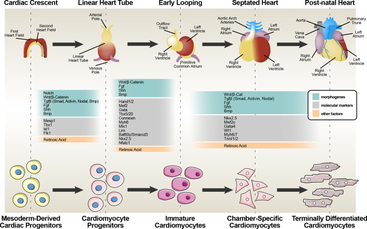
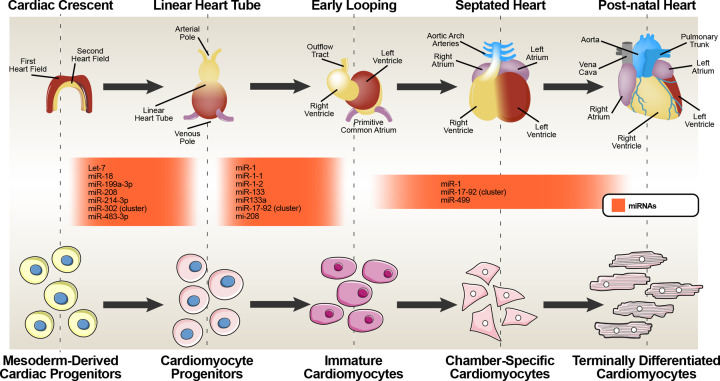
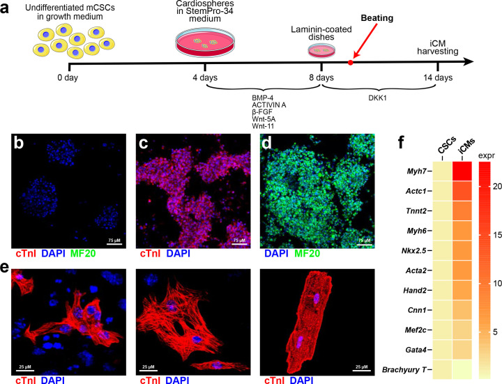
Cardiac muscle damage-induced loss of cardiomyocytes (CMs) and dysfunction of the remaining ones leads to heart failure, which nowadays is the number one killer worldwide. Therapies fostering effective cardiac regeneration are the holy grail of cardiovascular research to stop the heart failure epidemic. The main goal of most myocardial regeneration protocols is the generation of new functional CMs through the differentiation of endogenous or exogenous cardiomyogenic cells. Understanding the cellular and molecular basis of cardiomyocyte commitment, specification, differentiation and maturation is needed to devise innovative approaches to replace the CMs lost after injury in the adult heart. The transcriptional regulation of CM differentiation is a highly conserved process that require sequential activation and/or repression of different genetic programs. Therefore, CM differentiation and specification have been depicted as a step-wise specific chemical and mechanical stimuli inducing complete myogenic commitment and cell-cycle exit. Yet, the demonstration that some microRNAs are sufficient to direct ESC differentiation into CMs and that four specific miRNAs reprogram fibroblasts into CMs show that CM differentiation must also involve negative regulatory instructions. Here, we review the mechanisms of CM differentiation during development and from regenerative stem cells with a focus on the involvement of microRNAs in the process, putting in perspective their negative gene regulation as a main modifier of effective CM regeneration in the adult heart.
Keywords: cardiac stem cells; microRNA; myogenesis; regeneration.
© 2022 The Author(s).
Conflict of interest statement
The authors declare that there are no competing interests associated with the manuscript.
Figures
Similar articles
-
Small and long non-coding RNAs in cardiac homeostasis and regeneration.Biochim Biophys Acta. 2013 Apr;1833(4):923-33. doi: 10.1016/j.bbamcr.2012.08.010. Epub 2012 Aug 23. Biochim Biophys Acta. 2013. PMID: 22951218 Review.
-
The role of microRNAs in cardiac development and regenerative capacity.Am J Physiol Heart Circ Physiol. 2016 Mar 1;310(5):H528-41. doi: 10.1152/ajpheart.00181.2015. Epub 2015 Dec 23. Am J Physiol Heart Circ Physiol. 2016. PMID: 26702142 Free PMC article. Review.
-
The cardiac stem cell compartment is indispensable for myocardial cell homeostasis, repair and regeneration in the adult.Stem Cell Res. 2014 Nov;13(3 Pt B):615-30. doi: 10.1016/j.scr.2014.04.008. Epub 2014 Apr 29. Stem Cell Res. 2014. PMID: 24838077 Review.
-
Integrated transcriptomic and regulatory network analyses identify microRNA-200c as a novel repressor of human pluripotent stem cell-derived cardiomyocyte differentiation and maturation.Cardiovasc Res. 2018 May 1;114(6):894-906. doi: 10.1093/cvr/cvy019. Cardiovasc Res. 2018. PMID: 29373717
-
The promise of enhancer-associated long noncoding RNAs in cardiac regeneration.Trends Cardiovasc Med. 2015 Oct;25(7):592-602. doi: 10.1016/j.tcm.2015.01.014. Epub 2015 Feb 7. Trends Cardiovasc Med. 2015. PMID: 25753179 Review.
Cited by
-
Identification of miR-20b-5p as an inhibitory regulator in cardiac differentiation via TET2 and DNA hydroxymethylation.Clin Epigenetics. 2024 Mar 15;16(1):42. doi: 10.1186/s13148-024-01653-7. Clin Epigenetics. 2024. PMID: 38491513 Free PMC article.
-
miR-1 as a Key Epigenetic Regulator in Early Differentiation of Cardiac Sinoatrial Region.Int J Mol Sci. 2024 Jun 15;25(12):6608. doi: 10.3390/ijms25126608. Int J Mol Sci. 2024. PMID: 38928314 Free PMC article.
-
Epigenetic Regulation of Mammalian Cardiomyocyte Development.Epigenomes. 2024 Jun 29;8(3):25. doi: 10.3390/epigenomes8030025. Epigenomes. 2024. PMID: 39051183 Free PMC article. Review.
-
Polarizing Macrophage Functional Phenotype to Foster Cardiac Regeneration.Int J Mol Sci. 2023 Jun 28;24(13):10747. doi: 10.3390/ijms241310747. Int J Mol Sci. 2023. PMID: 37445929 Free PMC article. Review.
-
Anti-inflammatory, Anti-fibrotic and Pro-cardiomyogenic Effects of Genetically Engineered Extracellular Vesicles Enriched in miR-1 and miR-199a on Human Cardiac Fibroblasts.Stem Cell Rev Rep. 2023 Nov;19(8):2756-2773. doi: 10.1007/s12015-023-10621-2. Epub 2023 Sep 13. Stem Cell Rev Rep. 2023. PMID: 37700183 Free PMC article.
References
Publication types
MeSH terms
Substances
LinkOut - more resources
Full Text Sources
Medical