. 2022 Sep 14;96(17):e0074122.
doi: 10.1128/jvi.00741-22. Epub 2022 Aug 18.

UBR5 Acts as an Antiviral Host Factor against MERS-CoV via Promoting Ubiquitination and Degradation of ORF4b

Affiliations

UBR5 Acts as an Antiviral Host Factor against MERS-CoV via Promoting Ubiquitination and Degradation of ORF4b

Yuzheng Zhou et al. J Virol. .

Abstract

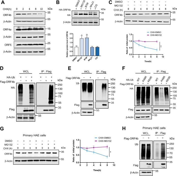
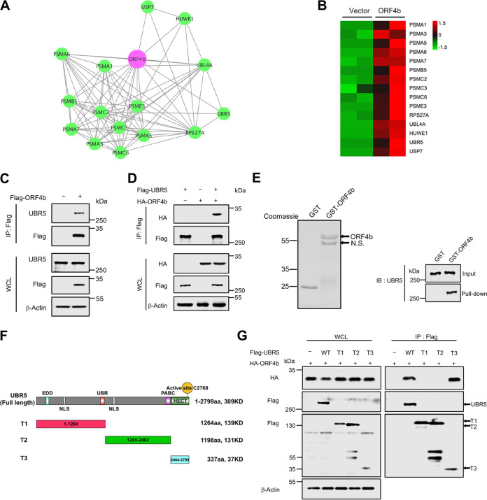
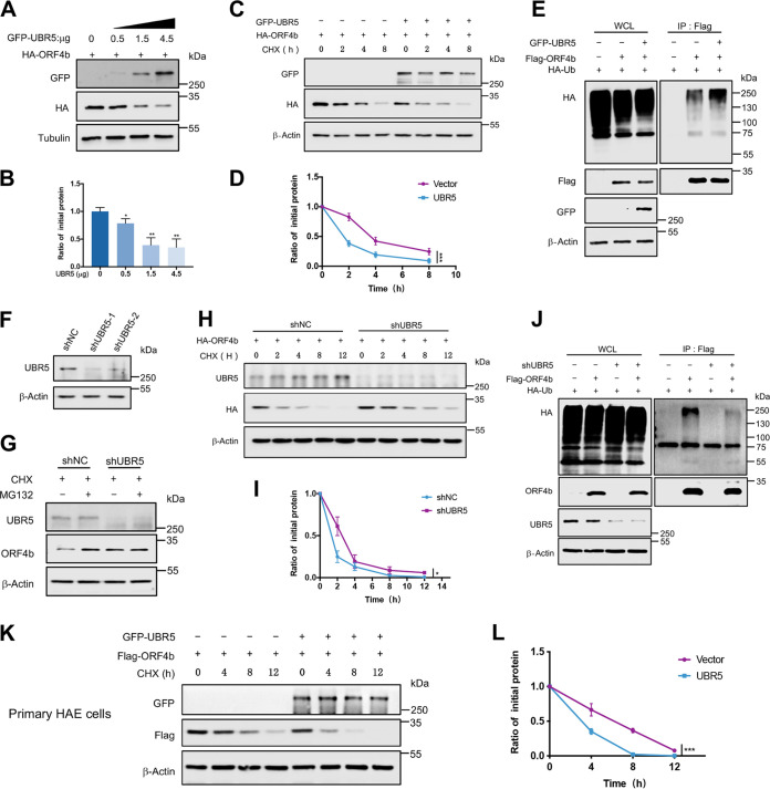
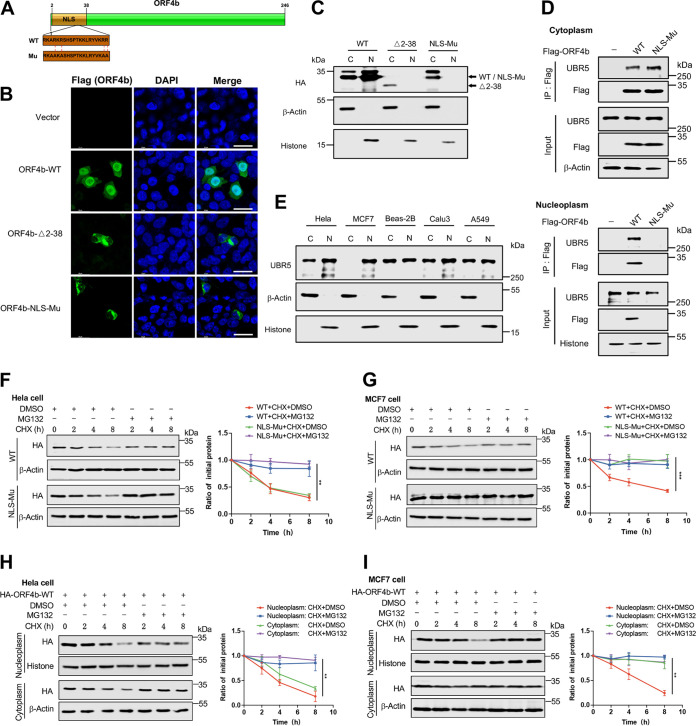
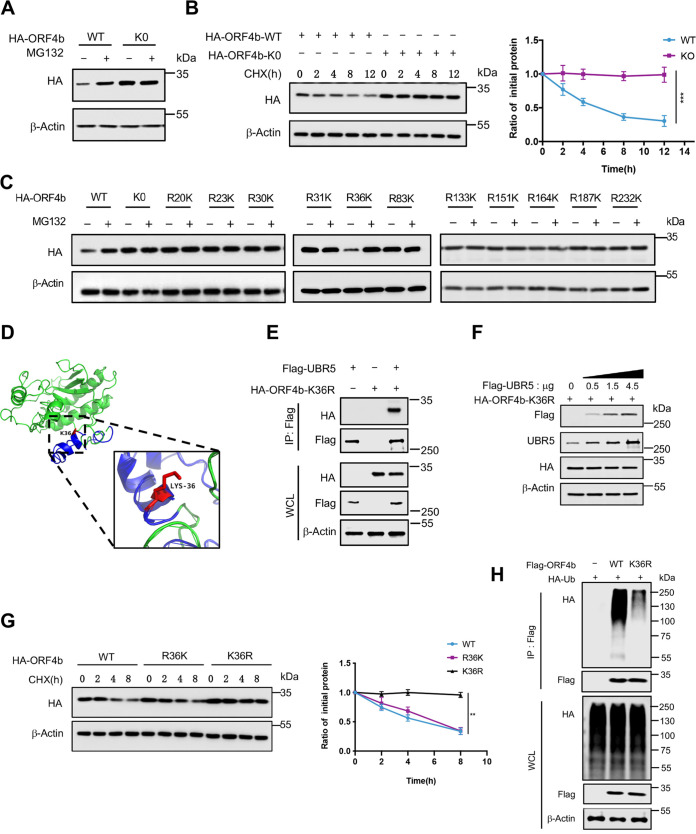
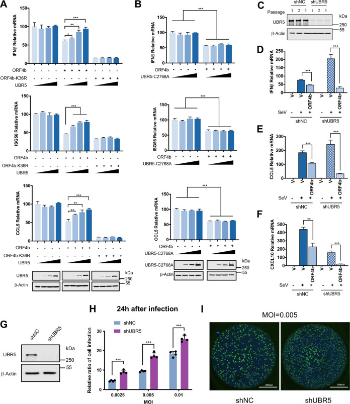
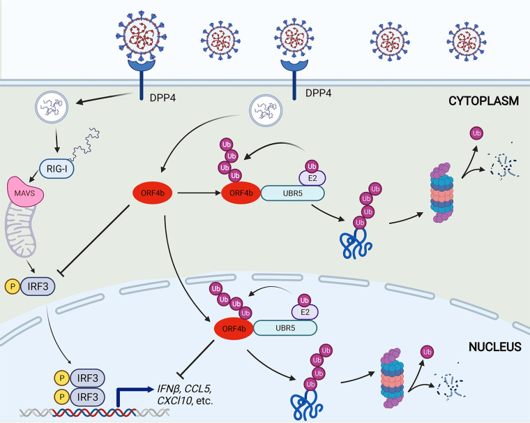
Within the past 2 decades, three highly pathogenic human coronaviruses have emerged, namely, severe acute respiratory syndrome coronavirus (SARS-CoV), Middle East respiratory syndrome coronavirus (MERS-CoV), and severe acute respiratory syndrome coronavirus 2 (SARS-CoV-2). The health threats and economic burden posed by these tremendously severe coronaviruses have paved the way for research on their etiology, pathogenesis, and treatment. Compared to SARS-CoV and SARS-CoV-2, MERS-CoV genome encoded fewer accessory proteins, among which the ORF4b protein had anti-immunity ability in both the cytoplasm and nucleus. Our work for the first time revealed that ORF4b protein was unstable in the host cells and could be degraded by the ubiquitin proteasome system. After extensive screenings, it was found that UBR5 (ubiquitin protein ligase E3 component N-recognin 5), a member of the HECT E3 ubiquitin ligases, specifically regulated the ubiquitination and degradation of ORF4b. Similar to ORF4b, UBR5 can also translocate into the nucleus through its nuclear localization signal, enabling it to regulate ORF4b stability in both the cytoplasm and nucleus. Through further experiments, lysine 36 was identified as the ubiquitination site on the ORF4b protein, and this residue was highly conserved in various MERS-CoV strains isolated from different regions. When UBR5 was knocked down, the ability of ORF4b to suppress innate immunity was enhanced and MERS-CoV replication was stronger. As an anti-MERS-CoV host protein, UBR5 targets and degrades ORF4b protein through the ubiquitin proteasome system, thereby attenuating the anti-immunity ability of ORF4b and ultimately inhibiting MERS-CoV immune escape, which is a novel antagonistic mechanism of the host against MERS-CoV infection. IMPORTANCE ORF4b was an accessory protein unique to MERS-CoV and was not present in SARS-CoV and SARS-CoV-2 which can also cause severe respiratory disease. Moreover, ORF4b inhibited the production of antiviral cytokines in both the cytoplasm and the nucleus, which was likely to be associated with the high lethality of MERS-CoV. However, whether the host proteins regulate the function of ORF4b is unknown. Our study first determined that UBR5, a host E3 ligase, was a potential host anti-MERS-CoV protein that could reduce the protein level of ORF4b and diminish its anti-immunity ability by inducing ubiquitination and degradation. Based on the discovery of ORF4b-UBR5, a critical molecular target, further increasing the degradation of ORF4b caused by UBR5 could provide a new strategy for the clinical development of drugs for MERS-CoV.

Keywords: IRF3; MERS-CoV; accessory protein; antiviral mechanism; coronavirus; posttranslational modification.


Conflict of interest statement

The authors declare no conflict of interest.

Figures

FIG 1
FIG 1
Identifications of MERS-CoV ORF4b functions and its host’s interacting proteins. (A to C) HEK293T cells were cotransfected with expression plasmids for MERS-CoV accessory proteins as indicated and pRL-TK plasmids, together with plasmids expressing firefly luciferase under the control of the IFN-β promoter (A), IRF3 responsive element (B), or NF-κB responsive element (C). Twenty-four hours after transfection, the cells were infected with Sendai virus (SeV) (100 HAU/mL) for 12 h and lysed for the dual-luciferase assay. (D) HEK293T cells were transfected with plasmids expressing HA-tagged MERS-CoV accessory proteins. Twenty-four hours after transfection, the cells were infected with SeV for 12 h and collected for Western blotting. The levels of the indicated proteins were detected with relevant antibodies. (E) HEK293T cells were transfected with empty vector or plasmids expressing the indicated viral proteins. At 24 h posttransfection, cells were infected with SeV (100 HAU/mL) for 12 h. Total RNA was extracted, reverse transcribed, and analyzed by real-time PCR with primers specific for IFN-β, CCL5, CXCL10, and ISG56. (F) HEK293T cells were transfected with an empty vector or increasing amounts of plasmids expressing ORF4b. Twenty-four hours later, cells were infected with SeV for 12 h. The cells were used to extract the total RNA for real-time PCR. (G) HEK293T cells were transfected with empty vector or Flag-ORF4b-expressing plasmid and collected at 48 h posttransfection. Cells were lysed, and whole-cell lysates were immunoprecipitated by anti-Flag beads. Proteins were eluted and detected by silver staining. (H) Cellular Component enrichment analysis of the interacting proteins of ORF4b; (I) KEGG enrichment analysis of the interacting proteins of ORF4b. Error bars indicate SD from technical triplicates. Statistical significance was calculated using an unpaired, two-tailed Student’s t test. *, P < 0.05; **, P < 0.01; ***, P < 0.001.
FIG 2
FIG 2
ORF4b is degraded by the ubiquitin-proteasome system. (A) HEK293T cells in a 6-cm dish were transfected with the indicated HA-tagged plasmids. Twelve hours later, the cells were split evenly into the wells of 12-well plates and treated with CHX (30 μg/mL) when they reached 100% confluence. Cells were collected at the indicated times to detect the level of viral protein by anti-HA antibody. (B) HEK293T cells transfected with the indicated plasmids were treated with dimethyl sulfoxide (DMSO), MG132 (20 μM), bortezomib (BTM) (10 μM), chloroquine (CQ) (20 μM), and NH4Cl (10 mM) for 8 h before collection. The protein level of ORF4b was detected by Western blotting (top). Quantification of ORF4b protein levels relative to β-actin is shown. Results are shown as mean ± SD (n = 3 independent experiments). *, P < 0.05, and **, P < 0.01, by Student's t test (bottom). (C) HEK293T cells transfected with HA-ORF4b expressing plasmid were cotreated with CHX (30 μg/mL) and DMSO or cotreated with CHX (30 μg/mL) and MG132 (20 μM). Cells were collected at the indicated times for Western blotting by anti-HA antibody (top). Quantification of ORF4b protein levels relative to β-actin is shown. Results are shown as mean ± SD (n = 3 independent experiments). ***, P < 0.001 by two-way analysis of variance (ANOVA) (bottom). (D and E) HEK293T cells cotransfected with HA-ubiquitin and Flag-ORF4b (D) or transfected with Flag-ORF4b alone (E) were treated with MG132 for 8 h before collection. The whole-cell lysates were incubated with anti-Flag beads and used for Western blotting with anti-HA or anti-ubiquitin antibodies to detect the polyubiquitination chain of ORF4b. (F) The ORF4b ubiquitination linkage was analyzed in HEK293T cells transfected with ORF4b and the indicated ubiquitin-WT, Lys-48→Arg, and Lys-63→Arg plasmids. The whole-cell lysates were subjected to pulldown with anti-Flag beads and Western blotting to detect the polyubiquitination chain of ORF4b. (G) The primary human airway epithelial (HAE) cells were transfected with Flag-ORF4b-expressing plasmids and treated and collected as indicated for Western blotting with anti-Flag antibody (left). Quantification of ORF4b protein levels relative to β-actin is shown as mean ± SD (n = 3 independent experiments). **, P < 0.01 by two-way ANOVA (right). (H) The primary HAE cells were transfected with Flag-ORF4b-expressing plasmids and lysed to analyze the polyubiquitination chain of ORF4b.
FIG 3
FIG 3
ORF4b interacts with UBR5. (A) The protein-protein interaction network of ORF4b and interacting proteasome subunits and E3 ligase candidates is shown, based on mass spectrometry results. (B) A heat map shows the ORF4b-interacting proteins’ enrichment in the proteasome pathway and E3 ligase family. (C) HEK293T cells transfected with empty vector or plasmid containing Flag-ORF4b were collected at 48 h after transfection. Cells were lysed and immunoprecipitated with anti-Flag beads. Proteins in the whole-cell lysates and immunoprecipitate were blotted with the indicated antibodies. (D) Flag-UBR5- and HA-ORF4b-expressing plasmids were cotransfected into HEK293T cells. Forty-eight hours after transfection, cells were lysed and immunoprecipitated with anti-Flag beads. The whole-cell lysates and immunoprecipitated proteins were detected with anti-HA and anti-Flag antibodies by Western blotting. (E) GST and GST-ORF4b were purified from E. coli and analyzed by Coomassie staining (left). The beads binding the target proteins were incubated with the whole-cell lysates from HEK293T cells, and the pulldown proteins were detected with anti-UBR5 antibody by immunoblotting (right). N.S., nonspecific. (F) The structural domains of UBR5 are diagrammed. (G) HEK293T cells were transfected with the indicated plasmids. Whole-cell lysates were precipitated with anti-Flag beads. Whole-cell lysates and precipitated proteins were analyzed by Western blotting with the indicated antibodies.
FIG 4
FIG 4
UBR5 mediates the ubiquitination and degradation of ORF4b. (A and B) HEK293T cells transfected with ORF4b-expressing plasmid were split into the wells of a 12-well plate and were then transfected with increasing amounts of plasmids containing UBR5. The cells were collected at 48 h posttransfection to analyze the protein level of ORF4b by Western blotting (A). Quantification of ORF4b protein levels relative to β-actin is shown. Results are shown as mean ± SD (n = 3 independent experiments). *, P < 0.05, and **, P < 0.01, by Student's t test (B). (C and D) The plasmids containing HA-ORF4b, together with empty vector or UBR5-expressing plasmids were transfected into HEK293T cells and were split into the wells of a 12-well plate. Twenty-four hours later, the cells were treated with CHX (30 μg/mL) and collected at the indicated times to detect the protein level of ORF4b (C). Quantification of ORF4b protein levels relative to β-actin is shown. Results are shown as mean ± SD (n = 3 independent experiments). ***, P < 0.001 by two-way ANOVA (D). (E) ORF4b ubiquitination was analyzed in HEK293T cells transfected with ORF4b together with UBR5 or not. (F) Immunoblots of UBR5 from HEK293T cells infected with lentivirus containing control or shRNA targeting UBR5 for 72 h. (G) HEK293T cells stably expressing shNC and shUBR5 were transfected with plasmid containing ORF4b and were treated with MG132 (20 μM) for 8 h. Cells were collected to detect the protein level of ORF4b by Western blotting. (H and I) HEK293T cells stably expressing UBR5 shRNA and shNC were transfected with ORF4b-expressing plasmid and split evenly into the wells of a 12-well plate. Cells were treated with CHX (30 μg/mL) and collected at the indicated times to test the protein level of ORF4b (H). Quantification of ORF4b protein levels relative to β-actin is shown. Results are shown as mean ± SD (n = 3 independent experiments). *, P < 0.05 by two-way ANOVA (I). (J) ORF4b ubiquitination was analyzed in UBR5 shRNA and shNC-transduced HEK293T cells, transfected with HA-ubiquitin together with Flag-ORF4b or not. (K and L) The plasmids containing Flag-ORF4b together with empty vector or UBR5-expressing plasmids were transfected into the primary HAE cells. The cells were treated and collected as indicated to test the half-lives of ORF4b (K). Quantification of ORF4b protein levels relative to β-actin is shown as mean ± SD (n = 3 independent experiments). ***, P < 0.001 by two-way ANOVA (L).
FIG 5
FIG 5
UBR5 induces the degradation of ORF4b in both cytoplasm and nucleoplasm. (A) The pattern diagram shows the mutations of arginine to alanine in the basic amino acid group of ORF4b nuclear localization signal. (B) HeLa cells transfected with the indicated plasmids were analyzed by confocal microscopy. The Flag-tagged ORF4b WT and mutants were labeled with anti-Flag antibody (green). Cell nuclei were stained using DAPI (blue). Representative images are shown. Scale bars, 25 μm. (C) HEK293T cells transfected with HA-tagged ORF4b and mutants were used for nuclear and cytoplasmic protein extraction. The indicated proteins in cytoplasm and nucleoplasm were detected by Western blotting. (D) The proteins of cytoplasm and nucleoplasm were extracted from HEK293T cells transfected with the indicated plasmids. The cytoplasmic and nuclear proteins were subjected to pulldown with anti-Flag beads. ORF4b and endogenous UBR5 were detected with the indicated antibodies by Western blotting. (E) The cytoplasmic and nuclear proteins of HeLa, MCF7, BEAS-2B, Calu3, and A549 cells were extracted. The protein levels of UBR5 were detected by Western blotting. (F and G) HeLa (F) and MCF7 (G) cells transfected with indicated plasmids expressing HA-ORF4b-WT and HA-ORF4b-NLS-Mu (nuclear location signal mutant) were cotreated with CHX (30 μg/mL) and DMSO or CHX (30 μg/mL) and MG132 (20 μM). Cells were collected to detect the indicated protein level by Western blotting. (H and I) HeLa (H) and MCF7 (I) cells transfected with HA-tagged ORF4b were cotreated with CHX and DMSO or CHX and MG132. Cells were collected and used for nuclear and cytoplasmic protein extraction. The levels of the indicated proteins in both cytoplasm and nucleoplasm were analyzed by Western blotting. Error bars indicate SD from technical triplicates. Quantification of ORF4b protein levels relative to β-actin is shown. **, P < 0.01, and ***, P < 0.001, by two-way ANOVA.
FIG 6
FIG 6
Ubiquitination-resistant mutation restores the stability of ORF4b. (A) The plasmid expressing ORF4b-WT or ORF4b-K0 was transfected into HEK293T cells. The cells were treated with MG132 (20 μM) or not for 8 h before collection. The protein level of ORF4b was detected with anti-HA antibody by Western blotting. (B) HEK293T cells transfected with plasmids containing ORF4b-WT and ORF4b-K0 (in which all lysine was mutated to arginine) were cotreated with CHX (30 μg/mL) and DMSO or CHX (30 μg/mL) and MG132 (20 μM). Cells were collected to detect the level of indicated protein (left). Quantification of the ORF4b protein level was normalized to β-actin. Results are shown as mean ± SD (n = 3 independent experiments). ***, P < 0.001 by two-way ANOVA (right). (C) HEK293T cells were transfected with ORF4b-WT, ORF4b-K0, and the ORF4b single mutant with arginine changed to lysine based on the K0 mutant. Cells with different transfection were split evenly and treated with MG132 (20 μM) or not for 8 h before collection. The indicated protein levels were detected by Western blotting. (D) A PDB file of ORF4b was modeled by the I-TASSER server from the Zhang laboratory. Visualization of the three-dimensional model of ORF4b was performed with PyMOL. K36 is located on the surface (red) and in the NLS (blue). (E) The plasmids expressing Flag-UBR5 and HA-ORF4b-K36R were cotransfected into HEK293T cells. The whole-cell lysates were immunoprecipitated with anti-Flag beads. The proteins were detected with the indicated antibodies by Western blotting. (F) HEK293T cells transfected with the ORF4b-K36R-expressing plasmid were split into the wells of a 12-well plate and then were transfected with increasing amounts of plasmids containing UBR5. The cells were collected at 48 h posttransfection to analyze the protein level of ORF4b by Western blotting. (G) Half-life analysis of ORF4b-WT, ORF4b-R36K, and ORF4b-K36R. HEK293T cells transfected with indicated plasmid were split evenly into the wells of a 12-well plate. Cells were treated with CHX (30 μg/mL) and collected at the indicated times to detect the level of target proteins (left). Quantification of the ORF4b protein level was normalized to β-actin. Results are shown as mean ± SD (n = 3 independent experiments). **, P < 0.01 by two-way ANOVA (right). (H) Ubiquitination analysis of ORF4b-WT and ORF4b-K36R was performed with HEK293T cells transfected with the indicated plasmids. The polyubiquitination chains were detected by anti-HA antibody.
FIG 7
FIG 7
UBR5 acts as an anti-MERS-CoV factor in host cells. (A) Increasing amounts of plasmids containing Flag-UBR5 were transfected into HEK293T cells expressing empty vector, HA-ORF4b-WT, or HA-ORF4b-K36R. Cells were infected with SeV (100 HAU/mL) for 12 h and collected. Thirty percent of the cells were lysed and used for Western blotting (bottom), and the rest were used for RNA extraction and analyzed by real-time PCR with primers specific for the indicated genes. (B) The plasmid containing Flag-UBR5-C2768A was transfected into HEK293T cells expressing empty vector or ORF4b protein in increasing gradients. After being treated with SeV (100 HAU/mL) for 12 h, 30% of the cells were collected to detect the indicated proteins (bottom). Total RNA of the rest cells was extracted, reverse transcribed, and analyzed by real-time PCR with primers specific for target genes. (C) Calu3 cells were infected with lentivirus containing UBR5 shRNA and were selected by puromycin (2 μg/mL) for 72 h. The cell lines were passaged for three generations, and each passage was collected to detect the protein level of endogenous UBR5 by Western blotting. (D to F) The plasmids expressing empty vector or ORF4b were transfected into shNC or shUBR5 Calu3 stable cell lines and infected with SeV (100 HAU/mL) for 12 h. Total RNA was extracted, and the relative mRNA level of IFN-β (D), CCL5 (E), and CXCL10 (F) was analyzed by real-time PCR with specific primers. (G) Huh7 cells were infected with lentivirus containing UBR5 shRNA and were selected by puromycin (2 μg/mL) for 72 h. The cells were collected to detect the protein level of endogenous UBR5 by Western blotting. (H) Huh7 cells were infected with MERS-CoV at the indicated MOIs of 0.0025, 0.005, and 0.01. Twenty-four hours later, an indirect immunofluorescence assay (IFA) was used to detect S protein expression in MERS-CoV-infected cells and the relative ratio of cell infection was analyzed. (I) Huh7 cells infected with MERS-CoV were analyzed by confocal microscopy. Cells were incubated with anti-MERS-CoV S protein antibody, and nuclei were stained with DAPI (blue). Representative images are shown. Scale bars, 2,000 μm. Error bars indicate SD from technical triplicates. Statistical significance was calculated using an unpaired, two-tailed Student’s t test: *, P < 0.05; **, P < 0.01; ***, P < 0.001.
FIG 8
FIG 8
Pattern diagram of UBR5 as an antiviral factor against MERS-CoV by promoting degradation of ORF4b. DPP4, dipeptidyl peptidase 4; RIG-I, retinoic acid-induced gene I; MDA5, melanoma differentiation-associated protein 5; MAVS, mitochondrial antiviral-signaling protein.

Similar articles

Cited by

References

    1. Ziv O, Price J, Shalamova L, Kamenova T, Goodfellow I, Weber F, Miska EA. 2020. The short- and long-range RNA-RNA interactome of SARS-CoV-2. Mol Cell 80:1067–1077.e5. 10.1016/j.molcel.2020.11.004. - DOI - PMC - PubMed
    1. Wu A, Peng Y, Huang B, Ding X, Wang X, Niu P, Meng J, Zhu Z, Zhang Z, Wang J, Sheng J, Quan L, Xia Z, Tan W, Cheng G, Jiang T. 2020. Genome composition and divergence of the novel coronavirus (2019-nCoV) originating in China. Cell Host Microbe 27:325–328. 10.1016/j.chom.2020.02.001. - DOI - PMC - PubMed
    1. Petrosillo N, Viceconte G, Ergonul O, Ippolito G, Petersen E. 2020. COVID-19, SARS and MERS: are they closely related? Clin Microbiol Infect 26:729–734. 10.1016/j.cmi.2020.03.026. - DOI - PMC - PubMed
    1. Zaki AM, van Boheemen S, Bestebroer TM, Osterhaus AD, Fouchier RA. 2012. Isolation of a novel coronavirus from a man with pneumonia in Saudi Arabia. N Engl J Med 367:1814–1820. 10.1056/NEJMoa1211721. - DOI - PubMed
    1. Assiri A, Al-Tawfiq JA, Al-Rabeeah AA, Al-Rabiah FA, Al-Hajjar S, Al-Barrak A, Flemban H, Al-Nassir WN, Balkhy HH, Al-Hakeem RF, Makhdoom HQ, Zumla AI, Memish ZA. 2013. Epidemiological, demographic, and clinical characteristics of 47 cases of Middle East respiratory syndrome coronavirus disease from Saudi Arabia: a descriptive study. Lancet Infect Dis 13:752–761. 10.1016/S1473-3099(13)70204-4. - DOI - PMC - PubMed

MeSH terms