

GABAB receptor agonist baclofen promotes central nervous system remyelination
- PMID: 35980256
- PMCID: PMC9804779
- DOI: 10.1002/glia.24262
GABAB receptor agonist baclofen promotes central nervous system remyelination
Abstract
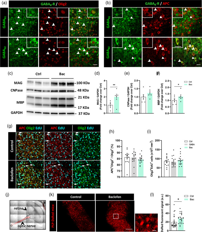
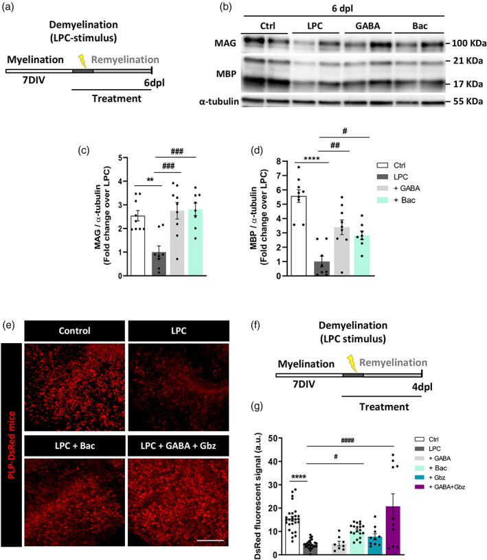
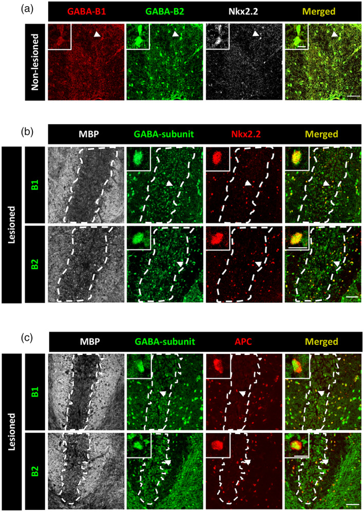
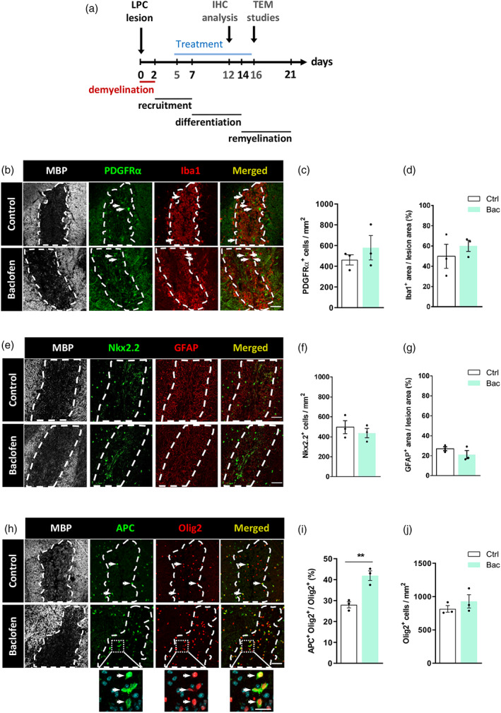
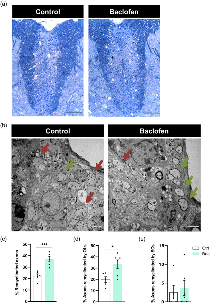
Promoting remyelination is considered as a potential neurorepair strategy to prevent/limit the development of permanent neurological disability in patients with multiple sclerosis (MS). To this end, a number of clinical trials are investigating the potential of existing drugs to enhance oligodendrocyte progenitor cell (OPC) differentiation, a process that fails in chronic MS lesions. We previously reported that oligodendroglia express GABAB receptors (GABAB Rs) both in vitro and in vivo, and that GABAB R-mediated signaling enhances OPC differentiation and myelin protein expression in vitro. Our goal here was to evaluate the pro-remyelinating potential of GABAB R agonist baclofen (Bac), a clinically approved drug to treat spasticity in patients with MS. We first demonstrated that Bac increases myelin protein production in lysolecithin (LPC)-treated cerebellar slices. Importantly, Bac administration to adult mice following induction of demyelination by LPC injection in the spinal cord resulted in enhanced OPC differentiation and remyelination. Thus, our results suggest that Bac repurposing should be considered as a potential therapeutic strategy to stimulate remyelination in patients with MS.
Keywords: GABAB receptor; baclofen; multiple sclerosis; myelin; oligodendrocyte; remyelination.
© 2022 The Authors. GLIA published by Wiley Periodicals LLC.
Conflict of interest statement
The authors declare no conflicts of interest.
Figures
Similar articles
-
Oligodendrocyte progenitor cell recruitment and remyelination in multiple sclerosis: the more, the merrier?Brain. 2022 Dec 19;145(12):4178-4192. doi: 10.1093/brain/awac307. Brain. 2022. PMID: 36093726 Review.
-
Drug-based modulation of endogenous stem cells promotes functional remyelination in vivo.Nature. 2015 Jun 11;522(7555):216-20. doi: 10.1038/nature14335. Epub 2015 Apr 20. Nature. 2015. PMID: 25896324 Free PMC article.
-
PARP1-mediated PARylation activity is essential for oligodendroglial differentiation and CNS myelination.Cell Rep. 2021 Oct 5;37(1):109695. doi: 10.1016/j.celrep.2021.109695. Cell Rep. 2021. PMID: 34610310 Free PMC article.
-
Loss of Tuberous Sclerosis Complex1 in Adult Oligodendrocyte Progenitor Cells Enhances Axon Remyelination and Increases Myelin Thickness after a Focal Demyelination.J Neurosci. 2017 Aug 2;37(31):7534-7546. doi: 10.1523/JNEUROSCI.3454-16.2017. Epub 2017 Jul 10. J Neurosci. 2017. PMID: 28694334 Free PMC article.
-
Nudging oligodendrocyte intrinsic signaling to remyelinate and repair: Estrogen receptor ligand effects.J Steroid Biochem Mol Biol. 2016 Jun;160:43-52. doi: 10.1016/j.jsbmb.2016.01.006. Epub 2016 Jan 14. J Steroid Biochem Mol Biol. 2016. PMID: 26776441 Free PMC article. Review.
Cited by
-
Dysregulated Cholinergic Signaling Inhibits Oligodendrocyte Maturation Following Demyelination.J Neurosci. 2024 Jul 10;44(28):e0051242024. doi: 10.1523/JNEUROSCI.0051-24.2024. J Neurosci. 2024. PMID: 38749703 Free PMC article.
-
Genetic Downregulation of GABAB Receptors from Oligodendrocyte Precursor Cells Protects Against Demyelination in the Mouse Spinal Cord.Cells. 2024 Dec 5;13(23):2014. doi: 10.3390/cells13232014. Cells. 2024. PMID: 39682762 Free PMC article.
-
β-carbolines that enhance GABAA receptor response expressed in oligodendrocytes promote remyelination in an in vivo rat model of focal demyelination.Front Cell Neurosci. 2024 Apr 17;18:1369730. doi: 10.3389/fncel.2024.1369730. eCollection 2024. Front Cell Neurosci. 2024. PMID: 38694535 Free PMC article.
-
Demyelination and Remyelination: General Principles.Adv Neurobiol. 2025;43:207-255. doi: 10.1007/978-3-031-87919-7_9. Adv Neurobiol. 2025. PMID: 40500500 Review.
-
Neuronal activity and remyelination: new insights into the molecular mechanisms and therapeutic advancements.Front Cell Dev Biol. 2023 Jul 26;11:1221890. doi: 10.3389/fcell.2023.1221890. eCollection 2023. Front Cell Dev Biol. 2023. PMID: 37564376 Free PMC article. Review.
References
-
- Abel, S. , Vavasour, I. , Lee, L. E. , Johnson, P. , Ristow, S. , Ackermans, N. , Chan, J. , Cross, H. , Laule, C. , Dvorak, A. , Schabas, A. , Hernández‐Torres, E. , Tam, R. , Kuan, A. J. , … Kolind, S. H. (2020). Associations between findings from myelin water imaging and cognitive performance among individuals with multiple sclerosis. JAMA Network Open, 3, e2014220. 10.1001/jamanetworkopen.2020.14220 - DOI - PMC - PubMed
-
- Arellano, R. O. , Sánchez‐Gómez, M. V. , Alberdi, E. , Canedo‐Antelo, M. , Chara, J. C. , Palomino, A. , Pérez‐ Samartín, A. , & Matute, C. (2016). Axon‐to‐glia interaction regulates GABAA receptor expression in oligodendrocytes. Molecular Pharmacology, 89, 63–74. 10.1124/mol.115.100594 - DOI - PubMed
Publication types
MeSH terms
Substances
LinkOut - more resources
Full Text Sources
Medical
