. 2022 Oct 18;81(11):885-899.
doi: 10.1093/jnen/nlac073.

Developmentally Arrested Basket/Stellate Cells in Postnatal Human Brain as Potential Tumor Cells of Origin for Cerebellar Hemangioblastoma in von Hippel-Lindau Patients

Affiliations

Developmentally Arrested Basket/Stellate Cells in Postnatal Human Brain as Potential Tumor Cells of Origin for Cerebellar Hemangioblastoma in von Hippel-Lindau Patients

Sharon Baughman Shively et al. J Neuropathol Exp Neurol. .

Abstract

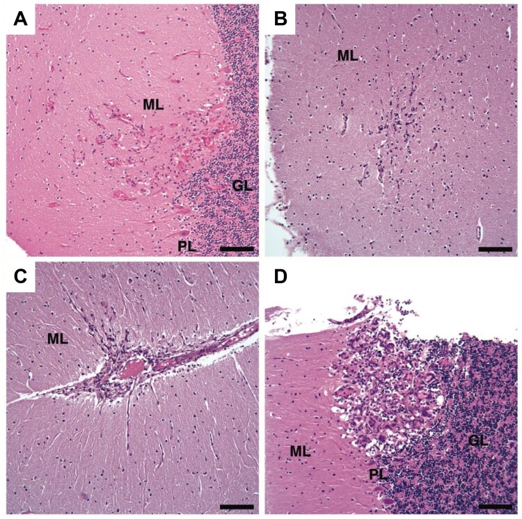
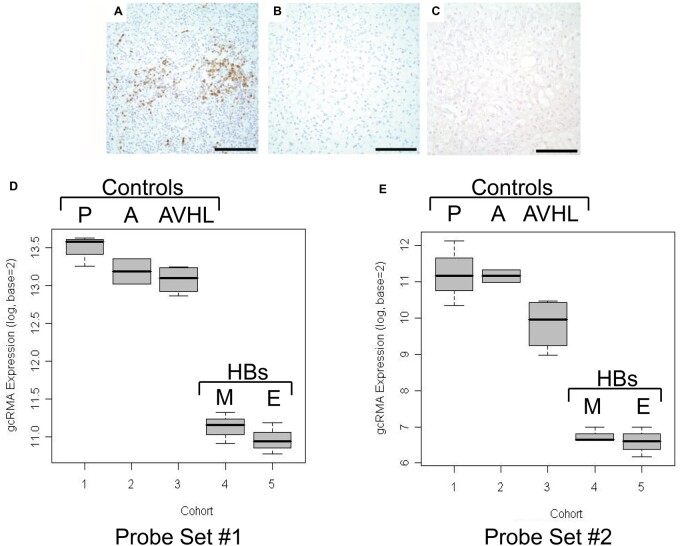
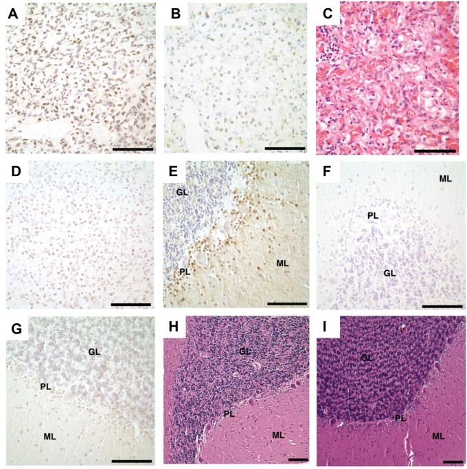
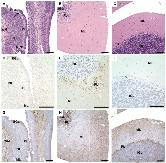
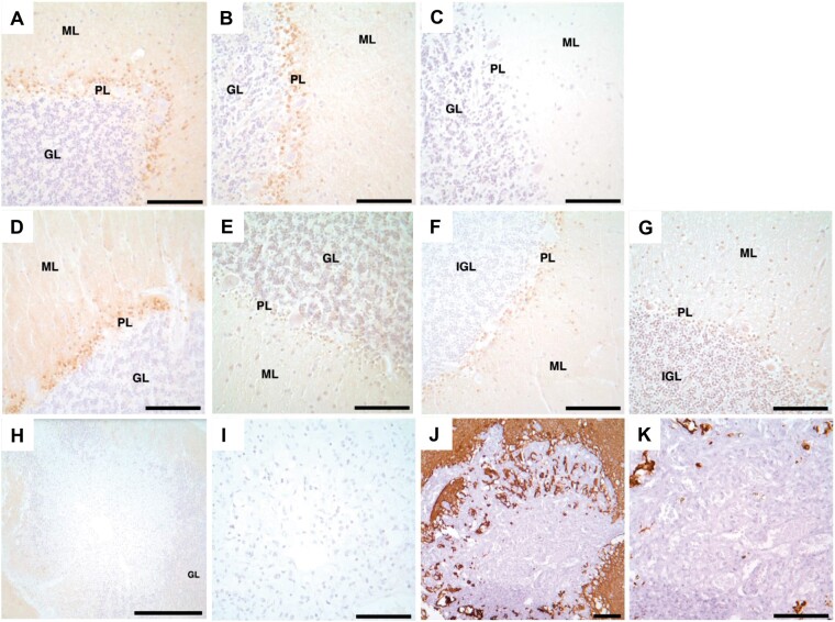
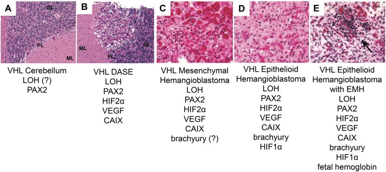
von Hippel-Lindau (VHL) disease is an autosomal dominant hereditary cancer disorder caused by a germline mutation in the VHL tumor suppressor gene. Loss of the wild-type allele results in VHL deficiency and the potential formation of cerebellar hemangioblastomas, which resemble embryonic hemangioblast proliferation and differentiation processes. Multiple, microscopic, VHL-deficient precursors, termed developmentally arrested structural elements (DASEs), consistently involve the cerebellar molecular layer in VHL patients, indicating the tumor site of origin. Unlike hemangioblastomas, however, cerebellar DASEs do not express brachyury, a mesodermal marker for hemangioblasts. In this study, neuronal progenitors occupying the molecular layer were investigated as tumor cells of origin. By immunohistochemistry, cerebellar DASEs and hemangioblastomas lacked immunoreactivity with antibody ZIC1 (Zic family member 1), a granule cell progenitor marker with concordance from oligonucleotide RNA expression array analyses. Rather, cerebellar DASEs and hemangioblastomas were immunoreactive with antibody PAX2 (paired box 2), a marker of basket/stellate cell progenitors. VHL cerebellar cortices also revealed PAX2-positive cells in Purkinje and molecular layers, resembling the histological and molecular development of basket/stellate cells in postnatal non-VHL mouse and human cerebella. These data suggest that VHL deficiency can result in the developmental arrest of basket/stellate cells in the human cerebellum and that these PAX2-positive, initiated cells await another insult or signal to form DASEs and eventually, tumors.

Keywords: Developmentally arrested basket/stellate cells; Hemangioblastoma; Paired box 2 (PAX2); Tumor suppressor syndrome; VHL human cerebellum tumor cell of origin; von Hippel-Lindau (VHL) disease.


Figures

FIGURE 1.
FIGURE 1.
H&E stains of the 4 smallest DASEs in cerebellar cortices from VHL patients. (A) VHL Patient #1, (B–D) VHL Patient #2. H&E, hematoxylin and eosin; DASEs, developmentally arrested structural elements; VHL, von Hippel-Lindau; ML, molecular layer; GL, granular layer; PL, Purkinje layer. Scale bars: A–D = 100 μm.
FIGURE 2.
FIGURE 2.
Lack of ZIC1 expression in VHL cerebellar HBs and DASEs. ZIC1 immunohistochemistry shows immunoreactivity in (A) medulloblastoma as positive control, but no immunoreactivity in (B) cerebellar HB or (C) cerebellar DASE. (D, E) Expression profile data analyses for ZIC1. Tukey box plot of gcRMA expression for (D) probe set #1 (Affymetrix probe set 206373_at) and (E) probe set #2 (Affymetrix probe set 236896_at) show decreased ZIC1 mRNA expression in mesenchymal and epithelioid VHL HB cohorts in comparison to the 3 cerebellar cortices cohorts. The y-axes show values above the designated noise threshold value of 6. Cohorts: pediatric non-VHL cerebellar cortex (P), adult non-VHL cerebellar cortex (A), adult VHL cerebellar cortex (AVHL), mesenchymal VHL HB (M), epithelioid VHL HB (E). ZIC1, Zic family member 1; VHL, von Hippel-Lindau; DASEs, developmentally arrested structural elements; HBs, hemangioblastomas. Scale bars: A-C= 100 μm.
FIGURE 3.
FIGURE 3.
PAX2 immunoreactivity in VHL cerebellar hemangioblastomas, DASEs and cortices. PAX2 immunohistochemistry shows immunoreactivity in (A) hemangioblastoma. Nonserial tissue sections of DASE show (B) PAX2 immunoreactivity and (C) H&E stain. Immunohistochemistry similarly shows immunoreactivity with the second PAX2 antibody in (D) hemangioblastoma. PAX2 immunohistochemistry shows immunoreactivity in (E) VHL cerebellar cortices from Patients #1–4 and 6, but no immunoreactivity in (F) VHL cerebellar cortex from Patient #5 and (G) adult non-VHL patients. H&E stains show no histological differences between adult (H) VHL and (I) non-VHL cerebellar cortices. PAX2, paired box 2; VHL, von Hippel-Lindau; DASEs, developmentally arrested structural elements; H&E, hematoxylin and eosin; ML, molecular layer; GL, granular layer; PL, Purkinje layer. Scale bars: A-I = 100 μm.
FIGURE 4.
FIGURE 4.
VHL cerebellar cortex shows normal vascularization and lack of HIF2α, HIF1α, and CAIX immunoreactivity. Immunohistochemistry shows immunoreactivity in VHL renal clear cell carcinoma as positive controls for (A) HIF2α, (B) HIF1α, and (C) CAIX. Serial tissue sections of VHL cerebellar cortex show no immunoreactivity with (D) HIF2α, (E) HIF1α, and (F) CAIX antibodies. VHL cerebellar cortex shows normal histology and vascularization (arrowhead) with (G) H&E stain and (H) CD34 and (I) CD31 immunohistochemistry. VHL, von Hippel-Lindau; HIF2α, hypoxia-inducible factor 2 alpha subunit; HIF1α, hypoxia-inducible factor 1 alpha subunit; CAIX, carbonic anhydrase IX; H&E, hematoxylin and eosin; ML, molecular layer, GL, granular layer, PL, Purkinje layer. Scale bars: A-I = 100 μm.
FIGURE 5.
FIGURE 5.
PAX2 immunohistochemistry in infant and pediatric non-VHL cerebellar cortices. H&E stains of cerebellar cortex tissues for (A) infant non-VHL patient (5 days old), (B) pediatric non-VHL patient (2 years, 4 days), and (C) pediatric non-VHL patient (5 years, 343 days). Serial tissue sections of PAX2 immunohistochemistry show immunoreactivity in the (E) pediatric patient of 2 years, but not the (D) infant or (F) eldest pediatric patient. Serial tissue sections of GFAP immunohistochemistry indicate tissue integrity (G-I). PAX2, paired box 2; VHL, von Hippel-Lindau; H&E, hematoxylin and eosin; EGL, external granular layer (arrow); IGL, internal granular layer; GL, granular layer; ML, molecular layer (arrowhead), PL, Purkinje layer; WM, white matter. Scale bars: A-I =100 μm.
FIGURE 6.
FIGURE 6.
NDRG2 immunoreactivity in adult VHL, adult non-VHL and pediatric non-VHL cerebellar cortices, but not VHL cerebellar DASEs. NDRG2 immunohistochemistry shows immunoreactivity in (A) adult VHL including (B) Patient #5, (D) adult non-VHL, and (F) pediatric non-VHL (1 year, 78 days) cerebellar cortices. In contrast, PAX2 immunohistochemistry shows no immunoreactivity in (C) VHL Patient #5, (E) adult non-VHL, and (G) early pediatric non-VHL (1 year, 78 days) cerebellar cortices. VHL cerebellar DASEs do not indicate (H, I) NDRG2 or (J, K) GFAP immunoreactivity. NDRG2, NMYC downstream-regulated gene 2; VHL, von Hippel-Lindau; DASEs, developmentally arrested structural elements; PAX2, paired box 2; GFAP, glial fibrillary acidic protein; GL, granular layer; ML, molecular layer; PL, Purkinje layer. Scale bars: A–G, I–K =100 μm; H =500 μm.
FIGURE 7.
FIGURE 7.
Structural and molecular progression model for VHL cerebellar hemangioblastoma. (A) VHL cerebellum with PAX2-positive developmentally arrested basket/stellate cells throughout cerebellar cortex. (B) VHL DASE with LOH, HIF2α, VEGF, and CAIX. (C) VHL mesenchymal hemangioblastoma possibly expressing brachyury. (D) VHL epithelioid hemangioblastoma with brachyury and HIF1α. (E) VHL epithelioid hemangioblastoma with EMH expressing fetal hemoglobin (arrow). This model is not comprehensive. VHL, von Hippel-Lindau; PAX2, paired box 2; DASEs, developmentally arrested structural elements; LOH, loss of heterozygosity; ?, not determined; HIF2α, hypoxia-inducible factor 2 alpha subunit; VEGF, vascular endothelial growth factor; CAIX, carbonic anhydrase IX; HIF1α, hypoxia-inducible factor 1 alpha subunit; EMH, extramedullary hematopoiesis.

Similar articles

Cited by

References

    1. Kim WY, Kaelin WG.. Role of VHL gene mutation in human cancer. J Clin Oncol 2004;22:4991–5004 - PubMed
    1. Knudson AG. Mutation and cancer: Statistical study of retinoblastoma. Proc Natl Acad Sci USA 1971;68:820–3 - PMC - PubMed
    1. Latif F, Tory K, Gnarra J, et al.Identification of the von Hippel-Lindau disease tumor suppressor gene. Science 1993;260:1317–20 - PubMed
    1. Lamiell JM, Salazar FG, Hsia YE.. von Hippel-Lindau disease affecting 43 members of a single kindred. Medicine (Baltimore) 1989;68:1–29 - PubMed
    1. Lindau A. Discussion on vascular tumours of the brain and spinal cord. Proc R Soc Med 1931;24:363–70 - PMC - PubMed

Publication types

MeSH terms