. 2022 Oct 17;82(20):3774-3784.
doi: 10.1158/0008-5472.CAN-22-0970.

Inhibition of Melanoma Cell-Intrinsic Tim-3 Stimulates MAPK-Dependent Tumorigenesis

Affiliations

Inhibition of Melanoma Cell-Intrinsic Tim-3 Stimulates MAPK-Dependent Tumorigenesis

Tobias Schatton et al. Cancer Res. .

Abstract

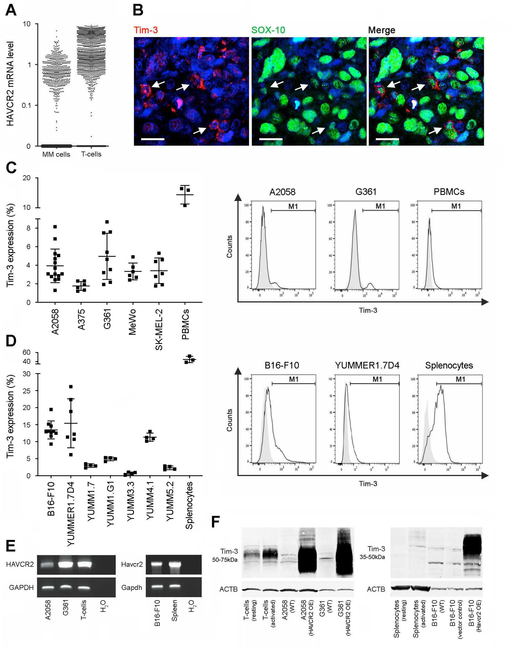
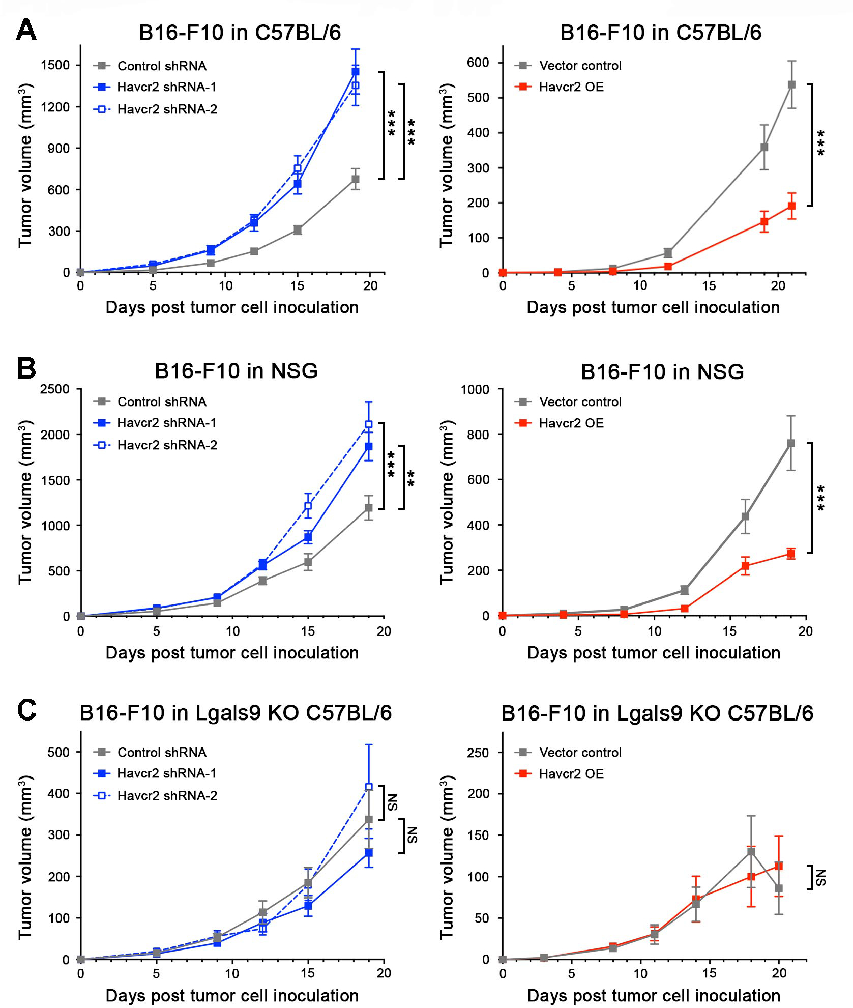
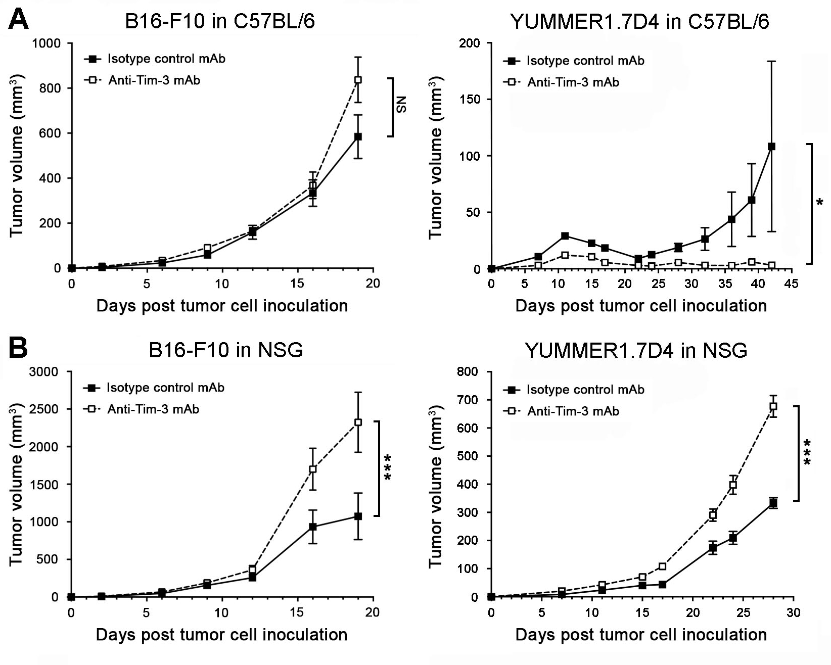
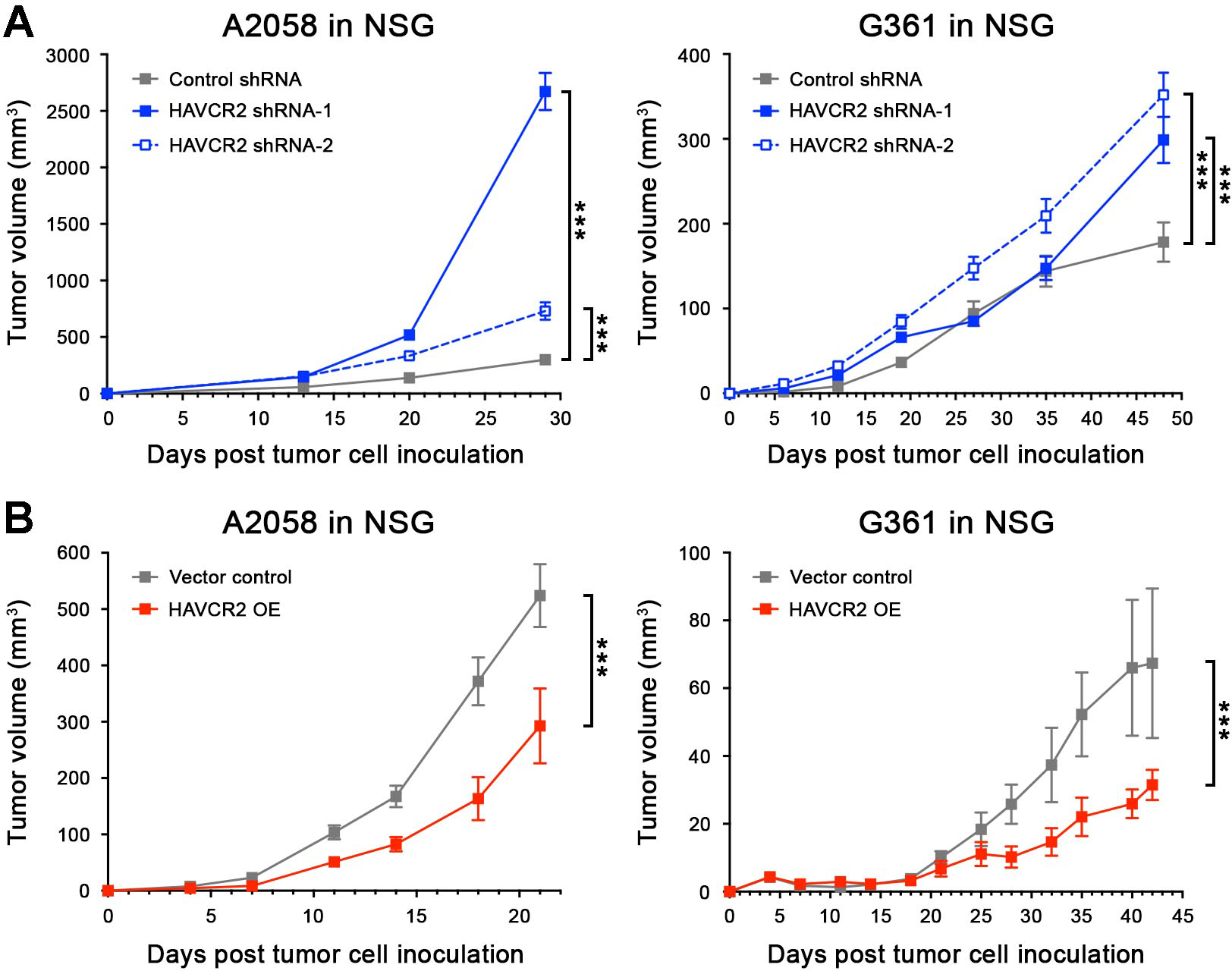
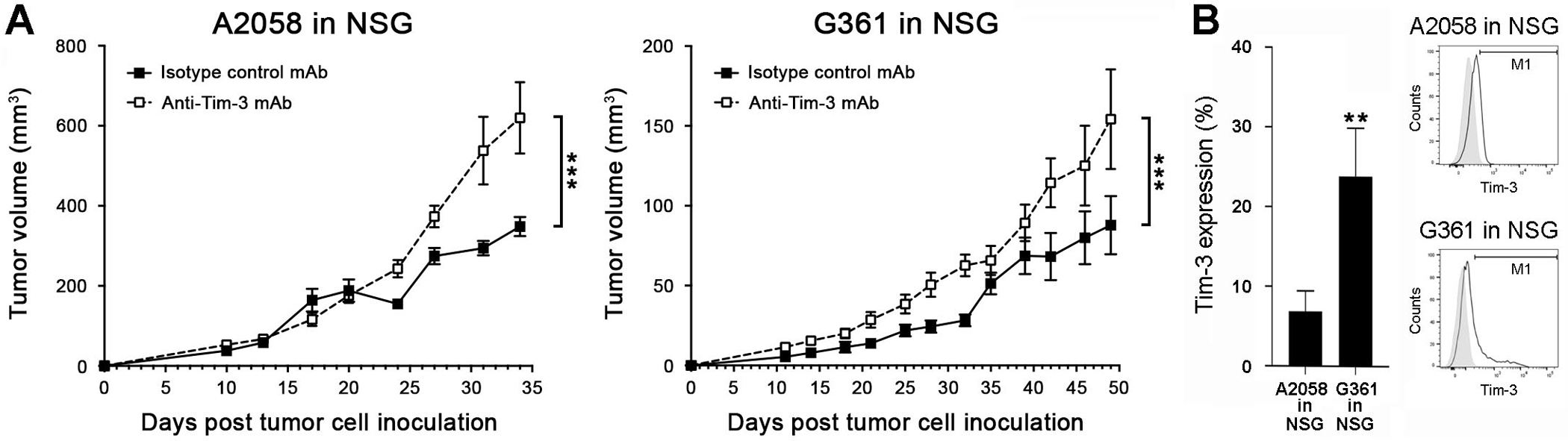
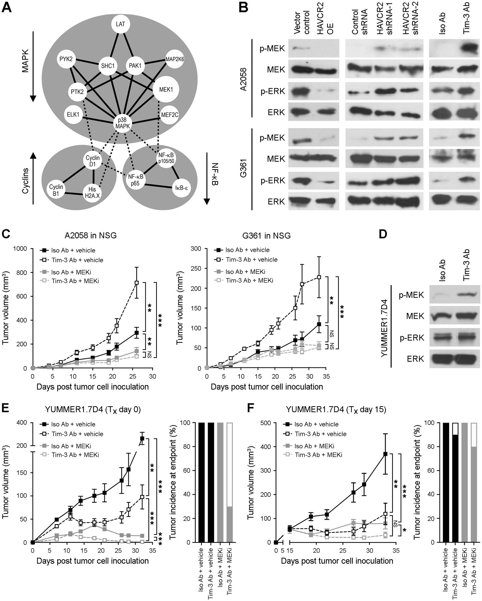
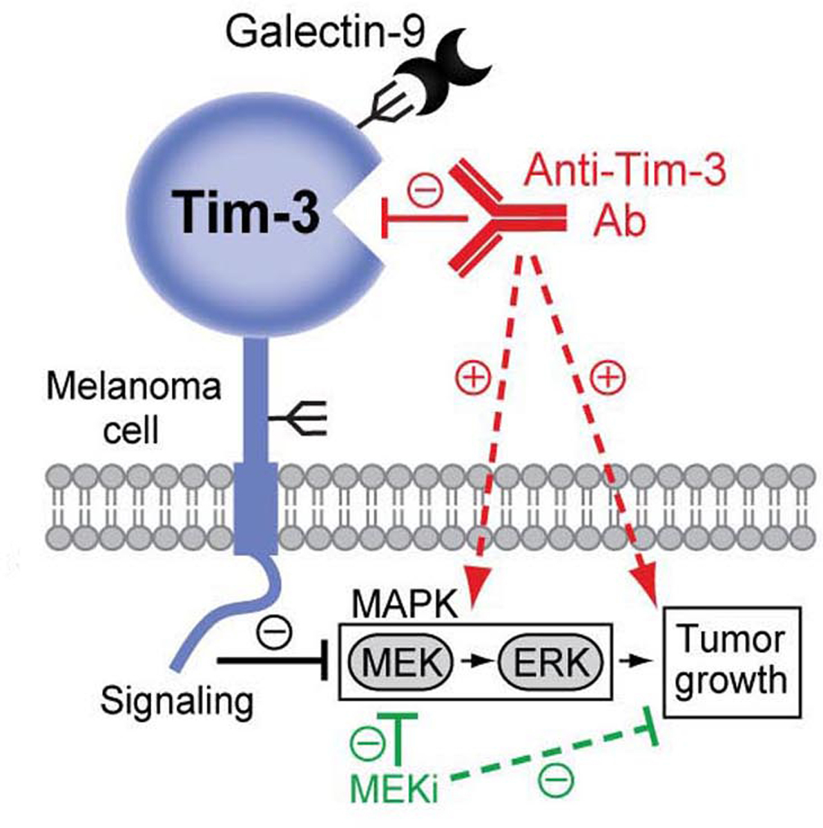
T-cell immunoglobulin mucin family member 3 (Tim-3) is an immune checkpoint receptor that dampens effector functions and causes terminal exhaustion of cytotoxic T cells. Tim-3 inhibitors are under investigation in immuno-oncology (IO) trials, because blockade of T-cell-Tim-3 enhances antitumor immunity. Here, we identify an additional role for Tim-3 as a growth-suppressive receptor intrinsic to melanoma cells. Inhibition of melanoma cell-Tim-3 promoted tumor growth in both immunocompetent and immunocompromised mice, while melanoma-specific Tim-3 overexpression attenuated tumorigenesis. Ab-mediated Tim-3 blockade inhibited growth of immunogenic murine melanomas in T-cell-competent hosts, consistent with established antitumor effects of T-cell-Tim-3 inhibition. In contrast, Tim-3 Ab administration stimulated tumorigenesis of both highly and lesser immunogenic murine and human melanomas in T-cell-deficient mice, confirming growth-promoting effects of melanoma-Tim-3 antagonism. Melanoma-Tim-3 activation suppressed, while its blockade enhanced, phosphorylation of pro-proliferative downstream MAPK signaling mediators. Finally, pharmacologic MAPK inhibition reversed unwanted Tim-3 Ab-mediated tumorigenesis in T-cell-deficient mice and enhanced desired antitumor activity of Tim-3 interference in T-cell-competent hosts. These results identify melanoma-Tim-3 blockade as a mechanism that antagonizes T-cell-Tim-3-directed IO therapeutic efficacy. They further reveal MAPK targeting as a combination strategy for circumventing adverse consequences of unintended melanoma-Tim-3 inhibition.

Significance: Tim-3 is a growth-suppressive receptor intrinsic to melanoma cells, the blockade of which promotes MAPK-dependent tumorigenesis and thus counteracts antitumor activity of T-cell-directed Tim-3 inhibition.


Conflict of interest statement

CONFLICT OF INTEREST: The authors declare no potential conflict of interest.

Figures

Figure 1.
Figure 1.. Tim-3 expression by melanoma cells.
A, Single-cell RNA-seq analysis of human Tim-3 gene (HAVCR2) expression in patient melanoma (MM) cells versus tumor-infiltrating T-cells. B, Representative dual immunofluorescence staining of a clinical melanoma biopsy for co-expression (arrows) of Tim-3 (red) and the melanocytic marker, SOX-10 (green). Nuclei were counterstained with DAPI (blue). Size bars, 20 μm. C-D, Percentages (mean ± SD, left) and representative flow cytometric histograms (right) of Tim-3 surface protein expression by C, human melanoma lines and PBMCs and by D, murine melanoma lines and C57BL/6-derived splenocytes (n = 3–10 independent experiments). E, RT-PCR expression analysis of the full-length HAVCR2 coding sequence by human A2058 and G361 (left) or murine B16-F10 (Havcr2, right) melanoma lines and respective T-cell or splenocyte positive controls. F, Immunoblot of Tim-3 protein expression by human (left) or murine (right) wild-type (WT), vector control and/or Tim-3 (HAVCR2/Havcr2)-overexpressing (OE) melanoma cells, resting versus activated T-cell or splenocyte positive controls. See also Figures S1–S6.
Figure 2.
Figure 2.. Melanoma cell-Tim-3 inhibits murine tumor growth in a Galectin-9-dependent fashion.
Tumor growth kinetics (mean ± SEM) of Tim-3 knockdown (Havcr2 shRNA-1/-2, left) and Tim-3-overexpressing (Havcr2 OE, right) versus shRNA or vector control B16-F10 cells, respectively, in A, immunocompetent C57BL/6, B, immunocompromised NSG, and C, Galectin-9 knockout (Lgals9−/− KO) C57BL/6 mice. All experiments were performed in n ≥ 10 mice per group on n = 2–3 independent occasions. **, p < 0.01; ***, p < 0.001; NS, not significant. See also Figure S7.
Figure 3.
Figure 3.. Antibody-mediated Tim-3 blockade inhibits growth of immunogenic melanomas in immunocompetent mice but promotes tumorigenesis in T-cell-deficient hosts.
Tumor growth kinetics (mean ± SEM) of B16-F10 (left) or YUMMER1.7D4 (right) wild-type cells in A, C57BL/6 and B, NSG mice treated with Tim-3 blocking versus isotype-matched control monoclonal antibodies (mAbs). All experiments were performed in n ≥ 10 mice per group on n = 2 independent occasions. *, p < 0.05; ***, p < 0.001; NS, not significant. See also Figures S7–S8.
Figure 4.
Figure 4.. Tim-3 expression by human melanoma cells inhibits tumorigenesis.
Tumor growth kinetics (mean ± SEM) in NSG mice of A, Tim-3 knockdown (HAVCR2 shRNA-1/-2) and B, Tim-3-overexpressing (HAVCR2 OE) versus respective control human A2058 (left) and G361 (right) melanoma cell inoculates (n = 10 each, representative of n = 3 independent experiments, respectively). ***, p < 0.001. See also Figure S9.
Figure 5.
Figure 5.. Antibody-mediated human melanoma-Tim-3 blockade promotes tumorigenesis.
Tumor growth kinetics (mean ± SEM) of wild-type A, A2058 (left) and G361 cells (right) in NSG mice treated with human-specific anti-Tim-3 blocking versus isotype-matched control monoclonal antibodies (mAbs, n = 4–10 each, representative of n = 3 independent experiments). B, In vivo reactivity (% positive cells, mean ± SEM, left) of the anti-human Tim-3 blocking mAb used in A-B with nuclear GFP-labeled A2058 and G361 melanoma cells isolated from tumor xenografts 3 weeks post tumor cell inoculation into NSG mice (n = 4–6 each). Representative histogram plots are shown on the right. **, p < 0.01; ***, p < 0.001. See also Figure S10.
Figure 6.
Figure 6.. MEK inhibition reverses melanoma-Tim-3 blockade-mediated growth stimulation.
A, Protein-protein interaction map (STRING) of differentially phosphorylated proteins (Phospho Explorer Antibody Array) in Tim-3 (HAVCR2/Havcr2)-overexpressing (OE) versus vector control human A2058 and G361 or murine B16-F10 melanoma cells. Arrows indicate pathway activation (up) or inhibition (down); solid lines, strong protein-protein interactions; dashed lines, weaker protein–protein interactions. B, Immunoblots of phosphorylated (p) and total MEK1/2 and ERK1/2 in vector control versus HAVCR2 OE (left), control shRNA versus HAVCR2 knockdown (shRNA-1/-2, middle), and Tim-3 blocking versus isotype control Ab-treated (right) human A2058 (top) and G361 (bottom) cells. C, Tumor growth kinetics (mean ± SEM) of human A2058 (left) and G361 (right) cells in NSG mice treated with human-specific Tim-3 blocking versus isotype control mAbs, with or without submaximal dosage (0.15 mg/kg/d, p.o.) of the MEK inhibitor, trametinib. D, Immunoblots of p- and total MEK1/2 and ERK1/2 in anti-Tim-3 versus isotype control Ab-treated murine YUMMER1.7D4 melanoma cells. E-F, Tumor growth kinetics (left, mean ± SEM) and tumor incidence at experimental endpoint (right) of murine YUMMER1.7D4 cells in C57BL/6 mice treated (Tx) with anti-murine Tim-3 blocking versus isotype control mAbs, with or without trametinib (as in C) starting on E, day 0 or F, day 15 (n = 10 mice per treatment group, respectively). * p < 0.05; **, p < 0.01; ***, p < 0.001; NS, not significant. See also Figure S11.
Figure 7.
Figure 7.. Therapeutic implications of the melanoma cell intrinsic-Tim-3 signaling axis.
The melanoma cell-Tim-3 receptor binds Galectin-9 and suppresses MAPK signaling to inhibit tumor growth (black, solid line). Ab-based melanoma-Tim-3 blockade (red, solid line) activates MAPK effectors, MEK and ERK, thereby unintentionally promoting tumorigenesis (red, dashed lines). Combination therapy with MEK inhibitors (MEKi) reverses Tim-3 Ab-mediated MEK activation (green, solid line), leading to desirable melanoma growth suppression (green, dashed line).

References

    1. Sharma P, Allison JP. Immune checkpoint targeting in cancer therapy: toward combination strategies with curative potential. Cell 2015;161:205–14 - PMC - PubMed
    1. Kalbasi A, Ribas A. Tumour-intrinsic resistance to immune checkpoint blockade. Nat Rev Immunol 2020;20:25–39 - PMC - PubMed
    1. Koyama S, Akbay EA, Li YY, Herter-Sprie GS, Buczkowski KA, Richards WG, et al. Adaptive resistance to therapeutic PD-1 blockade is associated with upregulation of alternative immune checkpoints. Nat Commun 2016;7:10501. - PMC - PubMed
    1. Shayan G, Srivastava R, Li J, Schmitt N, Kane LP, Ferris RL. Adaptive resistance to anti-PD1 therapy by Tim-3 upregulation is mediated by the PI3K-Akt pathway in head and neck cancer. Oncoimmunol 2017;6:e1261779 - PMC - PubMed
    1. Fourcade J, Sun Z, Benallaoua M, Guillaume P, Luescher IF, Sander C, et al. Upregulation of Tim-3 and PD-1 expression is associated with tumor antigen-specific CD8+ T cell dysfunction in melanoma patients. J Exp Med 2010;207:2175–86 - PMC - PubMed

Publication types