

Diagnostic value of water-fat-separated images and CT-like susceptibility-weighted images extracted from a single ultrashort echo time sequence for the evaluation of vertebral fractures and degenerative changes of the spine
- PMID: 35980430
- PMCID: PMC9889472
- DOI: 10.1007/s00330-022-09061-2
Diagnostic value of water-fat-separated images and CT-like susceptibility-weighted images extracted from a single ultrashort echo time sequence for the evaluation of vertebral fractures and degenerative changes of the spine
Abstract
Objectives: To evaluate the performance of single-echo Dixon water-fat imaging and computed tomography (CT)-like imaging based on a single ultrashort echo time (sUTE) MR sequence for imaging of vertebral fractures as well as degenerative bone changes of the spine in comparison to conventional CT and MR sequences.
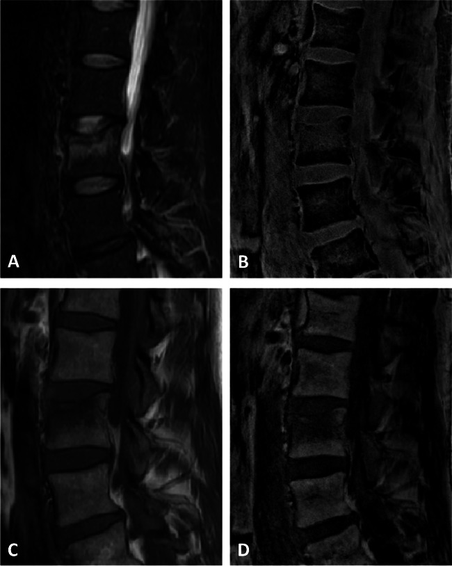
Methods: Thirty patients with suspected acute vertebral fractures were examined using a 3-T MRI, including an sUTE sequence as well as short-tau inversion recovery (STIR) and T1-weighted sequences. During postprocessing, water-fat separation was performed by solving the smoothness-constrained inverse water-fat problem based on a single-complex UTE image. By removing the unwanted low-frequency phase terms, additional MR-based susceptibility-weighted-like (SW-like) images with CT-like contrast were created. Two radiologists evaluated semi-quantitative and quantitative features of fractures and degenerative changes independently and separately on CT and MR images.
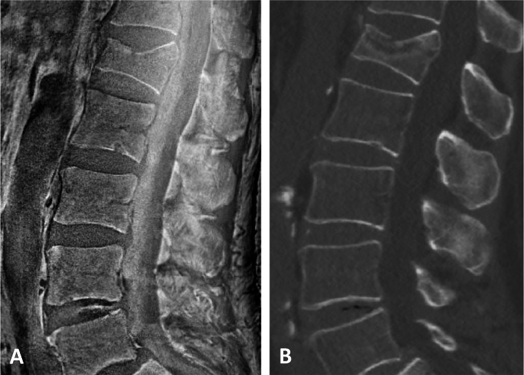
Results: In total, all 58 fractures were accurately detected of whom 24 were correctly classified as acute fractures with an edema detected on the water-fat-separated UTE images, using STIR and T1w sequences as standard of reference. For the morphological assessment of fractures and degenerative changes, the overall agreement between SW-like images and CT was substantial to excellent (e.g., Genant: κ 0.90 (95% confidence interval 0.54-1.00); AO/Magerl: κ 0.75 (95% confidence interval 0.43-1.00)). Overall inter-reader agreement for water-fat-separated UTE images and SW-like images was substantial to almost perfect.
Conclusion: Detection and assessment of vertebral fractures and degenerative bone changes of the spine were feasible and accurate using water-fat-separated images as well as SW-like images, both derived from the same sUTE-Dixon sequence.
Key points: • The detection of acute vertebral fractures was feasible using water-fat-separated images and CT-like images reconstructed from one sUTE sequence. • Assessment of the vertebral fractures using SW-like images with CT-like contrast was found to be comparable to conventional CT. • sUTE imaging of the spine can help reduce examination times and radiation exposure.
Keywords: Bone marrow edema; Magnetic resonance imaging (MRI); Ultrashort echo time (UTE) imaging; Vertebral fractures.
© 2022. The Author(s).
Conflict of interest statement
The authors declare no competing interests.
Figures


Similar articles
-
Magnetic resonance bone imaging: applications to vertebral lesions.Jpn J Radiol. 2023 Nov;41(11):1173-1185. doi: 10.1007/s11604-023-01449-4. Epub 2023 May 20. Jpn J Radiol. 2023. PMID: 37209299 Free PMC article. Review.
-
Assessment of vertebral fractures and edema of the thoracolumbar spine based on water-fat and susceptibility-weighted images derived from a single ultra-short echo time scan.Magn Reson Med. 2022 Apr;87(4):1771-1783. doi: 10.1002/mrm.29078. Epub 2021 Nov 9. Magn Reson Med. 2022. PMID: 34752650
-
CT-like images based on T1 spoiled gradient-echo and ultra-short echo time MRI sequences for the assessment of vertebral fractures and degenerative bone changes of the spine.Eur Radiol. 2021 Jul;31(7):4680-4689. doi: 10.1007/s00330-020-07597-9. Epub 2021 Jan 14. Eur Radiol. 2021. PMID: 33443599 Free PMC article.
-
Comparing CT-Like Images Based on Ultra-Short Echo Time and Gradient Echo T1-Weighted MRI Sequences for the Assessment of Vertebral Disorders Using Histology and True CT as the Reference Standard.J Magn Reson Imaging. 2024 May;59(5):1542-1552. doi: 10.1002/jmri.28927. Epub 2023 Jul 27. J Magn Reson Imaging. 2024. PMID: 37501387
-
Quantitative Ultrashort Echo Time (UTE) Magnetic Resonance Imaging of Bone: An Update.Front Endocrinol (Lausanne). 2020 Sep 18;11:567417. doi: 10.3389/fendo.2020.567417. eCollection 2020. Front Endocrinol (Lausanne). 2020. PMID: 33071975 Free PMC article. Review.
Cited by
-
mFFE CT-like MRI Sequences for the Assessment of Vertebral Fractures.Diagnostics (Basel). 2024 Oct 30;14(21):2434. doi: 10.3390/diagnostics14212434. Diagnostics (Basel). 2024. PMID: 39518401 Free PMC article.
-
Magnetic resonance bone imaging: applications to vertebral lesions.Jpn J Radiol. 2023 Nov;41(11):1173-1185. doi: 10.1007/s11604-023-01449-4. Epub 2023 May 20. Jpn J Radiol. 2023. PMID: 37209299 Free PMC article. Review.
-
Detection of caries lesions using a water-sensitive STIR sequence in dental MRI.Sci Rep. 2024 Jan 5;14(1):663. doi: 10.1038/s41598-024-51151-2. Sci Rep. 2024. PMID: 38182726 Free PMC article.
-
Bone injury imaging in knee and ankle joints using fast-field-echo resembling a CT using restricted echo-spacing MRI: a feasibility study.Front Endocrinol (Lausanne). 2024 Jul 11;15:1421876. doi: 10.3389/fendo.2024.1421876. eCollection 2024. Front Endocrinol (Lausanne). 2024. PMID: 39072275 Free PMC article.
References
MeSH terms
Substances
LinkOut - more resources
Full Text Sources
Medical
