Comment
. 2022 Oct 1;140(10):946-954.
doi: 10.1001/jamaophthalmol.2022.3131.

Association of Predominantly Peripheral Lesions on Ultra-Widefield Imaging and the Risk of Diabetic Retinopathy Worsening Over Time

Collaborators, Affiliations
Comment

Association of Predominantly Peripheral Lesions on Ultra-Widefield Imaging and the Risk of Diabetic Retinopathy Worsening Over Time

Dennis M Marcus et al. JAMA Ophthalmol. .

Erratum in

Abstract

Importance: Ultra-widefield (UWF) imaging improves the ability to identify peripheral diabetic retinopathy (DR) lesions compared with standard imaging. Whether detection of predominantly peripheral lesions (PPLs) better predicts rates of disease worsening over time is unknown.

Objective: To determine whether PPLs identified on UWF imaging are associated with increased disease worsening beyond the risk associated with baseline Early Treatment Diabetic Retinopathy Study (ETDRS) Diabetic Retinopathy Severity Scale (DRSS) score.

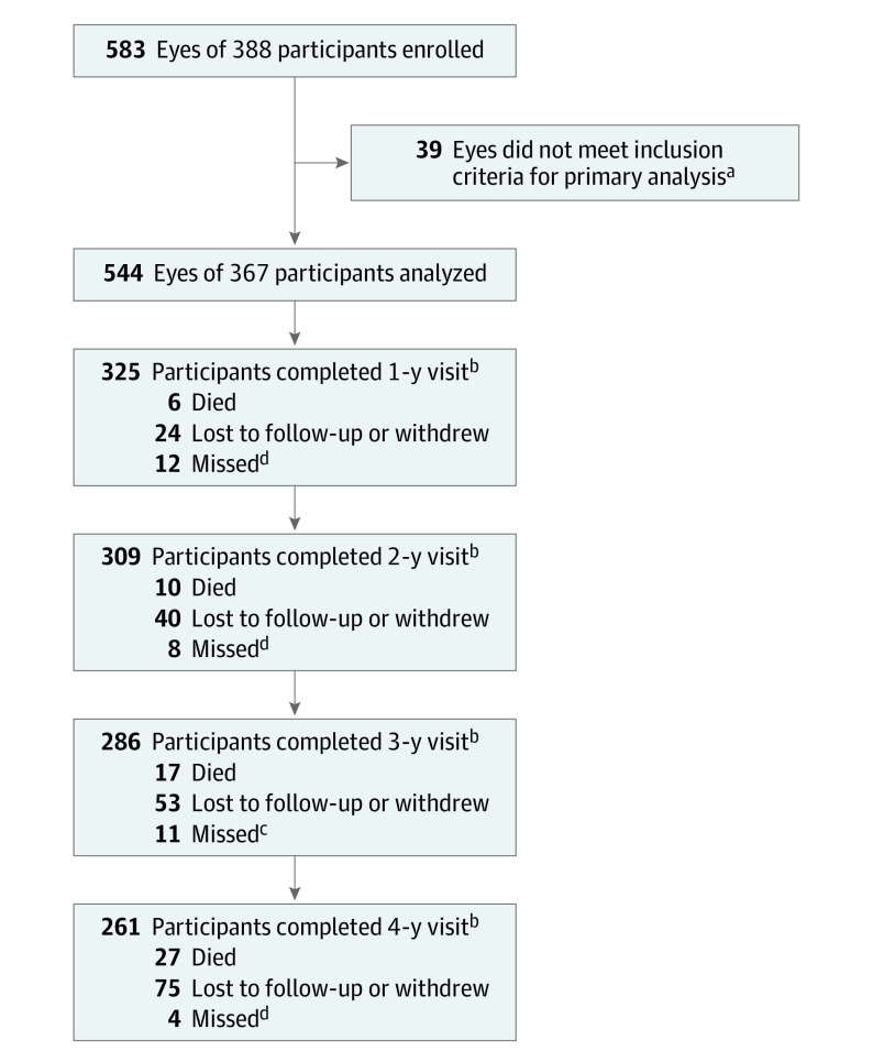
Design, setting, and participants: This cohort study was a prospective, multicenter, longitudinal observational study conducted at 37 US and Canadian sites with 388 participants enrolled between February and December 2015. At baseline and annually through 4 years, 200° UWF-color images were obtained and graded for DRSS at a reading center. Baseline UWF-color and UWF-fluorescein angiography (FA) images were evaluated for the presence of PPL. Data were analyzed from May 2020 to June 2022.

Interventions: Treatment of DR or diabetic macular edema was at investigator discretion.

Main outcomes and measures: Predominantly peripheral lesions were defined as DR lesions with a greater extent outside vs inside the 7 standard ETDRS fields. Primary outcome was disease worsening defined as worsening 2 steps or more on the DRSS or receipt of DR treatment. Analyses were adjusted for baseline DRSS score and correlation between 2 study eyes of the same participant.

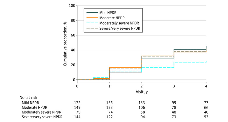
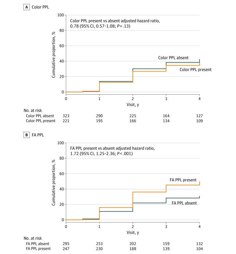
Results: Data for 544 study eyes with nonproliferative DR (NPDR) were analyzed (182 [50%] female participants; median age, 62 years; 68% White). The 4-year disease worsening rates were 45% for eyes with baseline mild NPDR, 40% for moderate NPDR, 26% for moderately severe NPDR, and 43% for severe NPDR. Disease worsening was not associated with color PPL at baseline (present vs absent: 38% vs 43%; HR, 0.78; 95% CI, 0.57-1.08; P = .13) but was associated with FA PPL at baseline (present vs absent: 50% vs 31%; HR, 1.72; 95% CI, 1.25-2.36; P < .001).

Conclusions and relevance: Although no association was identified with color PPL, presence of FA PPL was associated with greater risk of disease worsening over 4 years, independent of baseline DRSS score. These results suggest that use of UWF-FA to evaluate retinas peripheral to standard ETDRS fields may improve the ability to predict disease worsening in NPDR eyes. These findings support use of UWF-FA for future DR staging systems and clinical care to more accurately determine prognosis in NPDR eyes.


Conflict of interest statement

Conflict of Interest Disclosures: Dr Marcus reported fees from Jaeb Center for Health Research for serving as protocol chair during the conduct of the study; grants from Allergan, Amgen, Boehringer Inglheim, Alcon, Kalvista, Ionis, Xplore, Mylan, Samsung, Novartis, Opthea, Chenghdu, Clearside, Ophthotech/Iveric, Outlook, Gemini, Genentech, Graybug, Topcon, Optos, Gyroscope, Stealth Spiam, Apellis, Roche, Novartis, Xplore, Regenxbio, Kodiak, Zeiss, Annexon, Oculis, Alexion, and Regeneron Pharmaceuticals; and serving as a consultant for Regenxbio, Genentech/Roche, Regeneron, Clearside, and Vial outside the submitted work. Dr Silva reported grants from Optos, the Massachusetts Lions Eye Research Foundation, the Medical Research Council (United Kingdom), and the Philippine Council of Health Research and Development; personal fees and nonfinancial research support from Optos during the conduct of the study; personal fees and nonfinancial support from Optomed outside the submitted work; and professional fees from the American Diabetes Association outside the submitted work. Ms Liu reported grants from the National Institutes of Health (NIH) and JDRF; loaner ophthalmic cameras to clinical sites from Optos, Zeiss, and Optovue; and additional funding or devices outside of the submitted work from Regeneron, Helmsley Charitable Trust, PhotoOptx, and Roche. Dr Aiello reported professional fees from Guidepoint Global, Japan Society of Ophthalmology and Diabetes, Johns Hopkins, Novo Nordisk, Perfuse Therapeutics, Samsung, System Analytic, and Valo Health and equity stock and professional fees from Kalvista. Dr Antoszyk reported research support from Apellis, Gemini Therapeutics, Genentech, Kodiak, NGM Biopharmaceutical, Novartis, Novo Nordisk, Amgen, Regeneron, Roche, and Notal and serving on an advisory board for Allergan. Dr Elman reported grants from the Jaeb Center for Health Research during the conduct of the study. Dr Friedman reported research support from Amgen, Boehringer Ingelheim, Chengdu Kang Hong, Opthea, and Regeneron. Mr Glassman reported grants from the NIH and JDRF; loaner ophthalmic cameras to clinical sites from Optos, Zeiss, and Optovue; and additional funding or devices outside of the submitted work from Regeneron, Helmsley Charitable Trust, PhotoOptx, and Roche. Dr Jampol reported professional fees from Alkahest. Ms Melia reported grants from the NIH and JDRF; loaner ophthalmic cameras to clinical sites from Optos, Zeiss, and Optovue; and additional funding or devices outside of the submitted work from Regeneron, Helmsley Charitable Trust, PhotoOptx, and Roche. Ms Preston reported grants from the NIH and JDRF; loaner ophthalmic cameras to clinical sites from Optos, Zeiss, and Optovue; and additional funding or devices outside of the submitted work from Regeneron, Helmsley Charitable Trust, PhotoOptx, and Roche. Dr Wykoff reported grants and other research or professional fees from AbbVie, Adverum, Aerie, AGTC, Aldeyra, Alexion, Alimera Sciences, Alkahest, Allergan, Amgen, Allgenesis, Alnylam, Annexon Biosciences, Apellis, Arrowhead, Asclepix, Bausch and Lomb, Bayer, Boehringer Ingelheim, Bionic Vision, Chengdu Kang Hong, Cholgene Therapeutics, Clearside Biomedical, Curacle, EyePoint, Frontera, Gemini Therapeutics, Genentech, Graybug, Gyroscope Therapeutics, IACTA, Ionis Pharmaceuticals, Irenix, Iveric Bio, Janssen Pharmaceuticals, Kato Pharmaceuticals, Kiora, Kodiak, Kriya, Lowy Medical Research Institute, Nanoscope Technologies, Neurotech, NGM Biopharmaceuticals, Novartis, Ocular Therapeutix, Ocuphire Phara, OccuRx, OliX, ONL Therapeutics, Opthea, Oxurion, Palatin, Perfuse, Polyphotonix, Ray, Recens Medical, Regeneron, RegenXBio, Roche, SamChunDang Pharm, Sandoz, Stealth, Surrozen, THEA, Taiwan Liposome Company, Tissue Gen, Unity Biotechnology, Unity Valo Health, Vitranu, and Xbrane Biopharma and equity or stock in ONL Therapeutics, Polyphotonix Tissue, Gen Visgenx, and Vitranu. Dr Sun reported grants from Jaeb Center for Health Research during the conduct of the study; grants from Optovue, JDRF, Kalvista, Novartis, Novo Nordisk, Boerhinger Ingelheim, Genentech/Roche, Physical Sciences, Janssen, and the Massachusetts Lions Eye Research Foundation outside the submitted work; nonfinancial support from Optovue, Merck, Novartis, Novo Nordisk, Genentech/Roche, Boston Micromachines, and Adaptive Sensory Technologies outside the submitted work; and professional fees from the American Medical Association and American Diabetes Association outside the submitted work. No other disclosures were reported.

Figures

Figure 1.
Figure 1.. Visit Completion Over 4 Years
The primary analysis is a time-to-event analysis. Eyes that did not meet the outcome were censored at the last completed visit. aIncludes only participants who had at least 1 study eye with a baseline Diabetic Retinopathy Severity Scale score of 35-53. bCounts all participants who completed the visit within the following windows (±0.5 y from the target): 183-547 days for 1-year visit, 548-912 days for 2-year visit, 913-1277 days for 3-year visit, and 1278-1642 days for 4-year visit. Numbers of participants who died and were lost to follow-up or those who withdrew are cumulative. cIncludes participants who completed only a phone visit after missing the annual visit. Participants completing the visit outside the analysis window were considered as missing the visit: 1 at year 1, 1 at year 2, and 1 at year 3. dParticipants completing the visit outside the window were considered as missing the visit.
Figure 2.
Figure 2.. Cumulative Proportion of Disease Worsening Through 4 Years by Baseline Diabetic Retinopathy Severity Scale (DRSS) Score on Masked Ultra-Widefield (UWF) Color Images
Worsening was defined by either a worsening of 2 or more steps on the DRSS within Early Treatment Diabetic Retinopathy Study (ETDRS) fields on masked UWF-color images or receipt of diabetic retinopathy (DR) treatment. Eyes receiving treatment with anti–vascular endothelial growth factor, steroid, or vitrectomy for conditions other than DR were censored after treatment initiation. Eyes not meeting the primary outcome or receiving non-DR treatment were censored at the last visit. The cumulative proportion of primary outcome was 45% (95% CI, 37%-54%) in eyes with baseline mild nonproliferative diabetic retinopathy (NPDR), 40% (95% CI, 32%-49%) with moderate NPDR, 26% (95% CI, 17%-38%) with moderately severe NPDR, and 43% (95% CI, 34%-53%) with severe or very severe NPDR.
Figure 3.
Figure 3.. Cumulative Proportion of Disease Worsening Through 4 Years
Worsening was defined by either a worsening of 2 or more steps on the Diabetic Retinopathy Severity Scale (DRSS) within Early Treatment Diabetic Retinopathy Study (ETDRS) fields on masked ultra-widefield (UWF) color images or receipt of diabetic retinopathy (DR) treatment. Eyes receiving treatment with anti–vascular endothelial growth factor, steroid, or vitrectomy for conditions other than DR were censored after treatment initiation. Eyes not meeting the primary outcome or receiving non-DR treatment were censored at the last visit. The Cox proportional hazards model was adjusted for baseline DRSS score within the ETDRS fields on UWF-color images and the correlation between the 2 study eyes from the same participant. According to predominantly peripheral lesion (PPL) status on color images at baseline (A), the cumulative proportion for primary outcome of disease worsening was 38% (95% CI, 31%-45%) for eyes with color PPL and 43% (95% CI, 37%-49%) without color PPL. According to PPL status on fluorescein angiography (FA) at baseline (B), the cumulative proportion for primary outcome of disease worsening was 50% (95% CI, 44%-57%) in eyes with FA PPL and 31% (95% CI, 25%-38%) in eyes without FA PPL.

Comment on

References

    1. Early Treatment Diabetic Retinopathy Study Research Group . Grading diabetic retinopathy from stereoscopic color fundus photographs—an extension of the modified Airlie House classification: ETDRS report number 10. Ophthalmology. 1991;98(5)(suppl):786-806. doi:10.1016/S0161-6420(13)38012-9 - DOI - PubMed
    1. Early Treatment Diabetic Retinopathy Study Research Group . Fundus photographic risk factors for progression of diabetic retinopathy. ETDRS report number 12. Ophthalmology. 1991;98(5)(suppl):823-833. doi:10.1016/S0161-6420(13)38014-2 - DOI - PubMed
    1. Wilkinson CP, Ferris FL III, Klein RE, et al. ; Global Diabetic Retinopathy Project Group . Proposed international clinical diabetic retinopathy and diabetic macular edema disease severity scales. Ophthalmology. 2003;110(9):1677-1682. doi:10.1016/S0161-6420(03)00475-5 - DOI - PubMed
    1. Oishi A, Hidaka J, Yoshimura N. Quantification of the image obtained with a wide-field scanning ophthalmoscope. Invest Ophthalmol Vis Sci. 2014;55(4):2424-2431. doi:10.1167/iovs.13-13738 - DOI - PubMed
    1. Silva PS, Cavallerano JD, Sun JK, Noble J, Aiello LM, Aiello LP. Nonmydriatic ultrawide field retinal imaging compared with dilated standard 7-field 35-mm photography and retinal specialist examination for evaluation of diabetic retinopathy. Am J Ophthalmol. 2012;154(3):549-559.e2. doi:10.1016/j.ajo.2012.03.019 - DOI - PubMed

Publication types