. 2022 Sep;21(9):e13692.
doi: 10.1111/acel.13692. Epub 2022 Aug 18.

B cell-intrinsic changes with age do not impact antibody-secreting cell formation but delay B cell participation in the germinal centre reaction

Affiliations

B cell-intrinsic changes with age do not impact antibody-secreting cell formation but delay B cell participation in the germinal centre reaction

Jia Le Lee et al. Aging Cell. 2022 Sep.

Abstract

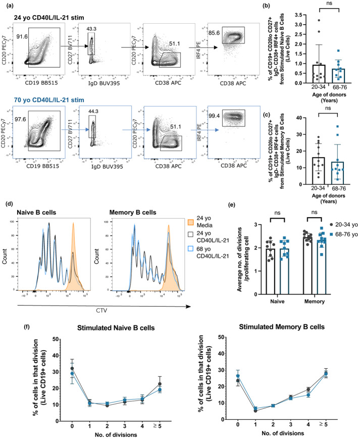
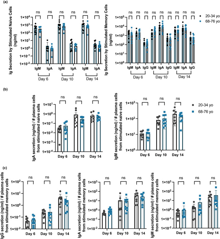
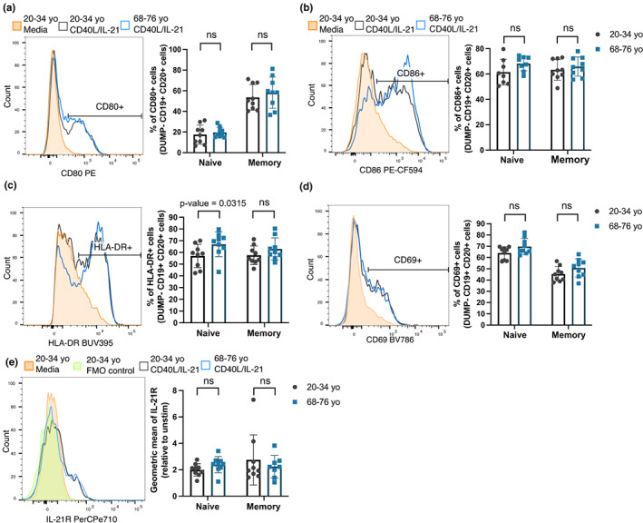
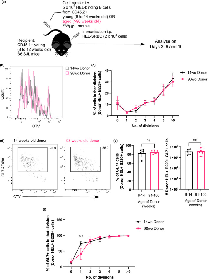
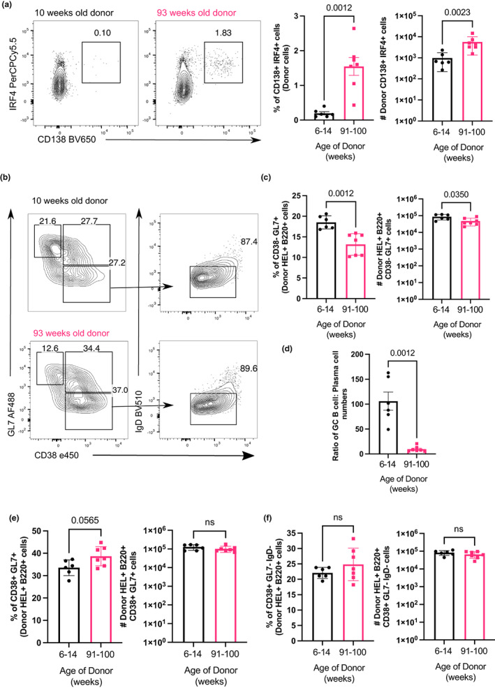
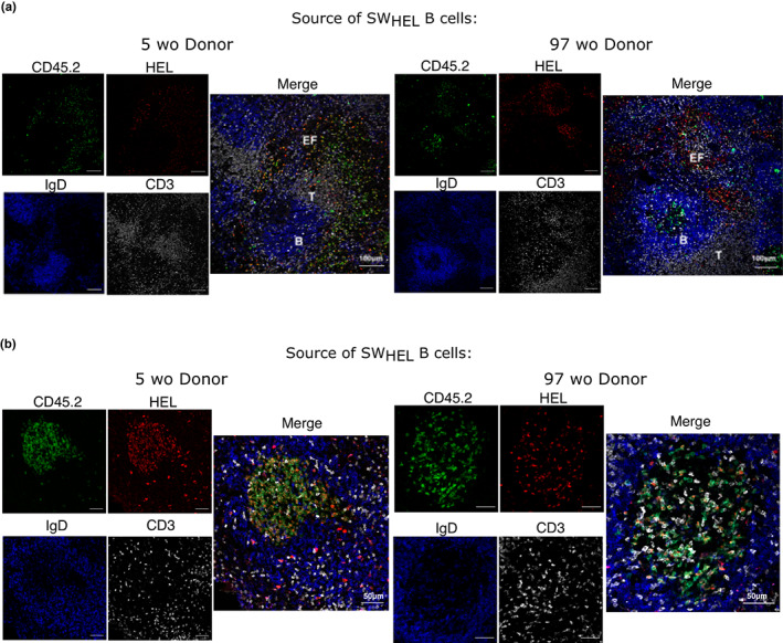
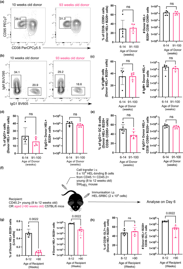
Vaccines typically protect against (re)infections by generating pathogen-neutralising antibodies. However, as we age, antibody-secreting cell formation and vaccine-induced antibody titres are reduced. Antibody-secreting plasma cells differentiate from B cells either early post-vaccination through the extrafollicular response or from the germinal centre (GC) reaction, which generates long-lived antibody-secreting cells. As the formation of both the extrafollicular antibody response and the GC requires the interaction of multiple cell types, the impaired antibody response in ageing could be caused by B cell intrinsic or extrinsic factors, or a combination of the two. Here, we show that B cells from older people do not have intrinsic defects in their proliferation and differentiation into antibody-secreting cells in vitro compared to those from the younger donors. However, adoptive transfer of B cells from aged mice to young recipient mice showed that differentiation into extrafollicular plasma cells was favoured at the expense of B cells entering the GC during the early stages of GC formation. In contrast, by the peak of the GC response, GC B cells derived from the donor cells of aged mice had expanded to the same extent as those from the younger donors. This indicates that age-related intrinsic B cell changes delay the GC response but are not responsible for the impaired antibody-secreting response or smaller peak GC response in ageing. Collectively, this study shows that B cells from aged individuals are not intrinsically defective in responding to stimulation and becoming antibody-secreting cells, implicating B cell-extrinsic factors as the primary cause of age-associated impairment in the humoral immunity.

Keywords: B cells; ageing; antibodies; vaccine response.


Conflict of interest statement

The authors declare that they have no conflicts of interest.

Figures

FIGURE 1
FIGURE 1
B cells from older human donors do not have defects in differentiating into plasma cells and proliferating upon stimulation. (a) Gating strategy for plasma cells (CD19+ CD27+ IgD CD20lo CD38+ IRF4+) 6 days after memory B cells were stimulated with CD40L and IL‐21 or unstimulated (cultured with media). Cells were pre‐gated for live cells and single cells. (b, c) Percentages of plasma cells out of live cells derived from naïve (b) and memory B cells (c) of younger (20–34 years old) and older donors (68–76 years old) after 6 days stimulation with CD40L and IL‐21. Data representative of four independent repeat experiments. (d) Representative flow cytometric histograms showing the cell trace violet stains of live naïve and memory CD19+ B cells from a 24 year old (grey) and 68 year old (blue) donor after 6 days incubation with CD40L and IL‐21 or media (orange). (e) Graph depicting the average number of divisions undergone by proliferating CD19+ naïve and memory B cells after 6 days stimulation with CD40L and IL‐21. (f) Graph showing the percentage of B cells in each division after 6 days stimulation with CD40L and IL‐21. Bar height corresponds to the mean, error bars indicate standard deviation, and each symbol represents values from independent donors. Statistics were calculated using the unpaired Mann–Whitney U test. Data were representative of three independent repeat experiments.
FIGURE 2
FIGURE 2
Stimulated naïve and memory B cells of older human donors have no defects in secreting antibodies. (a) Graphs showing the total amount of IgM, IgA and IgG secreted in the cultures of stimulated naïve (left) and memory (right) B cells 6, 10 and 14 days post‐stimulation with CD40L and IL‐21. (b, c) Graphs comparing the antibody‐secreting capacity (Ig concentration divided by number of antibody‐secreting cells) derived from stimulated (b) naive and (c) memory cells from younger and older donors after stimulation with CD40L and IL‐21. Number of antibody‐secreting cells was determined by flow cytometry with counting beads. Bar height corresponds to the mean, error bars indicate standard deviation, and each symbol represents values from independent donors. Statistics were calculated using the unpaired Mann–Whitney U test. Data were representative of three independent repeat experiments.
FIGURE 3
FIGURE 3
B cells from older human donors are able to upregulate costimulatory molecules and activation markers after stimulation. (a–e) Representative flow cytometric histograms showing the expression levels of costimulatory molecules by memory B cells from a 29 year old (grey) and 70 year old (blue) donor after 48 h stimulation with CD40L and IL‐21 or media (orange) and graphs showing the percentages of cells positive for the marker, or geometric mean of marker using the gating strategies shown, for CD80 (a), CD86 (b), HLA–DR (c), CD69 (d) and IL‐21R (e). These were gated on live CD19+ CD20+ cells. Fluorescent minus one (FMO) controls were included for the IL‐21R stain and are shown on the representative histogram. Bar height corresponds to the mean, error bars indicate standard deviation, and each symbol represents values from independent donors. Statistics were calculated using the unpaired Mann–Whitney U test. Data were representative of three independent repeat experiments.
FIGURE 4
FIGURE 4
B cells from aged donor mice do not have defects in proliferation after immunisation. (a) Schematic diagram of adoptive transfer experiments to compare intrinsic function of B cells from young and aged mice in young recipient mice post‐immunisation. (b) Representative flow cytometric histograms showing the cell trace violet stains of donor HEL+ B220+ cells from either a young adult (14 weeks old) or aged (98 weeks old) mouse 3 days post‐transfer and immunisation. (c) Graph showing the percentage of donor HEL+ B cells in each division in recipient spleens 3 days post‐transfer and immunisation. (d) Representative flow cytometric plots for gating of GL7+ cells. Numbers adjacent to gates indicate percentage of donor HEL+ B220+ cells. (e) Percentage and number of GL7+ cells derived from donor cells from young or aged mice in recipient spleens 3 days post‐transfer and immunisation. Bar graphs show the results of one of two independent experiments (n = 5–6 per group/experiment). Bar height corresponds to the mean, error bars indicate standard deviation, and each symbol represents one biological replicate. Statistics were calculated using the unpaired Mann–Whitney U test. (f) Graph showing the percentage of GL7+ out of HEL+ Donor B cells in each division. p‐Value shown was generated using two‐way ANOVA with the Sidak's multiple comparisons test. Data were representative of two independent repeat experiments.
FIGURE 5
FIGURE 5
B cells from aged donor mice have enhanced extrafollicular response and reduced GC formation early post‐vaccination. (a) Representative flow cytometric plots of CD138+ IRF4+ plasma cells gated on donor cells from 10‐ or 93‐week old donor mice in recipient spleens 6 days post‐transfer and immunisation. Numbers adjacent to gates indicate percentage of donor HEL+ B220+ cells. Percentage and number of donor‐derived CD138+ IRF4+ plasma cells are plotted on the graphs on the right. (b) Representative flow cytometric plots showing gating strategies for donor‐derived GC B cells (CD38 GL7+), activated B cells (CD38+ GL7+) and memory B cells (CD38+ GL7 IgD) from 10 weeks or 93 week old donor mice. Numbers adjacent to gates indicate percentage of donor HEL+ B220+ cells. (c) Percentage and number of HEL+ donor‐derived GC B cells in recipient spleen on day 6 post‐transfer and immunisation. (d) Graph showing the ratio of GC B cells: Plasma cells numbers derived from donor cells from 10 weeks or 93 weeks old donor mice. (e, f) Percentage and number of (e) CD38+ GL7+ B cells and (f) CD38+ GL7 IgD memory B cells on day 6 post‐transfer and immunisation. Bar graphs show the results of one of two independent experiments (n = 5–7 per group/experiment). Bar height corresponds to the mean, error bars indicate standard deviation, and each symbol represents one biological replicate. Statistics were calculated using the unpaired Mann–Whitney U test.
FIGURE 6
FIGURE 6
B cells from aged donor mice preferentially localize to the extrafollicular compartment early in the immune response. Confocal images of spleens of recipient mice taken 6 days post‐transfer of B cells from 5 or 97 weeks old SWHEL donor mouse and HEL–SRBC immunisation. 10 μm spleen sections were stained with anti‐CD45.2 (green), anti‐HEL (red), anti‐IgD (blue) and anti‐CD3 (white) antibodies and imaged at 20× to look at the localisation of transferred cells in the (a) extrafollicular compartment and the (b) germinal centre. Data were representative of two independent repeat experiments.
FIGURE 7
FIGURE 7
B cells from aged donor mice do not have defects in participating in the GC 10 days after immunization in young recipient mice and fewer B cells from young donor mice are recovered when transferred into aged recipient mice. (a) Representative flow cytometric plots of CD38 CD95+ GC B cells gated on donor cells from 10‐week‐old or 93‐week‐old donor mice in recipient spleens on day 10 post‐transfer and immunisation. Numbers adjacent to gates indicate percentage of donor HEL+ B220+ cells. Percentage and number of donor‐derived GC B cells (Donor HEL+ B220+ CD38 CD95+) are plotted on the graphs on the right. (b) Representative flow cytometric plots of IgM+ and IgG1+ donor HEL+ B cells. Numbers adjacent to gates indicate percentage of donor HEL+ B220+ cells. (c, d) Percentage and number of (c) IgM+ and (d) IgG1+ B cells derived from donor cells from young or aged mice in recipient spleens 10 days post‐transfer and immunisation. (e) Graph showing the percentage and number of IgG1+ GC B cells out of total Donor HEL+ B220+ CD38 CD95+ cells. (f) Schematic diagram of adoptive transfer of B cells from young SWHEL mice into young (8–12 weeks old) or aged (>90 weeks old) mice in which B cells response was analysed 6 days post‐transfer and immunisation. (g) Percentage and number of donor HEL+ B220+ cells in spleens of young or aged recipient mice 6 days post‐transfer and immunisation. (h) Percentage and number of donor‐derived GC B cells (Donor HEL+ B220+ CD38 GL7+) in spleens of young or aged recipient mice 6 days post‐transfer and immunisation. Bar graphs show the results of one of two independent experiments (n = 5–6 per group/experiment). Bar height corresponds to the mean, error bars indicate standard deviation and each symbol represents one biological replicate. Statistics were calculated using the unpaired Mann–Whitney U test.

Similar articles

Cited by

References

    1. Attridge, K. , Kenefeck, R. , Wardzinski, L. , Qureshi, O. S. , Wang, C. J. , Manzotti, C. , Okkenhaug, K. , & Walker, L. S. (2014). IL‐21 promotes CD4 T cell responses by phosphatidylinositol 3‐kinase‐dependent upregulation of CD86 on B cells. The Journal of Immunology, 192, 2195–2201. - PMC - PubMed
    1. Blaeser, A. , McGlauchlen, K. , & Vogel, L. A. (2008). Aged B lymphocytes retain their ability to express surface markers but are dysfunctional in their proliferative capability during early activation events. Immunity and Ageing, 5, 1–10. - PMC - PubMed
    1. Breitbart, E. , Wang, X. , Leka, L. S. , Dallal, G. E. , Meydani, S. N. , & Stollar, B. D. (2002). Altered memory B‐cell homeostasis in human aging. Journals of Gerontology – Series A Biological Sciences and Medical Sciences, 57A, 304–311. - PubMed
    1. Brink, R. , Paus, D. , Bourne, K. , Hermes, J. R. , Gardam, S. , Phan, T. G. , & Chan, T. D. (2015). The SWHEL system for high‐resolution analysis of in vivo antigen‐specific T‐dependent B cell responses. Methods in Molecular Biology, 1291, 103–123. - PubMed
    1. Bums, E. A. , Lum, L. G. , L'hommedieu, G. , & Goodwin, J. S. (1993). Specific humoral immunity in the elderly: In vivo and in vitro response to vaccination. Journal of Gerontology: Biological Sciences, 48, 231–236. - PubMed

Publication types