Observational Study
. 2022 Nov 1;31(11):854-859.
doi: 10.1097/IJG.0000000000002101. Epub 2022 Aug 11.

Frequency of Optical Coherence Tomography Testing to Detect Progression in Glaucoma

Affiliations
Observational Study

Frequency of Optical Coherence Tomography Testing to Detect Progression in Glaucoma

Bruna Melchior et al. J Glaucoma. .

Abstract

Prcis: With high specificity and less variability than perimetry, more frequent testing resulted in shorter time to detect progression, though a 6-month testing interval provides a reasonable trade-off for following glaucoma patients using optical coherence tomography (OCT).

Purpose: To investigate the time to detect progression in glaucomatous eyes using different OCT test intervals.

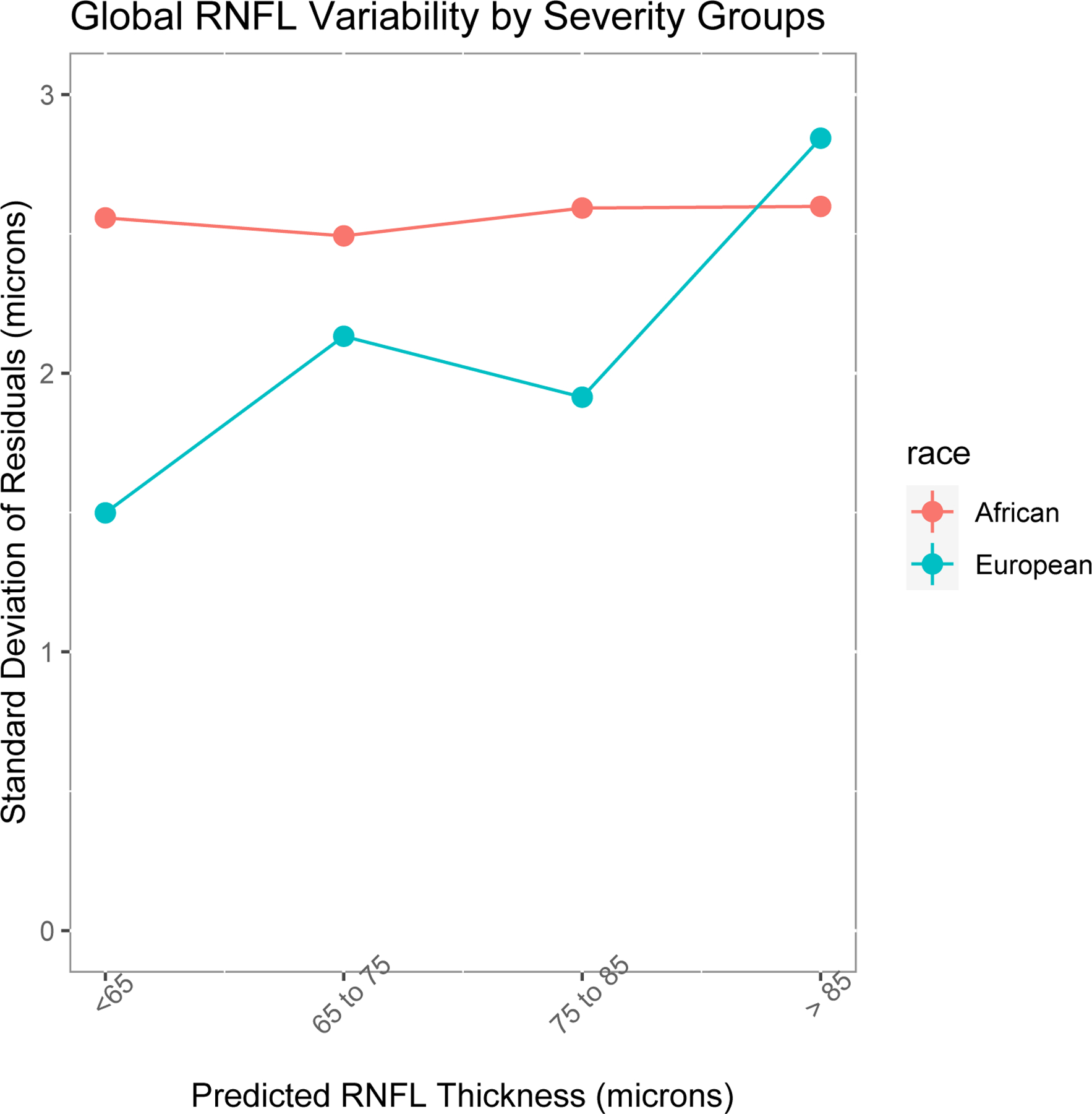
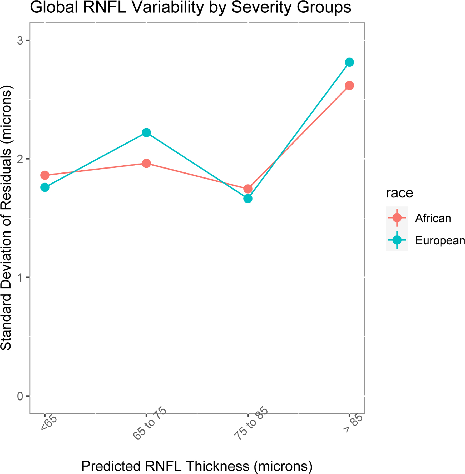
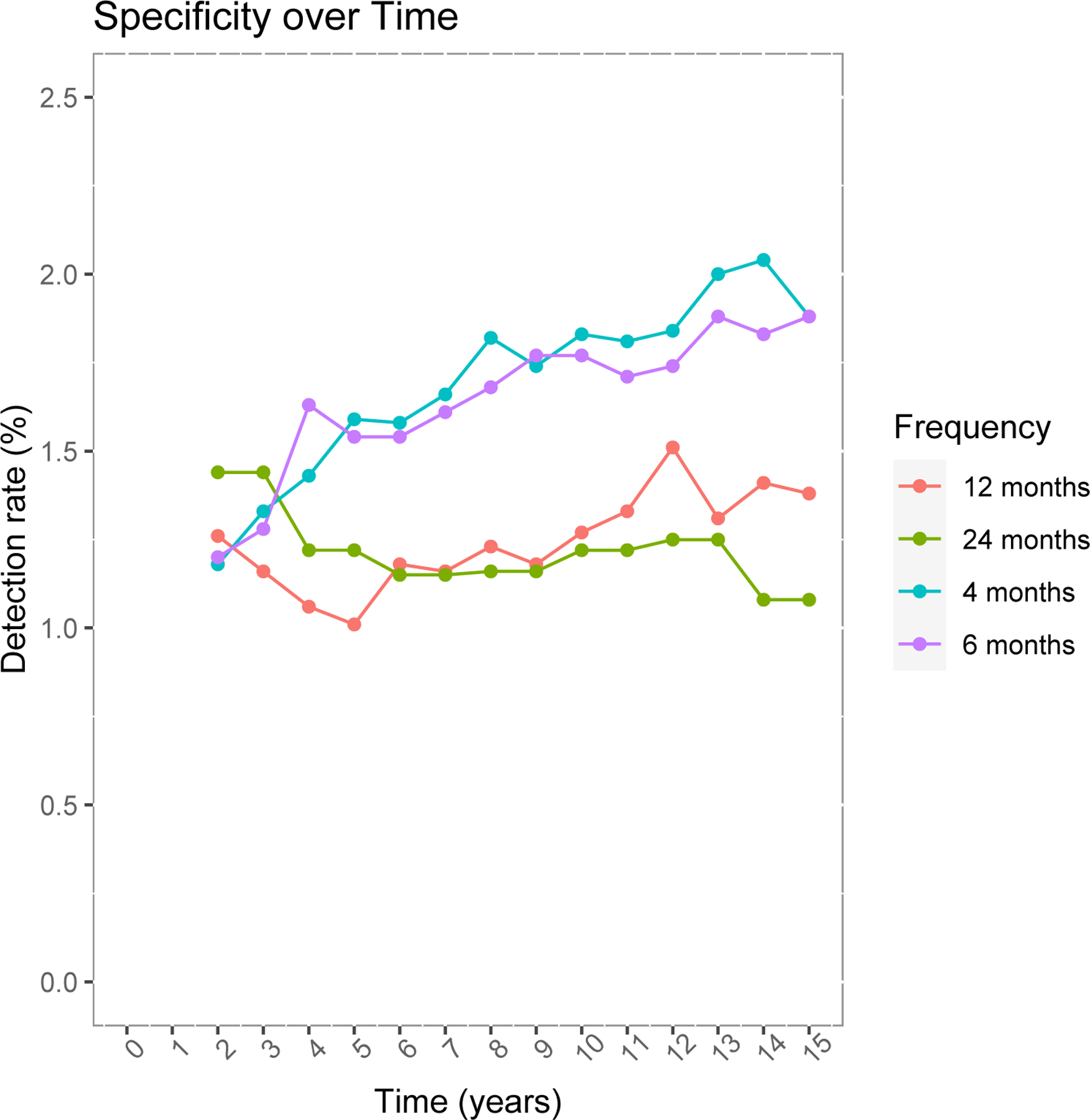
Materials and methods: Participants with manifest glaucoma from the African Descent and Glaucoma Evaluation Study (ADAGES), a multicenter, prospective, observational cohort study, were included. A total of 2699 OCT tests from 171 glaucomatous and 149 normal eyes of 182 participants, with at least 5 tests and 2 years of follow-up, were analyzed. Computer simulations (n=10,000 eyes) were performed to estimate time to detect progression of global circumpapillary retinal nerve fiber layer thickness (cpRNFL) measured with OCT tests. Simulations were based on different testing paradigms (every 4, 6, 12, and 24 mo) and different rates of change (µm/year). Time to detect significant progression ( P <0.05) at 80% and 90% power were calculated for each paradigm and rate of cpRNFL change.

Results: As expected, more frequent testing resulted in shorter time to detect progression. Although there was clear disadvantage for testing at intervals of 24 versus 12 months (~22.4% time [25 mo] increase in time to progression detection) and when testing 12 versus 6 months (~22.1% time [20 mo] increase), the improved time to detect progression was less pronounced when comparing 6 versus 4 months (~11.5% time [10 mo] reduction).

Conclusion: With high specificity and less variability than perimetry, a 6-month testing interval provides a reasonable trade-off for following glaucoma patients using OCT.


Conflict of interest statement

Disclosure: The authors declare no conflict of interest.

Figures

Figure 1.
Figure 1.
Variability of global circumpapillary retinal nerve fiber layer (cpRNFL) thickness measured with optical coherence tomography (OCT). The variability is described as the standard deviation (µm) of global cpRNFL thickness residuals at 4 levels of RNFL thickness, representing different stages of glaucoma severity. (A) Comparison in eyes with glaucoma by ancestry; (B) comparison in eyes without glaucoma by ancestry.
Figure 1.
Figure 1.
Variability of global circumpapillary retinal nerve fiber layer (cpRNFL) thickness measured with optical coherence tomography (OCT). The variability is described as the standard deviation (µm) of global cpRNFL thickness residuals at 4 levels of RNFL thickness, representing different stages of glaucoma severity. (A) Comparison in eyes with glaucoma by ancestry; (B) comparison in eyes without glaucoma by ancestry.
Figure 2.
Figure 2.
Power to detect progression (%) of retinal nerve fiber layer (RNFL) in stable eyes (slope of average RNFL thickness = 0 µm/year) with optical coherence tomography in different test intervals (4, 6, 12 and 24 months).

References

    1. Garcia-Valenzuela E, Shareef S, Walsh J, Sharma SC. Programmed cell death of retinal ganglion cells during experimental glaucoma. Experimental Eye Research 1995;61(1):33–44. doi:10.1016/S0014-4835(95)80056-5 - DOI - PubMed
    1. Sommer A, Miller NR, Pollack I, Maumenee AE, George T. The Nerve Fiber Layer in the Diagnosis of Glaucoma. Archives of Ophthalmology 1977;95(12):2149–2156. doi:10.1001/archopht.1977.04450120055003 - DOI - PubMed
    1. Weinreb RN, Aung T, Medeiros FA. The pathophysiology and treatment of glaucoma: A review. JAMA - Journal of the American Medical Association 2014;311(18):1901–1911. doi:10.1001/jama.2014.3192 - DOI - PMC - PubMed
    1. Dong ZM, Wollstein G, Schuman JS. Clinical utility of optical coherence tomography in glaucoma. Investigative Ophthalmology and Visual Science 2016;57(9Special Issue):OCT556–OCT567. doi:10.1167/iovs.16-19933 - DOI - PMC - PubMed
    1. Wollstein G, Schuman JS, Price LL, et al. Optical coherence tomography longitudinal evaluation of retinal nerve fiber layer thickness in glaucoma. Archives of Ophthalmology 2005;123(4):464–470. doi:10.1001/archopht.123.4.464 - DOI - PMC - PubMed

Publication types