. 2022 Aug 18;17(8):e0269140.
doi: 10.1371/journal.pone.0269140. eCollection 2022.

Acyloxyacyl hydrolase regulates microglia-mediated pelvic pain

Affiliations

Acyloxyacyl hydrolase regulates microglia-mediated pelvic pain

Afrida Rahman-Enyart et al. PLoS One. .

Abstract

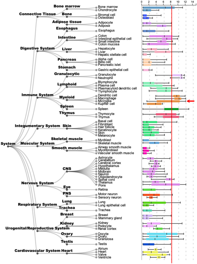
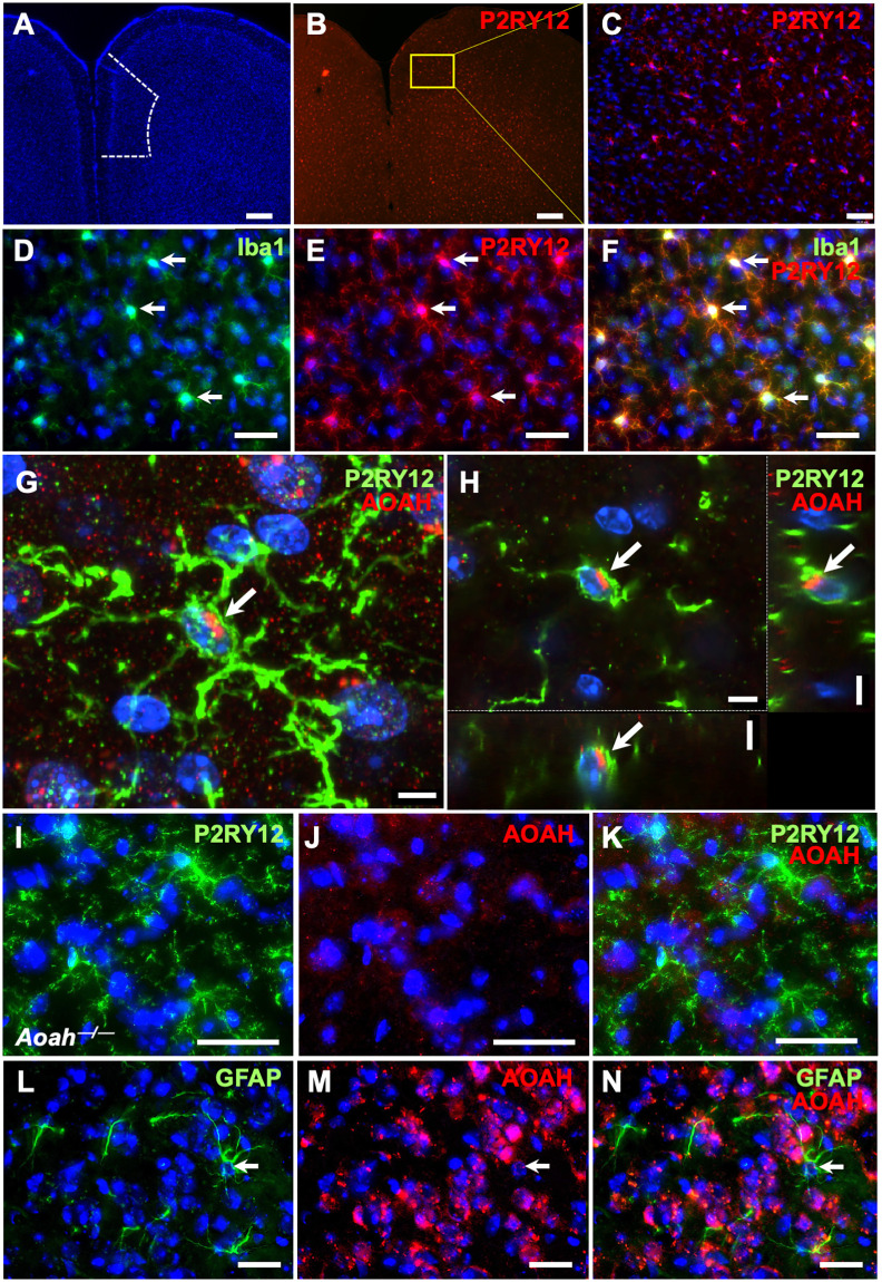
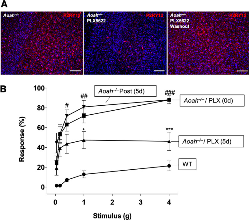
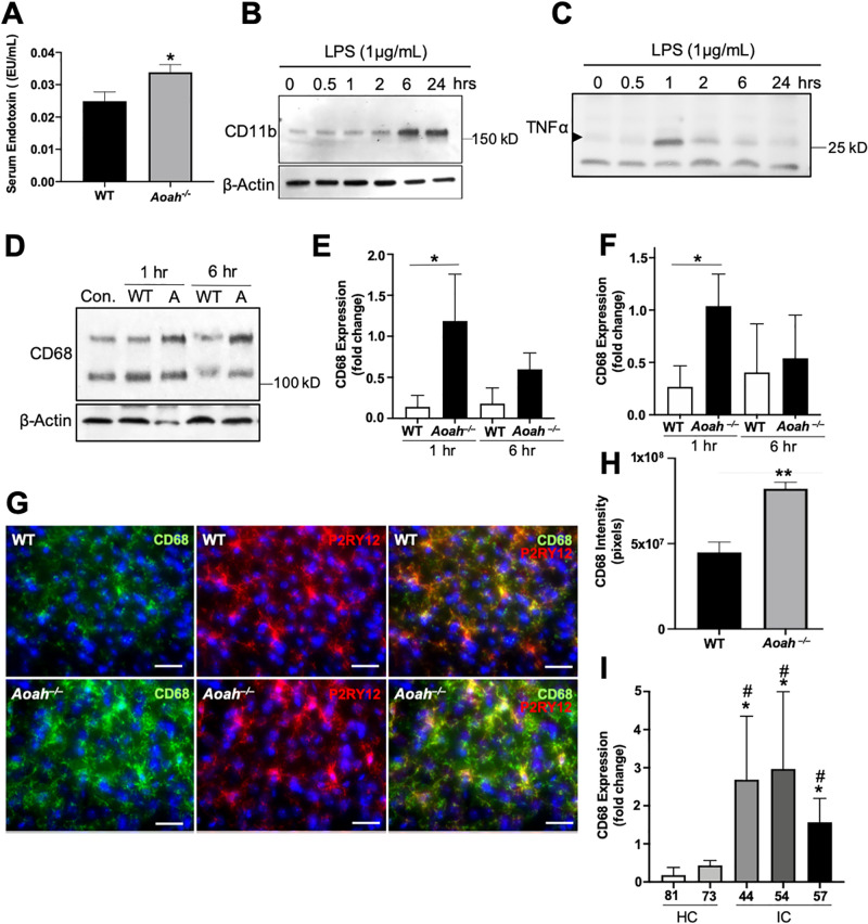
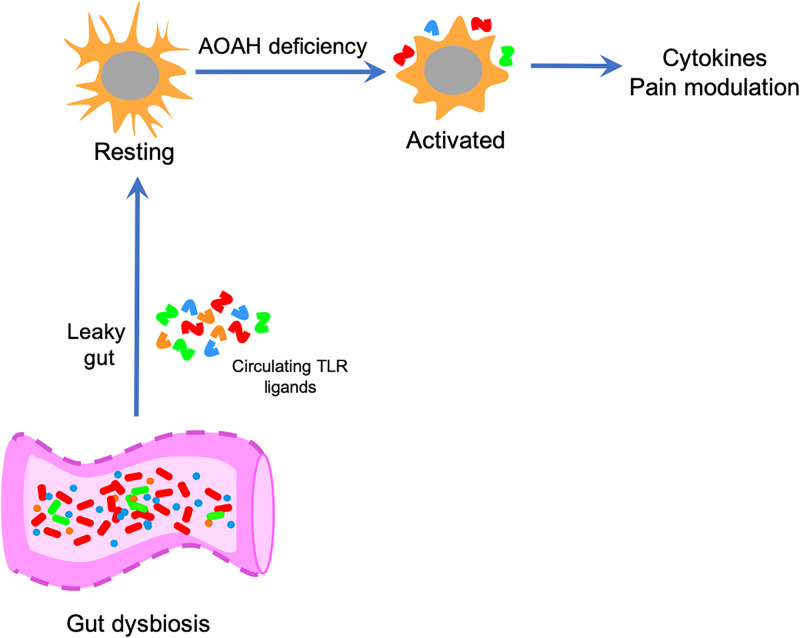
Chronic pelvic pain conditions such as interstitial cystitis/bladder pain syndrome (IC/BPS) remain clinical and mechanistic enigmas. Microglia are resident immune cells of the central nervous system (CNS) that respond to changes in the gut microbiome, and studies have linked microglial activation to acute and chronic pain in a variety of models, including pelvic pain. We have previously reported that mice deficient for the lipase acyloxyacyl hydrolase (AOAH) develop pelvic allodynia and exhibit symptoms, comorbidities, and gut dysbiosis mimicking IC/BPS. Here, we assessed the role of AOAH in microglial activation and pelvic pain. RNAseq analyses using the ARCHS4 database and confocal microscopy revealed that AOAH is highly expressed in wild type microglia but at low levels in astrocytes, suggesting a functional role for AOAH in microglia. Pharmacologic ablation of CNS microglia with PLX5622 resulted in decreased pelvic allodynia in AOAH-deficient mice and resurgence of pelvic pain upon drug washout. Skeletal analyses revealed that AOAH-deficient mice have an activated microglia morphology in the medial prefrontal cortex and paraventricular nucleus, brain regions associated with pain modulation. Because microglia express Toll-like receptors and respond to microbial components, we also examine the potential role of dysbiosis in microglial activation. Consistent with our hypothesis of microglia activation by leakage of gut microbes, we observed increased serum endotoxins in AOAH-deficient mice and increased activation of cultured BV2 microglial cells by stool of AOAH-deficient mice. Together, these findings demonstrate a role for AOAH in microglial modulation of pelvic pain and thus identify a novel therapeutic target for IC/BPS.


Conflict of interest statement

The authors have declared that no competing interests exist.

Figures

Fig 1
Fig 1. Tissue expression of Aoah/AOAH mRNA.
Boxplot obtained from the public RNAseq data analysis tool ARCHS4 showing Aoah/AOAH expression in different tissue types. For comparison of microglia (arrow) relative to other cell types, approximate mean microglial expression is indicated (red line) [41].
Fig 2
Fig 2. Immunostaining of AOAH in glial cells.
A-C: P2RY12 staining labels microglia in murine P2RY12+ cells (red) in and beyond the prefrontal cortex (A, prefrontal cortex outlined in a single hemisphere by dotted line). A and B captured with 2.5X objective (scale bar: 120μm). C captured with a 20X objective (scale bar: 15μm). D-F: Combined z-stack of WT mouse prefrontal cortex stained for the microglial proteins Iba1 (green) and P2RY12 (red). DAPI staining nuclei shown in blue (n = 3 mice, scale bar: 30 μm). Images were captured with a 40X objective. G and H: Confocal microscopy showing combined z-stack (G) and individual planes (H) obtained from WT mouse prefrontal cortex stained for microglial protein P2RY12 (green) and AOAH (red). DAPI staining nuclei shown in blue (n = 3 mice, scale bar: 5 μm). Images were captured with a 60X objective. I-K: Staining of AOAH-deficient prefrontal cortex with anti-AOAH antibodies revealed an absence of AOAH (red) immunoreactivity in P2RY12+ cells (green; scale bar: 30 μm). Z-stacks were captured with a 40X objective. n = 3 mice. L-N: Combined z-stack of WT mouse prefrontal cortex stained for the astrocyte protein glial fibrillary acidic protein (GFAP, green) and AOAH (red). DAPI staining nuclei shown in blue (n = 3 mice, scale bar: 30 μm). Images were taken with a 40X objective.
Fig 3
Fig 3. Pharmacologic ablation of microglia reduces pelvic allodynia of AOAH-deficient mice.
A: Immunostaining of P2RY12 in AOAH-deficient prefrontal cortex from mice that were untreated (left panel), treated with 90 mg/kg of PLX5622 for 5d (middle panel) to eliminate central nervous system (CNS) microglia, or treated with PLX5622 for 5d followed by washout for 5d (right panel). DAPI staining nuclei shown in blue (scale bar: 30 μm). Z-stacks were taken with a 10X objective. B: Stimulating the pelvic region with von Frey filaments revealed increased response in untreated and post-treated AOAH-deficient mice compared to WT and AOAH-deficient mice that were administered 90 mg/kg of PLX5622 for 5 days by oral gavage to eliminate CNS microglia (n = 7 mice for WT, n = 16 for baseline and PLX5622-treated AOAH-deficient mice, n = 11 mice for AOAH-deficient mice post-PLX5622 treatment; *P = 0.0365, ***P = 0.0003 PLX5622 treatment compared to AOAH-deficient baseline, #P = 0.0380, ##P = 0.0089, ###P = 0.0014 Post (5 days) PLX5622 treatment compared to AOAH-deficient baseline, One-Way ANOVA followed by post-hoc Tukey HSD). Data represented as average response (%) ± SEM.
Fig 4
Fig 4. Microglia in AOAH-deficient prefrontal cortex exhibit an activated phenotype.
A: Example of photomicrographs used for skeletal analyses obtained from the prefrontal cortex as defined in Fig 2A. Left column shows immunostaining of P2RY12 (green) in cortical microglial cells in WT (top) and AOAH-deficient (bottom) mice. DAPI staining nuclei shown in blue (scale bar: 30 μm). Middle column shows 8-bit grayscale images (scale bar: 30 μm). Right column shows example of skeletonized microglia used for quantification (scale bar: 30 μm). All z-stacks were taken with a 20X objective. B-G: Quantitative analyses of skeletonized microglia in the prefrontal cortex of female wild type and Aoah–/–mice (Student’s t test, two tailed. Data represented as average ± SEM; n = 15 fields from 3 mice). Aoah–/–mice exhibit significantly fewer branches (B, P = 0.0021), endpoints (C, P<0.001), mean process length (D, P = 0022), longest process (E, P = 0.0004), and microglia area (G, P = 0.0437). Microglia counts (F, P-0.192) were not significantly different.
Fig 5
Fig 5. Microbiome-dependent activation of BV2 cells.
A: Concentration of serum endotoxin in WT and AOAH-deficient mice (n = 6 mice for WT, n = 5 mice for Aoah—/—; *P = 0.0434, Student’s t-test, two tailed). B: Detergent extracts (50 μg of protein/lane) from BV2 cells activated with 1 μg/mL of LPS for 0, 0.5, 1, 2, 6, or 24 hours were analyzed by SDS-PAGE using 4–20% Tris-glycine gels followed by Western blotting. Blots were probed with CD11b (top panel, 1:2000) and β-Actin (bottom panel, 1:10000). n = 3 experiments. C: Cultured media (50 μg of protein/lane) from BV2 cells activated with 1μg/mL of LPS for 0, 0.5, 1, 2, 6, or 24 hours were analyzed by SDS-PAGE using 4–20% Tris-glycine gels followed by Western blotting. Blots were probed with TNFα (1:2000). n = 3 experiments. D: Detergent extracts (50 μg of protein/lane) from BV2 cells activated with 1 mg/mL of heat-killed stool slurry from WT (Con. and WT) or AOAH-deficient (A) stool for 0, 1, or 6 hours were analyzed by SDS-PAGE using 4–20% Tris-glycine gels followed by Western blotting. Blots were probed with CD68 (top panel, 1:2000) and β-Actin (bottom panel, 1:10000). n = 4 experiments. E and F: Band intensities were quantified by densitometric analysis and reported as relative levels from control baseline of the top (E) and bottom (F) bands of CD68 normalized to β-Actin (n = 4 experiments; *P = 0.05, Student’s t-test, two tailed). G: Immunostaining of CD68 in cortical microglial cells. Mouse prefrontal cortex from WT (top row) and AOAH-deficient (bottom row) mice were stained for the inflammatory marker CD68 (green) and the microglial marker P2RY12 (red). DAPI staining nuclei shown in blue (scale bar: 30 μm). Z-stacks were taken with a 40X objective. n = 3 mice. H: Quantification of CD68 immunostaining in microglia from prefrontal cortex (n = 5 for both conditions; **P = 0.0023, Student’s t-test, two tailed). Data represented as mean integrated density in pixels ± SEM. I: Detergent extracts (50 μg of protein/lane) from BV2 cells activated with 1 μg/mL of heat-killed stool slurry from healthy (HC) or IC/BPS (IC) patients for 1 hr were analyzed by SDS-PAGE using 4–20% Tris-glycine gels followed by Western blotting. Blots were probed with CD68 (1:2000) and β-Actin (1:10000). Band intensities were quantified by densitometric analysis and reported as relative levels from baseline of the bottom band of CD68 normalized to β-Actin (n = 3 experiments; *P<0.05 compared to 81 HC, #P<0.05 compared to 73 HC, One-Way ANOVA followed by post-hoc Tukey HSD).
Fig 6
Fig 6. Role for AOAH in microglial activation.
At baseline, microglia exhibit a ramified/resting phenotype. AOAH deficiency results in microglia with an activated phenotype. Microglial activation associated with AOAH deficiency could arise in direct response to circulating microbial ligands that the gut crossing the blood brain barrier and activating microglia, or through indirect mechanisms including enhanced CRF signaling, vagus nerve signaling to the CNS, or intrinsic hyper-responsiveness due to loss of AOAH-mediated arachidonic acid homeostasis. Prolonged microglial activation then modulates pelvic nociception.

Similar articles

Cited by

References

    1. Akiyama Y, Homma Y, Maeda D. Pathology and terminology of interstitial cystitis/bladder pain syndrome: A review. Histol Histopathol. 2019;34(1):25–32. Epub 2018/07/18. doi: 10.14670/HH-18-028 - DOI - PubMed
    1. Braundmeier-Fleming A, Russell NT, Yang W, Nas MY, Yaggie RE, Berry M, et al.. Stool-based biomarkers of interstitial cystitis/bladder pain syndrome. Sci Rep. 2016;6:26083. doi: 10.1038/srep26083 . - DOI - PMC - PubMed
    1. Kim HJ. Update on the Pathology and Diagnosis of Interstitial Cystitis/Bladder Pain Syndrome: A Review. International neurourology journal. 2016;20(1):13–7. Epub 2016/04/02. doi: 10.5213/inj.1632522.261 . - DOI - PMC - PubMed
    1. Mullins C, Bavendam T, Kirkali Z, Kusek JW. Novel research approaches for interstitial cystitis/bladder pain syndrome: thinking beyond the bladder. Transl Androl Urol. 2015;4(5):524–33. Epub 2016/01/28. doi: 10.3978/j.issn.2223-4683.2015.08.01 . - DOI - PMC - PubMed
    1. Belknap S, Blalock E, Erickson D. The Challenges of Interstitial Cystitis: Current Status and Future Prospects. Drugs. 2015;75(18):2057–63. Epub 2015/11/26. doi: 10.1007/s40265-015-0504-9 . - DOI - PubMed

Publication types

Substances