. 2022 Oct:56:102438.
doi: 10.1016/j.redox.2022.102438. Epub 2022 Aug 13.

CXCL1-CXCR2 signalling mediates hypertensive retinopathy by inducing macrophage infiltration

Affiliations

CXCL1-CXCR2 signalling mediates hypertensive retinopathy by inducing macrophage infiltration

Shuai Wang et al. Redox Biol. 2022 Oct.

Abstract

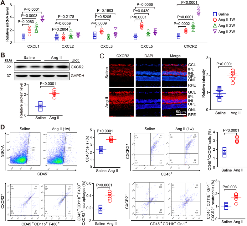
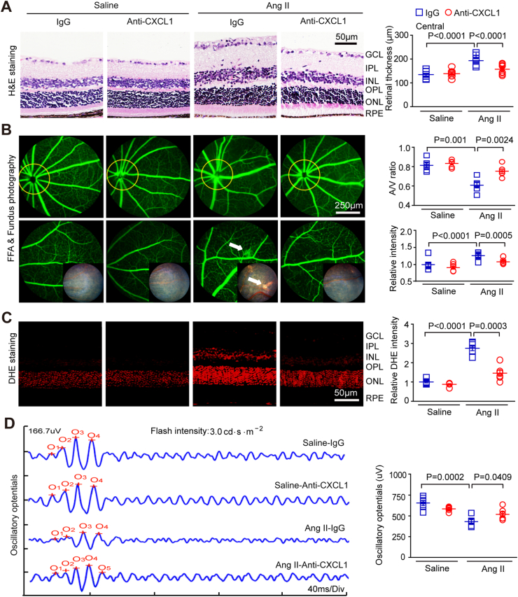
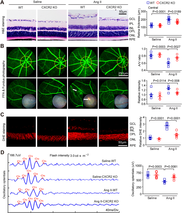
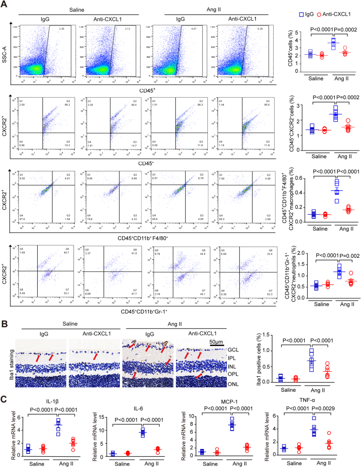
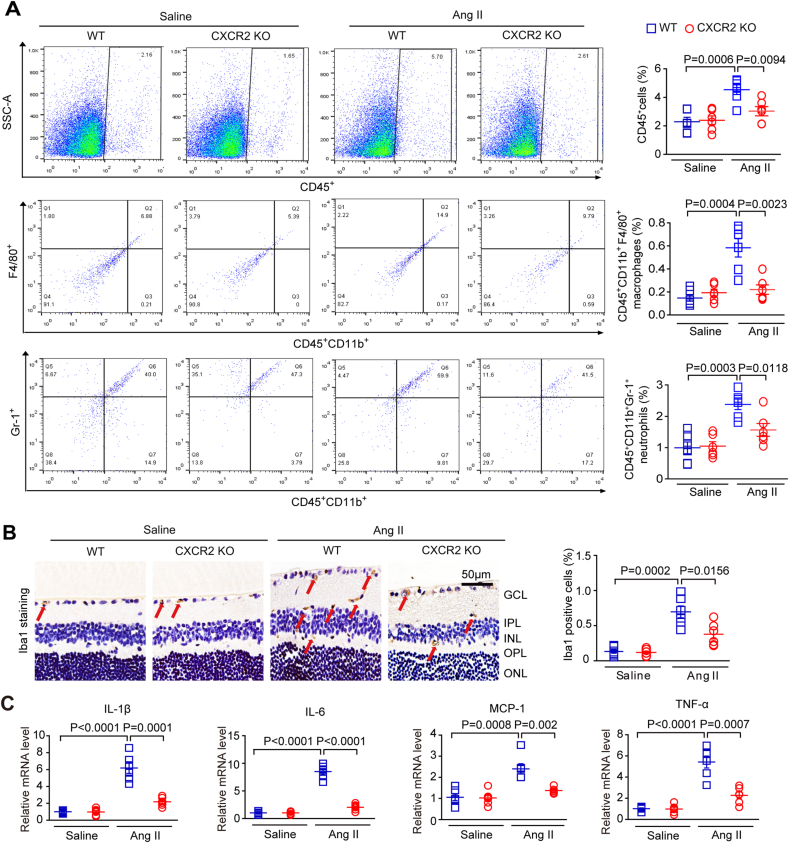
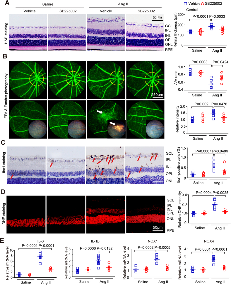
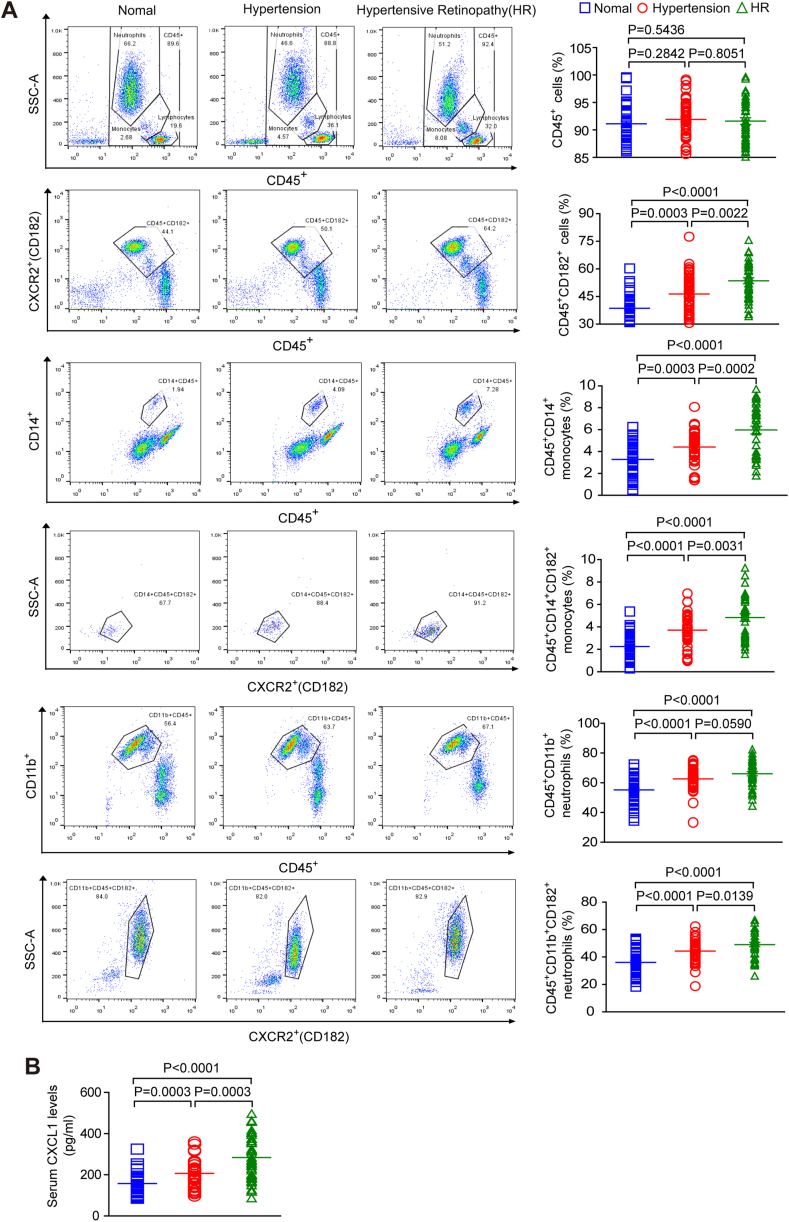
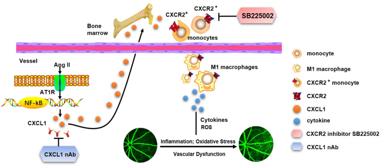
Inflammation plays an important role in hypertensive retinal vascular injury and subsequent retinopathy. Monocyte chemotaxis via CXCL1-CXCR2 binding has been implicated in various cardiovascular diseases, but the function of CXCL1-CXCR2 signalling involved in retinopathy, which was investigated as angiotensin II (Ang II)-induced retinopathy, is unclear. In our study, we established a hypertensive retinopathy (HR) model by Ang II infusion (3000 ng/min/kg) for 3 weeks. To determine the involvement of CXCR2 signalling, we used CXCR2 knockout (KO) mice or C57BL/6J wild-type (WT) mice as experimental subjects. The mice were treated with a CXCL1 neutralizing antibody or SB225002 (the specific CXCR2 inhibitor). Our results showed that after Ang II treatment, the mRNA levels of CXCL1 and CXCR2 and the number of CXCR2+ inflammatory cells were significantly elevated. Conversely, unlike in the IgG control group, the CXCL1 neutralizing antibody greatly reduced the increase in central retinal thickness induced by Ang II infusion, arteriolar remodelling, superoxide production, and retinal dysfunction in WT mice. Furthermore, Ang II infusion induced arteriolar remodelling, infiltration of Iba1+ macrophages, the production of oxidative stress, and retinal dysfunction, but the symptoms were ameliorated in CXCR2 KO mice and SB225002-treated mice. These protective effects were related to the reduction in the number of CXCR2+ immune cells, particularly macrophages, and the decrease in proinflammatory cytokine (IL-1β, IL-6, TNF-ɑ, and MCP-1) expression in Ang II-treated retinas. Notably, serum CXCL1 levels and the number of CXCR2+ monocytes/neutrophils were higher in HR patients than in healthy controls. In conclusion, this study provides new evidence that the CXCL1-CXCR2 axis plays a vital role in the pathogenesis of hypertensive retinopathy, and selective blockade of CXCL1-CXCR2 activation may be a potential treatment for HR.

Keywords: CXCL1; CXCR2; Hypertensive retinopathy; Inflammation; Macrophages.


Conflict of interest statement

Declaration of competing interest The authors declare no competing interests.

Figures

Fig. 1
Fig. 1
Ang II infusion promoted the upregulation of CXCL1 and CXCR2 expression and increased the number of myeloid-derived CXCR2-positive cells in retinas. (A) WT C57BL/6J mice were infused with Ang II or saline for 1, 2 and 3 weeks. Quantitative real-time polymerase chain reaction analyses of chemokine mRNA and CXCR2 mRNA expression in the retina (n = 6). (B) The expression of CXCR2 protein (right, upper) was analysed by Western blot, and the protein levels (right, lower; n = 6 in each group) were quantified. GAPDH was used as the internal control (n = 6). (C) Immunostaining of CXCR2 in retinal slices (left) with an anti-CXCR2 antibody and the quantification of the red fluorescence intensity in each group (middle; n = 6 in each group); (D) Flow cytometry analyses of CD45+ cells, CD45+ CXCR2+ cells, CD45+ CD11b+ F480+ CXCR2+ macrophages, and CD45+ CD11b+ Gr-1+ CXCR2+ neutrophils in the retina at one week after Ang II infusion. Percentage of each type of cell (right, n = 6). The data are presented as the mean ± SEM. (For interpretation of the references to colour in this figure legend, the reader is referred to the Web version of this article.)
Fig. 2
Fig. 2
Treatment with CXCL1 neutralizing antibody reduced retinal thickening, inflammation, ROS, and vascular dysfunction in Ang II-infused mice. (A) WT C57BL/6J mice were treated with IgG control or CXCL1 neutralizing antibody (anti-CXCL1, 100 μg/mouse/day) and then infused with Ang II for three weeks. Images of H&E staining of central retinal sections (left) and quantitation of the retinal thickness (right, n = 8). (B) Typical retinal angiograms and fundus photos (left); these white arrows indicate vascular fluorescein leakage and the corresponding area in the fundus photo. The ratio of retinal arteriovenous and fluorescence intensity was quantified (right; n = 6). (C) Retinal superoxide production was evaluated by DHE staining (left); the red fluorescence intensity of DHE was quantified (right; n = 6). (D) Waveforms of oscillatory potential (left) and amplitude quantification (right; n = 6). (For interpretation of the references to colour in this figure legend, the reader is referred to the Web version of this article.)
Fig. 3
Fig. 3
Genetic ablation of CXCR2 ameliorates Ang II-induced retinopathy in mice. (A) WT C57BL/6J mice and CXCR2 KO mice were both infused with angiotensin II or saline for three weeks. H&E-stained central retina sections (left). Retinal thickness was quantified (right, n = 8–10). (B) Representative angiograms and fundus photographs of retinal vessels (left); the arrows indicate leakage of fluorescein perivascularly and the corresponding fundus photograph area. The arteriovenous ratio and quantification of fluorescence intensity (right, n = 6). (C) DHE red fluorescence staining for analysis of superoxide production in the retinas of each group (left) and intensity quantification (right, n = 6). (D) Typical waveforms of oscillatory potentials and their amplitude quantification (n = 6). (For interpretation of the references to colour in this figure legend, the reader is referred to the Web version of this article.)
Fig. 4
Fig. 4
Treatment with a CXCL1-neutralizing antibody prevents Ang II-induced inflammatory cell infiltration. (A) WT C57BL/6J mice were pretreated with IgG control or anti-CXCL1 neutralizing antibody (100 μg/mouse/day) and then infused with angiotensin II for three weeks. Flow cytometry analyses of CD45+ cells, CD45+ CXCR2+ cells, CD45+ CD11b+ F480+ CXCR2+ macrophages and CD45+ CD11b+ Gr-1+ CXCR2+ neutrophils cells in each group (left). The percentage of gated cells among total cells (right, n = 6); (B) Representative images of Iba1 IHC staining in each group, red arrows indicate the Iba1-positive microglia/macrophages(scale bar: 50 μm, left); quantification of positive cells (right; n = 6 per group); (C) qPCR analyses of the mRNA levels of IL-1β, IL-6, tumour necrosis factor-α (TNF-α), and monocyte chemoattractant protein-1 (MCP-1) in each group (n = 6). GAPDH was used as an internal control. (For interpretation of the references to colour in this figure legend, the reader is referred to the Web version of this article.)
Fig. 5
Fig. 5
Knockout of CXCR2 attenuates inflammatory cell infiltration induced by Ang II. (A) Wild-type (WT) and CXCR2 KO mice were infused with Ang II (3000 ng/min/kg) or saline for 3 weeks. Flow cytometry analyses of CD45+ cells, CD45+ CD11b+ F480+ macrophages and CD45+ CD11b+ Gr-1+ neutrophils cells in retinas (left). The percentage of gated cells among total cells (right, n = 6). (B) Representative images of Iba1 IHC staining, red arrows indicate the Iba1-positive microglia/macrophages(scale bar: 50 μm, left); quantification of positive cells in each group (right, n = 6). (C) qPCR analyses of the mRNA levels of IL-1β, IL-6, TNF-α, and MCP-1 in the retinas (n = 6). GAPDH was used as an internal control. (For interpretation of the references to colour in this figure legend, the reader is referred to the Web version of this article.)
Fig. 6
Fig. 6
The CXCR2 inhibitor SB225002 alleviates Ang II-induced changes in retinal thickening, inflammation, ROS, and vascular dysfunction. (A) WT mice were treated with SB225002 or vehicle (DMSO) and then with angiotensin II infusion for three weeks. Central retinal sections stained with haematoxylin and eosin (left). Measurement of retinal section thickness (right, n = 8). (B) A representative retinal angiogram and fundus image (left); the white arrow indicates a fluorescein leak and the corresponding fundus image area. The ratio of retinal arteriovenous and fluorescence intensity was quantified (right, n = 6). (C) Representative images of Iba1 IHC staining, red arrows indicate the Iba1-positive microglia/macrophages(scale bar: 50 μm, left); quantification of positive cells in each group (right, n = 6). (D) Representative images of superoxide generation (left) and quantification of the fluorescence intensity (red) for DHE (right). (E) Analyses of IL-1β, IL-6, NOX1 and NOX4 transcript levels in the retina (n = 6). (For interpretation of the references to colour in this figure legend, the reader is referred to the Web version of this article.)
Fig. 7
Fig. 7
The number of CXCL1 and CXCR2+immunocytes in the serum is elevated in patients with hypertensive retinopathy. (A) Analysis of circulating immune cells based on flow cytometry, including CD45+ cells, CD45+CD182+ (CXCR2+) cells, CD45+CD14+ monocytes, CD45+CD14+CD182+ monocytes, CD45+CD11b+ neutrophils and CD45+CD11b+CD182+ neutrophils, in healthy controls (n = 40), hypertensive patients (HP) (n = 40) and hypertensive retinopathy patients (HR) (n = 40). (B) Analysis of serum CXCL1 levels in healthy controls (n = 40), HP patients (n = 40), and HR patients (n = 40) by ELISA.
Fig. 8
Fig. 8
A working model of the mechanism by which CXCL1 recruits CXCR2+ macrophages into the retina. These macrophages initiate and aggravate Ang II-induced changes in central retinal thickness, arterial remodelling, inflammatory cell infiltration, ROS production and vascular dysfunction. Conversely, suppressing CXCL1 with neutralizing antibodies (nAbs) or CXCR2 with SB225002 alleviates these negative consequences.

References

    1. Tsukikawa M., Stacey A.W. A review of hypertensive retinopathy and chorioretinopathy. Clin. Optom. 2020;12:67–73. - PMC - PubMed
    1. Dziedziak J., Zaleska-Zmijewska A., Szaflik J.P., Cudnoch-Jedrzejewska A. p impact of arterial hypertension on the eye: a review of the pathogenesis, diagnostic methods, and treatment of hypertensive retinopathy. Med. Sci. Monitor. 2022;28 - PMC - PubMed
    1. Wang L., Zhao X.C., Cui W., Ma Y.Q., Ren H.L., Zhou X., et al. Genetic and pharmacologic inhibition of the chemokine receptor CXCR2 prevents experimental hypertension and vascular dysfunction. Circulation. 2016;134(18):1353–1368. - PMC - PubMed
    1. Wenzel P., Knorr M., Kossmann S., Stratmann J., Hausding M., Schuhmacher S., et al. Lysozyme M-positive monocytes mediate angiotensin II-induced arterial hypertension and vascular dysfunction. Circulation. 2011;124(12):1370–1381. - PubMed
    1. Wang S., Li J., Bai J., Li J.M., Che Y.L., Lin Q.Y., et al. The immunoproteasome subunit LMP10 mediates angiotensin II-induced retinopathy in mice. Redox Biol. 2018;16:129–138. - PMC - PubMed