. 2023 Jan;64(1):137-144.
doi: 10.2967/jnumed.122.264284. Epub 2022 Aug 18.

Antigen-Dependent Inducible T-Cell Reporter System for PET Imaging of Breast Cancer and Glioblastoma

Affiliations

Antigen-Dependent Inducible T-Cell Reporter System for PET Imaging of Breast Cancer and Glioblastoma

Jaehoon Shin et al. J Nucl Med. 2023 Jan.

Abstract

For the past several decades, chimeric antigen receptor T-cell therapies have shown promise in the treatment of cancers. These treatments would greatly benefit from companion imaging biomarkers to follow the trafficking of T cells in vivo. Methods: Using synthetic biology, we engineered T cells with a chimeric receptor synthetic intramembrane proteolysis receptor (SNIPR) that induces overexpression of an exogenous reporter gene cassette on recognition of specific tumor markers. We then applied a SNIPR-based PET reporter system to 2 cancer-relevant antigens, human epidermal growth factor receptor 2 (HER2) and epidermal growth factor receptor variant III (EGFRvIII), commonly expressed in breast and glial tumors, respectively. Results: Antigen-specific reporter induction of the SNIPR PET T cells was confirmed in vitro using green fluorescent protein fluorescence, luciferase luminescence, and the HSV-TK PET reporter with 9-(4-18F-fluoro-3-[hydroxymethyl]butyl)guanine ([18F]FHBG). T cells associated with their target antigens were successfully imaged using PET in dual-xenograft HER2+/HER2- and EGFRvIII+/EGFRvIII- animal models, with more than 10-fold higher [18F]FHBG signals seen in antigen-expressing tumors versus the corresponding controls. Conclusion: The main innovation found in this work was PET detection of T cells via specific antigen-induced signals, in contrast to reporter systems relying on constitutive gene expression.

Keywords: CAR T; PET; SNIPR; cancer antigens; reporter.


Figures

None
Graphical abstract
FIGURE 1.
FIGURE 1.
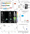
In vivo [18F]FHBG uptake in HER2+ tumors using activated anti-HER2 SNIPR T cells. (A) For PET/CT, we generated T cells by transducing 3 plasmids, including anti-HER2 SNIPR, anti-HER2 CAR, and inducible HSV-TK(SR39)-T2A-sIL2, followed by fluorescence-activated cell sorting using myc, GFP, and mCherry. (B) We repeated the in vitro radiotracer accumulation of activated SNIPR T cells also bearing CAR, following the experimental scheme shown in Supplemental Fig. 5B. We confirmed significantly higher [18F]FHBG accumulation in SNIPR T cells after coculturing with SKBR3 (HER2+) cells than in SNIPR T cells after coculturing with MB468 (HER2–) cells. (C) Double xenograft mouse models were generated by implanting SKBR3 (HER2+) and MD468 (HER2–) cells in left- and right-flank soft tissue. Four weeks after tumor implantation, SNIPR T cells were injected into tail veins. Small-animal PET/CT was performed 3, 6, 8, and 10 d after T-cell injection. (D) Representative CT images, [18F]FHBG PET images, [18F]FHBG PET/CT images, and maximum-intensity projection [18F]FHBG PET/CT images at day 8 demonstrated similar size of xenografts, with radiotracer enrichment only within SKBR3 (HER2+) xenograft and not within MD468 (HER2–) xenograft. (E) Quantitative ROI analyses of HER2+ and HER2– tumors and background (shoulder muscle) at day 8 demonstrated statistically significant radiotracer enrichment within HER2+ xenograft, 10 times and 13 times greater than within HER2– xenograft and background. (F) Time-dependent ROI analyses of radiotracer enrichment within HER2+ tumor demonstrated greatest radiotracer enrichment at day 8 after T-cell injection. Slightly decreased radiotracer enrichment was observed at day 10, at which point mice were killed for ex vivo analysis. (G) Biodistribution analysis (day 10) of [18F]FHBG enrichment within different organs demonstrated significantly greater [18F]FHBG enrichment within HER2+ xenograft than within HER2– xenograft. As seen on small-animal PET/CT, gastrointestinal system demonstrated high level of [18F]FHBG uptake. MIP = maximum-intensity projection. *P < 0.05. **P < 0.01. ***P < 0.001.
FIGURE 2.
FIGURE 2.
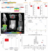
EGFRvIII SNIPR PET. (A) We generated SNIPR T cells with anti-EGFRvIII-SNIPR and inducible IL13-mutein (IL13m)-CAR-T2A-reporter constructs. We used 3 different reporters: BFP, nLuc, and HSV-TKSR39. In this system, SNIPR T cells express anti-EGFRvIII-SNIPR at baseline but do not express IL13m-CAR or reporters. When anti-EGFRvIII binds to EGFRvIII on target cells, SNIPR T cells induce expression of IL13m-CAR and reporters: BFP, nLuc, or HSV-TKSR39. Since most U87 cells express IL13 receptor α-2, T cells expressing IL13m-CAR secrete cytokines and growth factors that induce T-cell proliferation and survival. (B) SNIPR T cells incubated with EGFRvIII+ U87 cells demonstrated significantly higher level of BFP reporter expression, nLuc enzymatic activity, and HSV-TKSR39–mediated 18FHBG accumulation than did SNIPR T cells incubated with EGFRvIII– U87 cells. (C) Following a protocol similar to that used for HER2, EGFRvIII+ U87 and EGFRvIII– U87 cells were implanted into mouse flank subcutaneous tissues. At 4 wk after implantation, anti-EGFRvIII T cells with inducible anti-IL13-mutein-CAR-T2A-HSV-TK(SR39) were injected into tail veins. Representative maximum-intensity-projection [18F]FHBG PET/CT image (left) and cross-sectional [18F]FHBG PET/CT images (middle and right) at day 8 demonstrated high radiotracer enrichment within EGFRvIII+ U87 xenograft compared with EGFRvIII– U87 xenograft on contralateral side. (D) Time-dependent ROI analysis of radiotracer enrichment within EGFRvIII+ xenograft demonstrated greatest radiotracer enrichment at day 8 after T-cell injection, followed by slight decrease in PET signal at day 10, at which point animals were killed for ex vivo biodistribution analysis. (E) Quantitative ROI analysis of EGFRvIII+ and EGFRvIII– tumors and background (shoulder muscle) demonstrated statistically significant radiotracer enrichment within EGFRvIII+ xenograft, 14 times and 18 times greater than within EGFRvIII– xenograft and background. (F) Ex vivo analysis (day 10) of [18F]FHBG enrichment within different organs demonstrated significantly greater [18F]FHBG enrichment within EGFRvIII+ xenograft than within EGFRvIII– xenograft. As seen on small-animal PET/CT images, gastrointestinal system demonstrated high level of [18F]FHBG. *P < 0.05. **P < 0.01. ***P < 0.001.
FIGURE 3.
FIGURE 3.
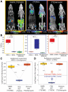
Comparison among SNIPR PET, [18F]FDG PET, and immuno-PET. (A) Representative images of [18F]FHBG SNIPR PET, [18F]FDG PET, and [89Zr]trastuzumab PET. SNIPR PET and trastuzumab immuno-PET demonstrated radiotracer enrichment within SKBR3 (HER2+) xenograft compared with MD468 (HER2–) tumor, whereas [18F]FDG PET demonstrated higher radiotracer enrichment within MD468 tumor. (B) ROI analysis demonstrated statistically significant, 9.9-fold greater enrichment of [18F]FHBG within HER2+ tumor than within HER2– tumor (left), nonstatistically significant enrichment (P > 0.05) of [18F]FDG within HER2– tumor compared with HER2+ tumor, and statistically significant 9.3 times greater enrichment of [89Zr]trastuzumab within HER2+ tumor than within HER2– tumor. (C and D) Fold enrichment of radiotracer within HER2+ tumor was significantly greater in SNIPR PET and immuno-PET than in [18F]FDG PET, when compared with background (C) and when compared with HER2– tumor (D). NS = not statistically significant. **P < 0.01. ***P < 0.001.

References

    1. Larson RC, Maus MV. Recent advances and discoveries in the mechanisms and functions of CAR T cells. Nat Rev Cancer. 2021;21:145–161. - PMC - PubMed
    1. Frey NV. Chimeric antigen receptor T cells for acute lymphoblastic leukemia. Am J Hematol. 2019;94(suppl 1):S24–S27. - PubMed
    1. Laetsch TW. Chimeric antigen receptor T-cell therapy for acute lymphocytic leukaemia: where are we in 2020? Lancet Haematol. 2020;7:e778–e779. - PubMed
    1. Tang X-J, Sun X-Y, Huang K-M, et al. . Therapeutic potential of CAR-T cell-derived exosomes: a cell-free modality for targeted cancer therapy. Oncotarget. 2015;6:44179–44190. - PMC - PubMed
    1. van der Stegen SJC, Hamieh M, Sadelain M. The pharmacology of second-generation chimeric antigen receptors. Nat Rev Drug Discov. 2015;14:499–509. - PMC - PubMed

Publication types