. 2022 Aug 18;13(1):4861.
doi: 10.1038/s41467-022-32609-1.

Chemical zymogens for the protein cysteinome

Affiliations

Chemical zymogens for the protein cysteinome

Mireia Casanovas Montasell et al. Nat Commun. .

Abstract

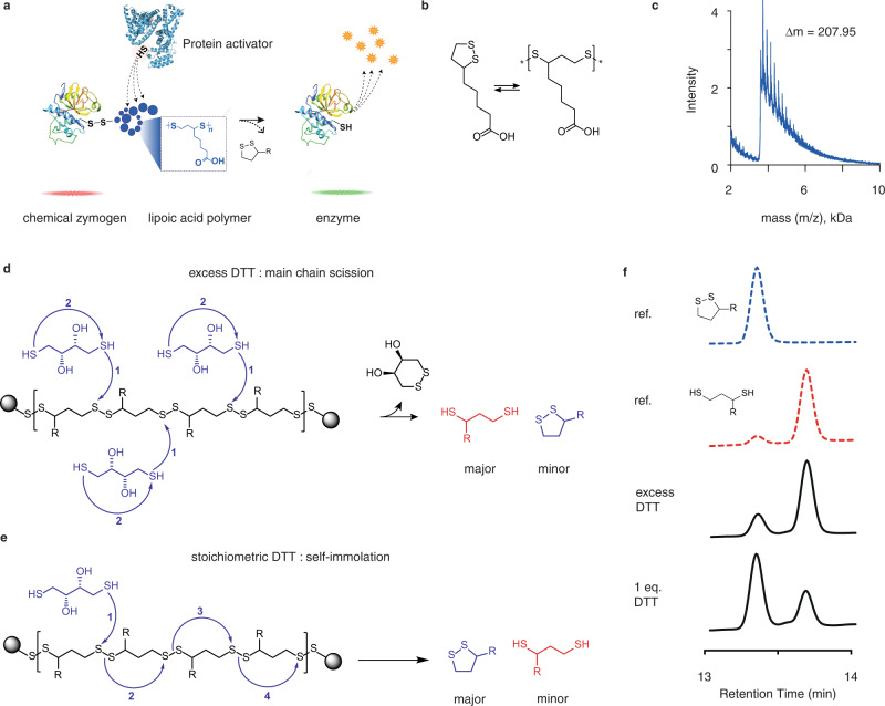
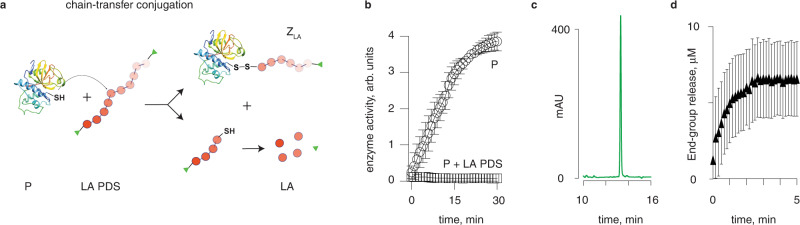
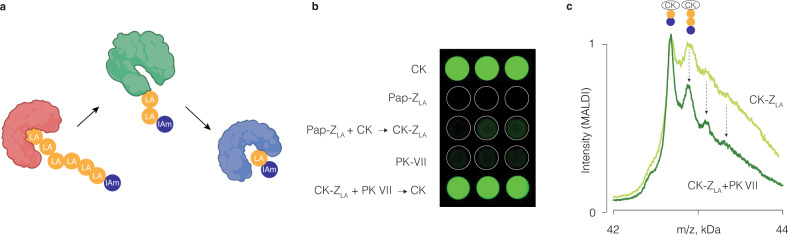
We present three classes of chemical zymogens established around the protein cysteinome. In each case, the cysteine thiol group was converted into a mixed disulfide: with a small molecule, a non-degradable polymer, or with a fast-depolymerizing fuse polymer (ZLA). The latter was a polydisulfide based on naturally occurring molecule, lipoic acid. Zymogen designs were applied to cysteine proteases and a kinase. In each case, enzymatic activity was successfully masked in full and reactivated by small molecule reducing agents. However, only ZLA could be reactivated by protein activators, demonstrating that the macromolecular fuse escapes the steric bulk created by the protein globule, collects activation signal in solution, and relays it to the active site of the enzyme. This afforded first-in-class chemical zymogens that are activated via protein-protein interactions. We also document zymogen exchange reactions whereby the polydisulfide is transferred between the interacting proteins via the "chain transfer" bioconjugation mechanism.


Conflict of interest statement

The authors declare no competing interests.

Figures

Fig. 1
Fig. 1. Polydisulfide polymer based on lipoic acid.
a Schematic illustration of the proposed self-immolative polydisulfide as a tool to collect activating chemical signal in solution and propagate it to the sterically hindered active site of an enzyme; b Chemical formula of lipoic acid and schematic illustration of its reversible polymerization into a linear disulfide; c MALDI spectrum of the lipoic acid polymer that shows inter-peak spacing of 207.95 Da, in agreement with the structure of lipoic acid as the monomer unit; d, e schematic illustration of mechanisms of depolymerization for LA PDS via main-chain scission (d) or self-immolation (e), depending on stoichiometry of added reducing agent; f reference HPLC traces of oxidized and reduced lipoic acid monomers, and experimental data on depolymerization of LA PDS in excess DTT and with equimolar DTT, illustrating main-chain scission or self-immolation as predominant mechanisms of depolymerization, depending on stoichiometry of reaction.
Fig. 2
Fig. 2. Design, synthesis, and characterization of the polydisulfide-based zymogen of papain.
a Schematic illustration of zymogen preparation via the “grafting from” polymerization route; P protein, M monomer; Z zymogen; b End-point measurements of enzymatic activity for P and Z, illustrating successful deactivation of papain by LA PDS and reactivation of catalysis via polymer decomposition; c Kinetic data for enzymatic catalysis in solutions of zymogens obtained from papain via polymerization of lipoic acid capped with thiol traps: IAm = iodoacetamide, DTDP = 2,2’ dithiodipyridine, Mal = 4-maleimidobutyric acid; Sulfone = phenyl vinyl sulfone; –ve = non-capped cryo-polymerization); d Enzymatic kinetics data in solutions of papain exposed to iodoacetamide with or without polymerized LA; e pH dependence of kinetics of zymogen reactivation. Experimental conditions; [P, Z] = 2 μM; enzyme substrate: N-α-benzoyl-L-arginine-7-amido-4-methylcoumarin hydrochloride, 5 μM; 25 mM borate buffer pH 8 (for buffer details in panel e: see experimental section); DTT 2 mM. Panel c: shown representative results from individual runs; panels b, d, e shown are results from three independent experiments as mean ± S.D. Statistics in panel b one-way ANOVA with the Sidak’s multiple comparisons test, *p < 0.05; ***p < 0.001.
Fig. 3
Fig. 3. Design and characterization of zymogens for cysteinome.
a Schematic illustration of the zymogens design, the chemical formulas of the corresponding disulfides, the reactivation data, and the MALDI characterization data for the zero-length zymogen Z0, PEG-based zymogen ZPEG, and the zymogen with the macromolecular fuse ZLA; experimental data are shown as mean ± S.D. based on three independent experiments; b gel electrophoresis characterization of the creatine kinase zymogens Z0, ZPEG, and ZLA; c luminescence end-point imaging of the coupled kinase/luciferase assay illustrating activity of CK masked in the composition of the three zymogens and recovered with the use of DTT (2 mM) as a reducing agent. Pap papain, Bro bromelain, CK creatine kinase.
Fig. 4
Fig. 4. Reactivation of zymogens using thiol-containing proteins.
a Kinetics of zymogen reactivation using protein activators (25 mM borate buffer pH 8, 25 mM NaCl, 1 mM EDTA; 10 µM papain, 1 µM protein activators); for reactivation with creatine kinase and pyruvate kinase VII, concentrations were Z0, ZPEG = 1 µM, ZLA = 0.3 µM; for reactivation with transglutaminase and pyruvate kinase II: Z0, ZPEG = 10 µM, ZLA = 3 µM; protein activators are shown alongside their computed surface accessibility of the thiols (R, Å2). b, c End-point fluorescence imaging (b) and kinetic measurements (c) for reactivation of papain zymogens using creatine kinase illustrating that papain activity is masked by the three zymogens and revealed by DTT for Z0, ZPEG, and ZLA, whereas recovery with the protein activator is possible only for ZLA; data were recorded using albumin as a protease substrate (blocked Cys-34, labeled with fluorescein isocyanate over the level of self-quenching); In panels a, c data shown are an average of three independent experiments (starting with independent zymogen syntheses) shown as mean ± st.dev., statistical analysis was carried via the two-way ANOVA test with statistical significance indicating the time-point at which ZLA reactivation becomes statistically significant compared to both, Z0 and ZPEG, p < 0.05; d gel electrophoresis analysis of proteolytic processing of albumin (Cys-34 blocked) by papain, native or recovered from its zymogens ZLA and Z0 by added pyruvate kinase (PK VII). In this panel, *Indicates a treatment with albumin by native papain.
Fig. 5
Fig. 5. Chain-transfer conjugation reaction between the polydisulfide polymer and papain.
a Schematic illustration of the reaction between LA PDS and a protein; b enzymatic activity of papain before and after reaction with LA PDS, c quantification of lipoic acid released during conjugation of LA PDS to papain via the chain transfer mechanism; d UV–Vis spectroscopy kinetics measurement of release of LA PDS thiopyridine terminal groups, resulting from a conjugation reaction between the polymer and papain. Panels b, d shows results are an average of three independent experiments, error bars signify st. dev.
Fig. 6
Fig. 6. Zymogen exchange reactions.
a Schematic illustration of a zymogen exchange reaction; b coupled luminescence-based read-out for activity of CK, which illustrates a loss of activity for the kinase upon an addition of papain ZLA as a result of the zymogen exchange reaction; shown are the results from three independent experiments as mean ± S.D; statistical analysis was carried via the two-way ANOVA test with statistical significance indicating the time-point at which the kinase activity in the presence of ZLA becomes statistically significant compared to both, ZLA and CK, p < 0.001; c MALDI-based characterization of the chain transfer reaction in solutions of papain ZLA and CK, PK VII, or transglutaminase; d MALDI data illustrating a “trans-PEGylation”: a chain transfer reaction between papain ZLA and CK, whereby ZLA contained a 5 kDa PEG as a terminal group. For reaction conditions, see Experimental section.
Fig. 7
Fig. 7. Two rounds of transfer of the polydisulfide polymer between proteins.
a Schematic illustration of the two-step chain transfer; b visualization of luminescence (read-out for activity of creatine kinase via a coupled luciferase-based assay) in solutions of CK, papain ZLA, and PK VII, as well as in solutions of papain ZLA with CK, and CK-ZLA with PK VII; c MALDI-based characterization of the two-step zymogen exchange reaction whereby CK acts first as an LA PDS acceptor and then as a polydisulfide donor. Peak intensities are normalized to that of the simplest zymogen (CK-LA-IAm).

References

    1. Karin M, Hunter T. Transcriptional control by protein phosphorylation: signal transmission from the cell surface to the nucleus. Curr. Biol. 1995;5:747–757. doi: 10.1016/S0960-9822(95)00151-5. - DOI - PubMed
    1. Bergeron JJM, Guglielmo GMD, Dahan S, Dominguez M, Posner BI. Spatial and temporal regulation of receptor tyrosine kinase activation and intracellular signal transduction. Annu. Rev. Biochem. 2016;85:573–597. doi: 10.1146/annurev-biochem-060815-014659. - DOI - PubMed
    1. Klein T, Eckhard U, Dufour A, Solis N, Overall CM. Proteolytic cleavage—mechanisms, function, and “Omic” approaches for a near-ubiquitous posttranslational modification. Chem. Rev. 2018;118:1137–1168. doi: 10.1021/acs.chemrev.7b00120. - DOI - PubMed
    1. Lu J, Wang H, Tian Z, Hou Y, Lu H. Cryopolymerization of 1,2-dithiolanes for the facile and reversible grafting-from synthesis of protein–polydisulfide conjugates. J. Am. Chem. Soc. 2020;142:1217–1221. doi: 10.1021/jacs.9b12937. - DOI - PubMed
    1. Liew SS, et al. Intracellular delivery of therapeutic proteins through N-terminal site-specific modification. Chem. Commun. 2020;56:11473–11476. doi: 10.1039/D0CC04728G. - DOI - PubMed

Publication types