. 2022 Sep;28(9):1944-1955.
doi: 10.1038/s41591-022-01908-x. Epub 2022 Aug 18.

Programmable antivirals targeting critical conserved viral RNA secondary structures from influenza A virus and SARS-CoV-2

Affiliations

Programmable antivirals targeting critical conserved viral RNA secondary structures from influenza A virus and SARS-CoV-2

Rachel J Hagey et al. Nat Med. 2022 Sep.

Abstract

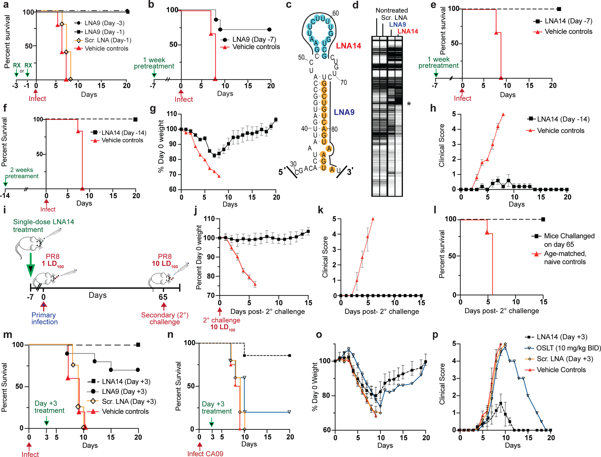
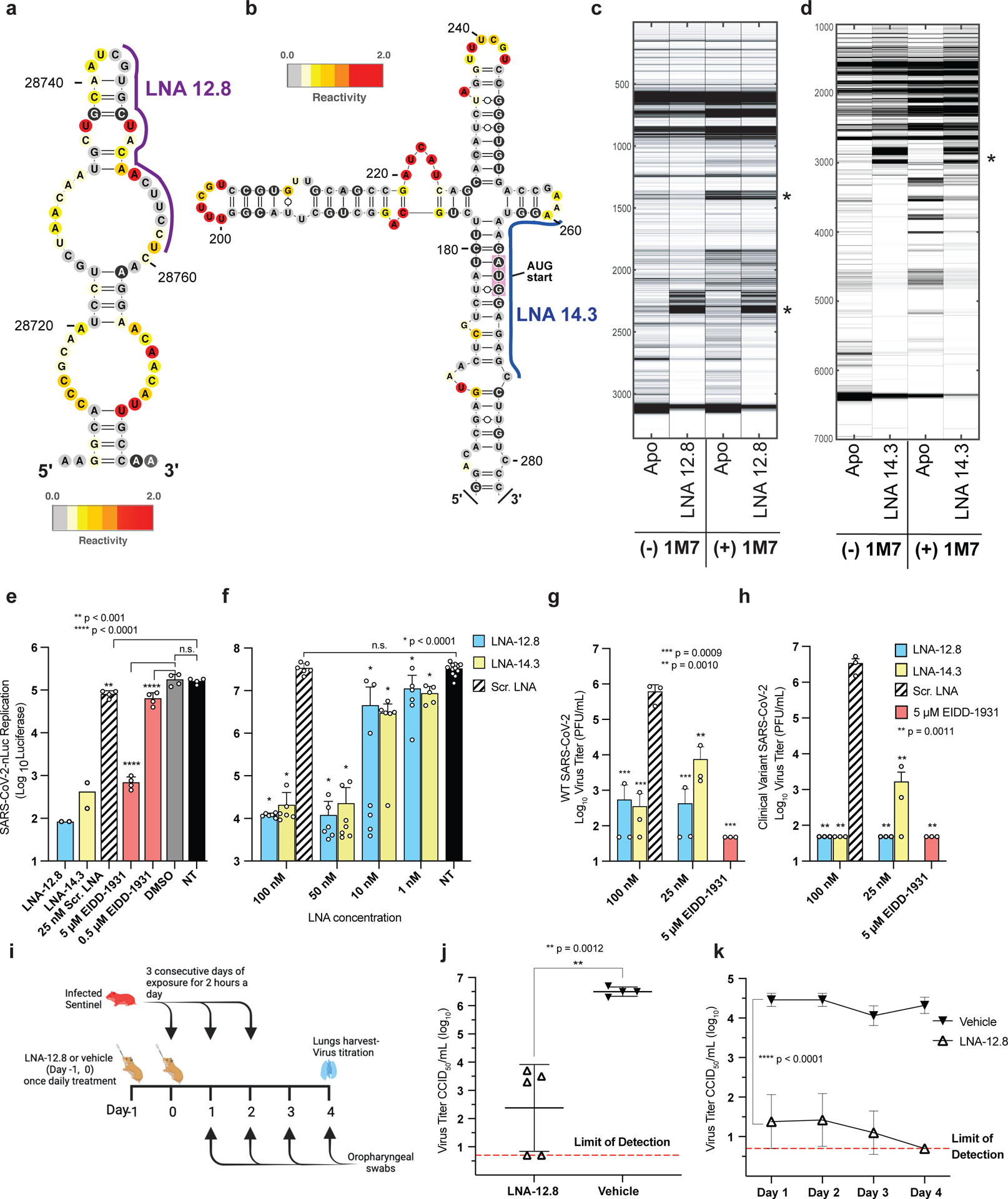
Influenza A virus's (IAV's) frequent genetic changes challenge vaccine strategies and engender resistance to current drugs. We sought to identify conserved and essential RNA secondary structures within IAV's genome that are predicted to have greater constraints on mutation in response to therapeutic targeting. We identified and genetically validated an RNA structure (packaging stem-loop 2 (PSL2)) that mediates in vitro packaging and in vivo disease and is conserved across all known IAV isolates. A PSL2-targeting locked nucleic acid (LNA), administered 3 d after, or 14 d before, a lethal IAV inoculum provided 100% survival in mice, led to the development of strong immunity to rechallenge with a tenfold lethal inoculum, evaded attempts to select for resistance and retained full potency against neuraminidase inhibitor-resistant virus. Use of an analogous approach to target SARS-CoV-2, prophylactic administration of LNAs specific for highly conserved RNA structures in the viral genome, protected hamsters from efficient transmission of the SARS-CoV-2 USA_WA1/2020 variant. These findings highlight the potential applicability of this approach to any virus of interest via a process we term 'programmable antivirals', with implications for antiviral prophylaxis and post-exposure therapy.


Conflict of interest statement

Competing Interests Statement

J.S.G., R.J.H., E.P. and M.E. are inventors on a patent pertaining to the materials presented in this article that has been filed with the U.S. Patent and Trademark Office by Stanford University.

Figures

Figure 1.
Figure 1.. SHAPE-determined RNA secondary structures of wild-type PB2 and packaging mutant vRNAs.
SHAPE-chemical mapping performed on full-length (−)-sense PB2 vRNAs. Colors denote SHAPE reactivity, which is proportional to the probability that a nucleotide is single-stranded. All structures are truncated to highlight the 5′ termini sequence structure. (a) SHAPE-predicted wild-type PB2 RNA secondary structure from strain A/Puerto Rico/8/1934 “PR8” (H1N1). Color-coded circles correspond to nucleotides sites where synonymous mutations were reported to affect PB2 packaging,. (b) Packaging efficiency of synonymous mutants in (a), determined by RT-qPCR. Results representative of two independent experiments with biological replicates, each performed in triplicate. Statistical analysis was performed using one-way ANOVA with Dunnett’s multiple comparisons test against the WT mean by GraphPad Prism 9 software (n=4). Error bars represents mean ± standard deviation (s.d.); **** p < 0.0001; * p = 0.0454, n.s. = not significant. Box below indicates mutant name and corresponding nucleotide change. Nucleotide numbering shown in the genomic (−)-sense orientation. (c) SHAPE-mapped structures of PB2 packaging-defective mutant vRNAs, m757 (G44C) and m745 (A80U) indicating loss of PSL2’s RNA secondary structure. Black arrowheads and boxed nucleotides denote site(s) of synonymous mutation. (d) Web-logo representation of the PSL2 region conservation across IAV strains and diverse influenza A viral subtypes (weblogo.berkeley.edu). The overall height represents sequence conservation at that nucleotide position, while the symbol height within each position indicates the relative frequency of each nucleotide at that site. Black box denotes PSL2 region. Sequences included in the alignment: pandemic A/Brevig Mission/1/1918 (H1N1), pandemic A/California/04/2009 (H1N1), seasonal human A/New York/470/2004 (H3N2), A/Puerto Rico/8/1934 (H1N1), highly pathogenic avian A/Vietnam/03/2004 (H5N1), avian A/mallard/Maryland/14OS1154/2014 (H6N1), pandemic A/Hong Kong/8/1968 (H3N2), and seasonal human A/New York/312/2001 (H1N1) (see Supplementary Fig. 1d). RNA nucleotides are numbered in (−) -sense orientation. (e) SHAPE-mapped structures of full-length wild-type PB2 vRNA from pandemic and highly pathogenic strains, including different subtypes to modern human strains: 1918 pandemic (A/Brevig Mission/1/1918 (H1N1)), highly-pathogenic avian (A/Vietnam/1203/2004 (H5N1)), 2009 pandemic ‘swine’ (A/California/04/2009 (H1N1)), and Fujian-like human seasonal virus, A/New York/470/2004 (H3N2)
Figure 2.
Figure 2.. 2-Dimensional Mutate-and-Map (M2) analysis and empiric validation of PSL2 motif.
(a) Systematic single nucleotide mutation and mapping of resulting chemical accessibility reveals interactions in the three-dimensional structure of the RNA. Chemical accessibilities, plotted in grayscale (black, highest SHAPE reactivity), across 59 single mutations at single-nucleotide resolutions of PSL2 element from PR8 strain segment PB2. Reactivity peaks (left to right) correspond to nucleotides from the 5′ to 3′ end of the PB2 RNA. Nucleotides corresponding to known packaging mutation sites are indicated on left in blue. Red bolded mutations denote packaging-defective mutant sites predicted by M2 analysis. Green bolded mutations indicate synonymous mutant sites analyzed in (b). (b) Packaging efficiencies of M2-identified synonymous mutants read out by RT-qPCR. Packaging efficiency represents the percentage of mutant PB2 vRNA packaging relative to parental wild-type PB2. Results from two independent experiments in biological duplicate and technical triplicate (n=4). **** p < 0.0001, * p = 0.0321. (c) Previously described synonymous mutants (m757, m745, m55c) are mapped onto PSL2 structure. Compensatory, non-synonymous mutations m55c-comp, m745-comp, and m757-comp were designed at sites predicted to restore wild-type PSL2 structure based on SHAPE and mutate-and-map chemical analyses. Black boxed nucleotides denote compensatory mutation sites. (−)-sense vRNA orientation is shown. (d) Packaging efficiencies of packaging-defective and compensatory mutant viruses. For compensatory mutations where a non-synonymous change was required, a wild-type PB2 protein expression plasmid was co-transfected during virus rescue. Values given as percentage of PB2 vRNA packaging relative to wild-type PR8 virus. Results from three independent experiments (n=3), assays performed in triplicate. **** p < 0.0001, *** p < 0.0005, n.s. = not significant. (e) Virus titer determined by plaque assay. Results in PFU / mL, plaque assays in triplicate (n=3). **** p < 0.0001, *** p = 0.0009, ** p = 0.0149. All error bars represent mean ± s.d. All statistical analysis were performed by ordinary one-way ANOVA using Dunnett’s multiple comparisons test against WT computed in GraphPad Prism 9 software.
Figure 3.
Figure 3.. Mutate-Map-Rescue analysis reveals novel PB2 packaging-defective and compensatory mutant partners.
(a) Electropherograms from systematic single nucleotide mutation SHAPE chemical mapping with rescue (Mutate-Map-Rescue) analysis of individual and compensatory double mutations to test base-pairings from 1D-data-guided models and to identify predicted successful synonymous PSL2-defective and compensatory mutant pairs. Chemical accessibilities, plotted in grayscale (black = highest SHAPE reactivity), across 59 single mutations at single-nucleotide resolutions of PSL2 element from PR8 strain segment PB2. Reactivity peaks (left to right) correspond to nucleotides from the 5′ to 3′ end of the PB2 RNA. See Supplementary Fig. 7 for complete set of Mutate-Rescue pairs. (b) Electropherogram of successful double synonymous mutant pair determined by Mutate-Map-Rescue analysis. (c) Mutational design of single mutants m52 (G52U) and m65 (C65A), and the double m52/65-comp rescue pair on the PSL2 structure. (d) Packaging efficiency of the synonymous single and double mutant Mutate-Rescue pair. Values given as a percentage of PB2 vRNA packaging relative to WT PR8 virus. Results represent two independent experiments with biological duplicates performed in technical triplicate (n=4), except for m65 performed in biological triplicate (n=6). Statistical analysis by ordinary one-way ANOVA using Dunnett’s multiple comparisons test against WT; * p < 0.0001, n.s. = not significant. (e) Viral titer of the supernatants collected in (d) in PFU / mL, plaque assays in biological duplicate (n=2), except for m65 (n=3), and performed in technical triplicate. (f-g) Percent Day 0 weight and survival of mice infected with single PSL2-disrupting, and compensatory PSL2-restoring double-mutant viruses. Six- to eight-week-old female BALB/c mice (n=6) were intranasally infected with PR8 wild-type (WT) virus, packaging-defective single mutant viruses, m52 and m745, compensatory double mutant viruses, m52/65-comp and m745-comp, or PBS control. (f) Percent Day 0 weight. (g) Kaplan-Meier survival plot of the individual cohorts depicted in (f). All error bars represent mean ± s.d. Statistics and graphs for all figures were generated in GraphPad Prism 9 software.
Figure 4.
Figure 4.. Locked Nucleic Acids (LNAs) targeting PSL2 RNA structure display potent antiviral activity in vitro.
(a) Regions of PSL2 targeted by indicated LNAs. (b) Antiviral screen of LNAs transfected into MDCK cells and infected 4 hours later with PR8 (H1N1) or A/Hong Kong/8/68 (H3N2) virus (0.01 MOI) and viral titers determined 48 hours post infection (n=3). Statistics performed by unpaired ordinary one-way ANOVA (PR8 and HK68) with Dunnett’s multiple comparisons. (c) Antiviral efficacy as a function of time of LNA addition (n=3), analyzed as in (b). Statistics by 2-way ANOVA with Dunnett’s multiple comparisons test against non-treated +Lipo3k (N.T.). (d) PB2 vRNA (PR8) packaging efficiency of viruses treated with 100 nM LNA9 or Scr. LNA control. Values given as a percentage of PB2 vRNA packaging in comparison to non-treated wild-type PR8 virus; readout by qPCR. Results from two biological replicates (n=2), assays performed in technical triplicates. (n=6). (e) LNA9 efficacy against multiple IAV strains in MDCK cells pretreated with 100 nM of indicated LNAs. Analyzed as in (b). Statistics as described in (c) against the N.T. control. (f) In vitro selection for drug resistance to LNA9 with escalating concentrations of LNA and the sensitivity of passaged virus in response to drug treatment. EC50s determined at the indicated passage (P) numbers. Results expressed as a percentage of nontreated virus titer. (g) In vitro selection of PR8 virus selected with oseltamivir carboxylate (OSLT). OSLT-treated PR8 virus and drug sensitivity determined by plaque reduction assay. The number of viral plaques with each drug concentration was normalized against the nontreated control to determine the EC50. (h, i) In vitro sensitivity of wild-type WSN33 (H1N1) and neuraminidase (NA) inhibitor-resistant (WSN H275Y NA mutant) virus to LNA9 (h) or OSLT (i). EC50 values were computed using a nonlinear regression model with variable slope. Statistics for all graphs performed in GraphPad Prism 9 software. All error bars represent mean ± s.d. * Statistical P value as indicated in each panel.
Figure 5.
Figure 5.. PSL2-targeted LNAs demonstrate potent antiviral activity in vivo.
(a-b) Kaplan-Meier survival plots of mice intranasally administered a single dose of LNA9, Scr. LNA, or Vehicle (mock-treated) on the indicated days with respect to a subsequent lethal inoculum of wild-type PR8 virus (PR8); (a) 20 μg LNA9 (n=5); (b) 30 μg LNA9 (n=7) or vehicle control (n=5). (c) Target sites of LNA9 and newly designed, LNA14, mapped to the PSL2 structure. (d) Electrophoretic profile of SHAPE analysis performed on PR8 PB2 vRNA in the presence of non-treated, or 100nM Scr. LNA, LNA9, or LNA14 Asterisk marks site of reverse transcription (RT) stops indicating strength of LNA – RNA binding. (e-f) Kaplan-Meier survival plots of mice pretreated with a single IN dose of (e) 30 μg LNA14 (n=7) or vehicle (n=5), Day −7; (f) 40 μg of LNA14 or vehicle (n=6), Day −14, before receiving lethal PR8 inoculum. (g) Percent day 0 weights, and (h) clinical score, of mice from (f). Error bars show mean ± s.e.m. (i-l) Mice (n=7) from (e), who had received a single 30 μg intranasal dose of LNA14 one week prior to a first lethal inoculum (1 LD100) of PR8 and that all survived, were inoculated 65 days post-initial infection, along with age-matched naïve controls (n=10), with a 10-times lethal dose (10 LD100). (i) Challenge study timeline. (j) Weight loss, (k) clinical score; (l) Kaplan-Meier survival curve; error bars, mean ± s.d.. (m) Kaplan-Meier survival plot of mice (n=10/group) infected with a lethal dose of PR8, followed 3 days later with, a single intravenous dose of 30 μg of LNA14, LNA9, Scr. LNA, or vehicle control. (n-p) Mice were infected with a lethal dose of mouse-adapted A/California/04/2009 (pH1N1) virus (CA09), then treated intranasally with a single 30 μg dose of LNA14 (n=7/group), Scr. LNA (n=5), or Vehicle (n=4) on Day +3 post-infection. Mice given OSLT (n= 5) were treated by oral gavage BID for 5 days starting at Day +3. (n) Kaplan-Meier survival curve. (o) Percent day 0 weight. (p) Clinical score. All studies performed with 6–8 week-old female BALB/c mice Error bars in (o, p) indicate mean ± s.e.m.
Figure 6.
Figure 6.. Antisense targeting of highly conserved RNA secondary structures in SARS-CoV-2.
(a, b) Two SHAPE reactivity-derived prediction of RNA secondary structures within (+)-sense SARS-CoV-2 vRNA and LNAs designed against these targets. (a) nucleotide (nt) region 28743 – 28827; LNA-12.8 and (b) nt region 258 – 276; LNA-14.3 (c, d) Electrophoretic profile of SHAPE analysis performed in the presence or absence of the corresponding LNAs (e) In vitro antiviral activity of 25 nM LNAs against SARS-CoV-2-Nluc virus in Huh-7 cells (n=4). The nucleoside analog, EIDD-1931, was included as a positive control at 5 μM and 0.5 μM. (f) LNA dose response against SARS-CoV-2-Nluc virus in ACE2-TMPRSS2-Huh-7.5 cells, in biological replicates (n=6–7 for all treatment groups, except for NT controls where n=12). (g, h) Virus titers of supernatant collected from A549-hACE2 cells treated with LNA or EIDD-1931 followed by infection with either (g) wild-type SARS-CoV-2 or (h) a patient-isolated clinical variant of SARS-CoV-2 containing multiple mutations in the spike protein region including D614G, E484K, and N501Y present in variants of concern (VOC) (n=3). Statistical analysis in e, f, g and h was performed using ordinary one-way ANOVA with Dunnett’s multiple comparison tests between the controls as indicated. Error bars represent mean ± s.d., p values as indicated. (i-k) Prevention of SARS-CoV-2 transmission in Syrian hamsters. Ten-week-old female Syrian hamsters were pretreated with either 100 μg of LNA-12.8 (n=5) or vehicle (n=4) on Day −1 and Day 0 before exposure to SARS-CoV-2-infected sentinel hamsters for 2 hours per day for 3 consecutive days. Four days after the initial exposure, lungs were harvested, and virus titers determined by CCID50 in triplicate. (i) Experimental timeline. (j) Virus titer of SARS-CoV-2 in lungs (two-sided unpaired student’s t test) and (k) oropharyngeal swabs of LNA- or vehicle-treated hamsters (2-way ANOVA with multiple comparisons against the vehicle controls). The assay method detection limit is 0.7 log10 CCID50. Error bars represent mean ± s.d. Figure (i) was created with BioRender.com.

Comment in

References

    1. Memoli MJ, Hrabal RJ, Hassantoufighi A, Eichelberger MC & Taubenberger JK Rapid selection of oseltamivir- and peramivir-resistant pandemic H1N1 virus during therapy in 2 immunocompromised hosts. Clin Infect Dis 50, 1252–1255 (2010). - PMC - PubMed
    1. Hai R, et al. Influenza A(H7N9) virus gains neuraminidase inhibitor resistance without loss of in vivo virulence or transmissibility. Nature communications 4, 2854 (2013). - PMC - PubMed
    1. Hayden FG & de Jong MD Emerging influenza antiviral resistance threats. The Journal of infectious diseases 203, 6–10 (2011). - PMC - PubMed
    1. Romero-Lopez C & Berzal-Herranz A Unmasking the information encoded as structural motifs of viral RNA genomes: a potential antiviral target. Reviews in medical virology 23, 340–354 (2013). - PMC - PubMed
    1. Pang PS, et al. Structural map of a microRNA-122: hepatitis C virus complex. J Virol 86, 1250–1254 (2012). - PMC - PubMed

Publication types