. 2022 Aug 19;20(1):180.
doi: 10.1186/s12915-022-01354-8.

Buoyancy and hydrostatic balance in a West Indian Ocean coelacanth Latimeria chalumnae

Affiliations

Buoyancy and hydrostatic balance in a West Indian Ocean coelacanth Latimeria chalumnae

Henrik Lauridsen et al. BMC Biol. .

Abstract

Background: Buoyancy and balance are important parameters for slow-moving, low-metabolic, aquatic organisms. The extant coelacanths have among the lowest metabolic rates of any living vertebrate and can afford little energy to keep station. Previous observations on living coelacanths support the hypothesis that the coelacanth is neutrally buoyant and in close-to-perfect hydrostatic balance. However, precise measurements of buoyancy and balance at different depths have never been made. RESULTS: Here we show, using non-invasive imaging, that buoyancy of the coelacanth closely matches its depth distribution. We found that the lipid-filled fatty organ is well suited to support neutral buoyancy, and due to a close-to-perfect hydrostatic balance, simple maneuvers of fins can cause a considerable shift in torque around the pitch axis allowing the coelacanth to assume different body orientations with little physical effort.

Conclusions: Our results demonstrate a close match between tissue composition, depth range and behavior, and our collection-based approach could be used to predict depth range of less well-studied coelacanth life stages as well as of deep sea fishes in general.

Keywords: Bone mineral density; Computed tomography; Depth regulation; Ecophysiology; Fatty organ; Headstand; Lipid accumulation; Magnetic resonance imaging; Magnetic resonance spectroscopy.


Conflict of interest statement

The authors declare that they have no competing interests.

Figures
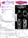
Fig. 1
Fig. 1
Distribution of bone mineral, lipid and muscle tissue in the coelacanth. a Volume rendering of a three-dimensional bone mineral density (BMD) map of the coelacanth and three extant species of lungfishes. b,c Distribution of bone mineral content (BMC) fraction of total BMC along the total length (TL) in the coelacanth and lungfishes. The dashed gray square in b is magnified in c. d Putative time calibrated phylogeny of extant non-tetrapod sarcopterygians. e Volume rendering of a three-dimensional lipid fraction map of the coelacanth. Same color scale as in a is used for lipid fraction ranging from 0 to 100%. f,g Sagittal (f) and transversal (g) sections of lipid fraction (f and leftmost sections in g) and water fraction (rightmost sections in g). Yellow isosceles triangles point to the lipid-filled fatty organ, yellow equilateral triangle points to the lipid-filled pericerebral space, yellow arrows point to the lipid-filled post ocular space, yellow arrow head points to the lipid-filled perivertebral space, purple isosceles triangle points to the water-rich muscle, purple arrow points to the water-rich notochord. h Distribution of mass of different tissue types along the total length in the coelacanth. Here, all water-rich lean tissues are included in “muscle”
Fig. 2
Fig. 2
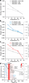
Effect of temperature, salinity, and pressure on density of seawater and lipids and on buoyancy of the coelacanth. a Density of Comoran seawater as a function of temperature with and without salinity (s) and pressure (p) adjustments (adj.). b Density of swim bladder lipid of Hoplostethus atlanticus (left vertical axis) as a function of temperature with and without pressure adjustment compared to density of Comoran seawater (right vertical axis). Notice that the density of the swim bladder lipid is more sensitive to pressure than seawater. Both vertical axes span 0.03 kg/l. c Density of oleyl oleate, the most prevalent wax ester in coelacanth (left vertical axis) as a function of temperature with and without pressure adjustment compared to density of Comoran seawater (right vertical axis). Notice that the density of oleyl oleate is more sensitive to pressure and temperature than seawater. Both vertical axes span 0.03 kg/l. d Absolute (left vertical axis) and relative (right vertical axis) buoyancy of the coelacanth at varying depths in Comoran waters. Prevalence (% of time) of coelacanths at different depths previously determined by ultrasonic telemetry [4] is presented in shades of red in the background of the graph. In its natural habitat at 190–400 m of depth, the coelacanth is close to neutrally buoyant. Venturing deeper, the coelacanth will become negatively buoyant. If brought rapidly to the surface (e.g., line fishing), the relatively cold coelacanth will initially be negatively buoyant but gradually become positively buoyant as it heats up to surface water temperature (exemplified from 190, 400, and 1000 m of depth to surface with increasingly dark blue circles and dashed lines)
Fig. 3
Fig. 3
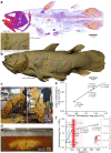
Hydrostatic balance of the coelacanth at different depths. a,b Modeled overall center of gravity (COG) (red reticle), overall center of buoyancy (COB) (blue reticle), bone mineral COG (gray reticle), lipid COG (yellow reticle), and muscle COG (purple reticle) overlaid on bone mineral volume reconstruction (a) and photogrammetry reconstructed surface model (b) of the coelacanth. c Physical measurement of COG using the plumb line method at surface pressure and room temperature of 70% v/v EtOH preserved coelacanth. The intersection of the extrapolated vertical lines from two different anchor points (left photo: ventral side, caudal to the pelvic fins; right photo: dorsal side, caudal to the 2nd dorsal fin) reveal the COG (under the assumption of a negligible buoyancy force provided by the displaced atmospheric air). d Balance point of coelacanth immersed in its 70% EtOH preservation fluid. A line that was adjustable in the long axis of the specimen was tied around the circumference of the specimen and the specimen was hung from two vertical lines with adjustable connections to the circumferential line in the dorsoventral (short) axis. Long and short axis position of the anchor points were adjusted until the specimen was in balance indicating the balance point for the specimen in the preservation fluid between the COG and COB. e Distances between COG and COB relative to total length (TL) and total height (TH) in the long and short axis at different depths with varying temperatures, pressures, and salinities. f Absolute and relative torque (relative to torque at surface) at different depths with varying temperatures, pressures, and salinities. Distance between the COG and COB in both long and short axis increases slightly with depth in the modeled depth range resulting in increased torque and the need to produce extra hydrodynamic force to counteract the hydrostatic force acting to push COB directly above COG. If brought rapidly to the surface, the GOG to GOB distance will gradually decrease as the coelacanth heats up to surface temperatures resulting in a decreased torque (exemplified from 190, 400, and 1000 m of depth to surface with increasingly dark blue circles and dashed lines)
Fig. 4
Fig. 4
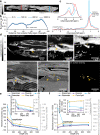
Hydrostatic balance in the coelacanth in different body postures. a Segmental model of the coelacanth from lateral (top) and dorsal (bottom) viewpoints consisting of 35 segments: main body, pectoral fins, pelvic fins, 1st and 2nd dorsal fins, anal fin, 26 transversal caudal fin segments, and fatty organ. b,c Stick figures of the segmental coelacanth model from lateral (top) and dorsal (bottom) viewpoints. Colored lines show long axis of segments with corresponding colors as in a. Colored circles with black centers show anchor points of fins. The coelacanth is displayed in two body postures: with straight caudal fin and remaining fins in extreme backward position (b) and with bent caudal fin and remaining fins in extreme forward position (c) (extreme fin positions according to live observation and anatomical measurements [44]). d Absolute (left vertical axis) and relative torque (right vertical axis). Relative to torque at the surface with straight tail and remaining fins backward at different body postures. At all modeled depths, the coelacanth can increase torque by ~ 131% by changing body posture
Fig. 5
Fig. 5
Fatty organ and vestigial lung composition and buoyancy contribution. a Sagittal section in the fatty organ imaged for lipid fraction with magnetic resonance imaging (MRI) using the Dixon method. Lipid fraction is visibly elevated in the caudal portion of the fatty organ. b Distribution of net buoyancy provided by the fatty organ (mass of tissue subtracted from mass of displaced seawater) along its long axis at four different depths in Comoran seawater. The caudal portion of the fatty organ provides more buoyancy than the cranial portion. c Magnetic resonance spectra acquired at the cranial (red graph and red line in a) and caudal (blue graph and blue line in a) portion of the fatty organ with the same scanning parameters (i.e., signal intensity reflects proton density). The spectrum at the caudal portion has a more pronounced methylene peak, whereas the cranial portion has a slightly more pronounced water peak, both supporting differences in lipid fraction along the craniocaudal axis of the fatty organ. d Tissue composition of coelacanth vestigial lung completely imbedded in the fatty organ. Low-resolution lipid (top left and top middle) and water fraction (top right) images of the cranial portion of the fatty organ (yellow isosceles triangles) containing the vestigial lung (yellow arrows). The vestigial lung contains very little lipid (see Table 1) and is instead water rich. A high-resolution T2-weighted MR image (bottom left) allows for better visualization of the vestigial lung within the fatty organ. X-ray computed tomography (CT) can be used to visualize the bony plates (yellow arrow heads) surrounding the vestigial lung (bottom middle) allowing for three-dimensional surface rendering of bony plates (bottom right). e,f Modeled absolute and relative buoyancy (e) and torque (f) (relative to surface torque with normal fatty organ) experienced by the coelacanth at surface, 190, 400, and 1000 m of depth in Comoran seawater if the fatty organ consisted of either seawater, muscle (or other water-rich lean tissues), pure lipid (oleyl oleate with no water content), or atmospheric air from inhalation. At the surface, an air-filled structure of similar size as the fatty organ would provide a marked increase in buoyancy and torque. However, as this air volume would compress at increasing depths under the assumption of a non-rigid wall, the lift provided by an air-filled bladder at depths would be less than that of a lipid-filled fatty organ. Magnifications in e and f contain the normal depth range of Comoran coelacanths
Fig. 6
Fig. 6
Prey items in gastrointestinal tract and prey with a swim bladder. a–d Mineralized remains of prey items in the distal portion of the digestive tract of the coelacanth imaged by X-ray computed tomography (CT) and magnetic resonance imaging (MRI). Dashed gray square in a is magnified in b and shown in a similar view plane using T2-weighted MRI (c). A three-dimensional surface rendering of the fecal matter is shown in d. e,f Sagittal T2-weighted MRI slice (e) and three component (skin, bones, swim bladder) model (f) made from CT and MRI of Beryx decadactylus, a known prey item of the coelacanth. This species contains an air-filled swim bladder (light blue segment in f) that under the assumption of neutral buoyancy displaces a volume of seawater with the same mass as the net weight of the fish (excluding the swim bladder) in seawater

References

    1. Erdmann MV, Caldwell RL, Kasim Moosa M. Indonesian ‘king of the sea’ discovered. Nature. 1998;395:335. doi: 10.1038/26376. - DOI
    1. Forey PL. History of the Coelacanth Fishes. London, United Kingdom: Chapman and Hall; 1998.
    1. Fricke H, Hissmann K, Schauer J, Erdmann M, Moosa MK, Plante R. Biogeography of the Indonesian coelacanths. Nature. 2000;403:38. doi: 10.1038/47400. - DOI - PubMed
    1. Hissmann K, Fricke H, Schauer J. Patterns of time and space utilisation in coelacanths (Latimeria chalumnae), determined by ultrasonic telemetry. Mar Biol. 2000;136:943–952. doi: 10.1007/s002270000294. - DOI
    1. Hissmann K, Fricke H, Schauer J, Ribbink AJ, Roberts M, Sink K, Heemstra PC. The South African coelacanths – an account of what is known after three submersible expeditions. S Afr J Sci. 2006;102:491–500.

Publication types

LinkOut - more resources