[Preprint]. 2022 Nov 21:2022.07.29.502029.
doi: 10.1101/2022.07.29.502029.

A multispecific antibody prevents immune escape and confers pan-SARS-CoV-2 neutralization

Affiliations

A multispecific antibody prevents immune escape and confers pan-SARS-CoV-2 neutralization

John Misasi et al. bioRxiv. .

Update in

  • A multispecific antibody against SARS-CoV-2 prevents immune escape in vitro and confers prophylactic protection in vivo.
    Misasi J, Wei RR, Wang L, Pegu A, Wei CJ, Oloniniyi OK, Zhou T, Moliva JI, Zhao B, Choe M, Yang ES, Zhang Y, Boruszczak M, Chen M, Leung K, Li J, Yang ZY, Andersen H, Carlton K, Godbole S, Harris DR, Henry AR, Ivleva VB, Lei QP, Liu C, Longobardi L, Merriam JS, Nase D, Olia AS, Pessaint L, Porto M, Shi W, Wallace SM, Wolff JJ, Douek DC, Suthar MS, Gall JG, Koup RA, Kwong PD, Mascola JR, Nabel GJ, Sullivan NJ. Misasi J, et al. Sci Transl Med. 2024 Oct 9;16(768):eado9026. doi: 10.1126/scitranslmed.ado9026. Epub 2024 Oct 9. Sci Transl Med. 2024. PMID: 39383243

Abstract

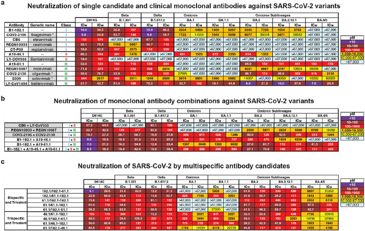
Despite effective countermeasures, SARS-CoV-2 persists worldwide due to its ability to diversify and evade human immunity1. This evasion stems from amino-acid substitutions, particularly in the receptor-binding domain of the spike, that confer resistance to vaccines and antibodies 2-16. To constrain viral escape through resistance mutations, we combined antibody variable regions that recognize different receptor binding domain (RBD) sites17,18 into multispecific antibodies. Here, we describe multispecific antibodies, including a trispecific that prevented virus escape >3000-fold more potently than the most effective clinical antibody or mixtures of the parental antibodies. Despite being generated before the evolution of Omicron, this trispecific antibody potently neutralized all previous variants of concern and major Omicron variants, including the most recent BA.4/BA.5 strains at nanomolar concentrations. Negative stain electron microscopy revealed that synergistic neutralization was achieved by engaging different epitopes in specific orientations that facilitated inter-spike binding. An optimized trispecific antibody also protected Syrian hamsters against Omicron variants BA.1, BA.2 and BA.5, each of which uses different amino acid substitutions to mediate escape from therapeutic antibodies. Such multispecific antibodies decrease the likelihood of SARS-CoV-2 escape, simplify treatment, and maximize coverage, providing a strategy for universal antibody therapies that could help eliminate pandemic spread for this and other pathogens.


Figures

Figure 1.
Figure 1.. Neutralization of SARS-CoV-2 variants by monoclonal antibodies, antibody cocktails and CODV formatted multispecific antibodies.
Neutralization of candidate and expanded access monoclonal antibodies (a) and cocktails (b) against D614G and the indicated SARS-CoV-2 variants: Beta (B.1.351), Delta (B.1.617.2), Omicron (B.1.1.529 or BA.1) and Omicron sub-lineages. When appropriate, generic names are indicated. Generic names with * indicate the presence of Fc domain mutations in the clinical product that are not found in the experimental versions used in this paper. Class indicates the Barnes RBD classification: class I antibodies bind to the ACE2 binding site when RBD is in the up position; class II bind to the ACE2 binding site when RBD is in the up or down position; class III bind outside the ACE2 binding site when RBD is in the up or down position; and class IV bind outside of the ACE2 binding site when RBD is in the up position. c, Neutralization of candidate multispecific antibodies against D614G and the indicated SARS-CoV-2 variants, including Beta (B.1.351), Delta (B.1.617.2), Omicron (B.1.1.529 or BA.1) and Omicron sub-lineages. Neutralization in pM is shown. Ranges are indicated with light blue (>67,000 pM), yellow (>10,000 to ≤67,000 pM), orange (>1,000 to ≤10,000 pM), red (>100 to ≤1,000 pM), maroon (>10 to ≤100 pM), and purple (≤10 pM).
Figure 2.
Figure 2.. Identification of potential binding modes of CODV to SARS-CoV-2 spike by negative stain-electron microscopy
a, Combination of RBD mutations on the SARS-CoV-2 spike designed to distinguish different binding modes of CODV. Mutations were made in spike in the spike trimer. The domains are colored green, blue, red and gray for Fv182.1, Fv46.1, Fv61.1 and constant domain, respectively. b-e, Evaluation of CODV Fv182 (b and c) binding to K444E/L452R or CODV Fv46 (d and e) binding to K444E/F486S. Shown at left in each panel is a schematic of the CODV domain being evaluated in the panel. The center-left subpanel indicates the Fv and position being evaluated (i.e., inner or outer). For clarity, the Fv domain that is not able to bind to the mutant is colored black in the subpanels. The center-right subpanel shows the negative stain electron micrograph class averages. Fv182 are indicated with a green arrow and Fv46 with a blue arrow. The white scale bar represents 10 nm in panel c and applies to each micrograph. The rightmost subpanel is a representative model of the binding mode observed in the micrograph. Panels b and c show that irrespective of position, both 182.1-46.1 and 46.1-182.1 are able to bind to spike protein that only allows binding by the Fv182. Panel d, shows that 182.1-46.1 (46.1 outer) is unable to bind spike protein that only allows binding by the Fv46.1. Panel e, shows that 46.1-182.1 (46.1 inner) is able to bind spike that only allows binding by Fv46. f, CODV 46.1-182.1 induced aggregation of the Omicron spike. Large clusters of aggregation were observed in the negative stain EM field, suggesting the CODV 46.1-182.1 can efficiently promote inter-spike crosslinking.
Figure 3.
Figure 3.. Mitigation of rcVSV-SARS-CoV-2 escape by 61.1/46.1-182.1 and an antibody cocktail
Replication competent VSV (rcVSV) bearing SARS-CoV-2 WA-1 spike protein was incubated with the indicated antibodies at 5-fold increasing concentrations (34 × 10−3 to 333,333 pM) and added to Vero cells. The appearance of viral growth, as indicated by the presence of >20% CPE, was determined after 3 days and supernatant from the highest antibody concentration with viral growth was passaged forward into a new selection round under the same conditions. Once viral growth appeared at 333,333 pM, the antibody was considered fully escaped and supernatant was no longer passaged forward. Data is graphed as the highest concentration of antibody at which viral growth was noted in each selection round. Each graph represents an independent experiment.
Figure 4.
Figure 4.. A trispecific antibody protects Syrian hamsters from SARS-CoV-2 variant BA.2
a, 46.1-182.1v/61.1-182.1v was passively transferred to Syrian hamsters intraperitoneally 24 hours prior to challenge with either 1x10⁵ PFU of BA.1, 2x10⁴ PFU of BA.2 or 1x10⁵ PFU of BA.5 SARS-CoV-2 Omicron variants of concern. For each virus, an additional group of hamsters received an equal volume of PBS in the same manner and served as controls. All animals were weighed daily to monitor weight loss and 4 animals per group were euthanized at days 2, 4, 6, and 10 to assess viral loads in the lung. (b, d, f) Hamsters were monitored daily for weight loss over 10 days. Symbols and error bars represent means and SEM, respectively. An unpaired, two-tailed student t-test was used to determine significance at each timepoint between treated and untreated animals. Significant p-values are indicated as: * (≤0.05), (≤0.01), (≤0.001) and ¥ (≤0.0001). (c, e, g) Viral load in the lung of BA.1 (c), BA.2 (e) and BA.5 (g) challenged hamsters were quantified by TCID50 per gram. Each symbol represents an individual animal and may overlap for equivalent values. Boxes and horizontal bars denote the IQR and medians, respectively; whisker end points are equal to the maximum and minimum values. Dotted lines indicate the assay’s lower limit of detection. An unpaired, two-tailed student t-test was used to determine significance at each timepoint between treated and untreated animals. Significant p-values are indicated on graph.

References

Main References

    1. Wu F. et al. A new coronavirus associated with human respiratory disease in China. Nature 579, 265–269 (2020). - PMC - PubMed
    1. Jones B. E. et al. The neutralizing antibody, LY-CoV555, protects against SARS-CoV-2 infection in nonhuman primates. Sci Transl Med 13, (2021). - PMC - PubMed
    1. Shi R. et al. A human neutralizing antibody targets the receptor-binding site of SARS-CoV-2. Nature 584, 120–124 (2020). - PubMed
    1. Hansen J. et al. Studies in humanized mice and convalescent humans yield a SARS-CoV-2 antibody cocktail. Science 369, 1010–1014 (2020). - PMC - PubMed
    1. Zost S. J. et al. Potently neutralizing and protective human antibodies against SARS-CoV-2. Nature 584, 443–449 (2020). - PMC - PubMed

Methods References

    1. Merchant A. M. et al. An efficient route to human bispecific IgG. Nat Biotechnol 16, 677–681 (1998). - PubMed
    1. Zalevsky J. et al. Enhanced antibody half-life improves in vivo activity. Nat Biotechnol 28, 157–159 (2010). - PMC - PubMed
    1. Barouch D. H. et al. A Human T-Cell Leukemia Virus Type 1 Regulatory Element Enhances the Immunogenicity of Human Immunodeficiency Virus Type 1 DNA Vaccines in Mice and Nonhuman Primates. J Virol 79, 8828–8834 (2005). - PMC - PubMed
    1. Catanzaro A. T. et al. Phase I clinical evaluation of a six-plasmid multiclade HIV-1 DNA candidate vaccine. Vaccine 25, 4085–92 (2007). - PubMed
    1. Doria-Rose N. A. et al. Booster of mRNA-1273 Strengthens SARS-CoV-2 Omicron Neutralization. medRxiv 2021.12.15.21267805 (2021) doi:10.1101/2021.12.15.21267805. - DOI

Publication types