. 2022 Aug 17:2:103.
doi: 10.1038/s43856-022-00167-8. eCollection 2022.

Temporal lineage replacements and dominance of imported variants of concern during the COVID-19 pandemic in Kenya

Affiliations

Temporal lineage replacements and dominance of imported variants of concern during the COVID-19 pandemic in Kenya

Gathii Kimita et al. Commun Med (Lond). .

Abstract

Background: Kenya's COVID-19 epidemic was seeded early in March 2020 and did not peak until early August 2020 (wave 1), late-November 2020 (wave 2), mid-April 2021 (wave 3), late August 2021 (wave 4), and mid-January 2022 (wave 5).

Methods: Here, we present SARS-CoV-2 lineages associated with the five waves through analysis of 1034 genomes, which included 237 non-variants of concern and 797 variants of concern (VOC) that had increased transmissibility, disease severity or vaccine resistance.

Results: In total 40 lineages were identified. The early European lineages (B.1 and B.1.1) were the first to be seeded. The B.1 lineage continued to expand and remained dominant, accounting for 60% (72/120) and 57% (45/79) in waves 1 and 2 respectively. Waves three, four and five respectively were dominated by VOCs that were distributed as follows: Alpha 58.5% (166/285), Delta 92.4% (327/354), Omicron 95.4% (188/197) and Beta at 4.2% (12/284) during wave 3 and 0.3% (1/354) during wave 4. Phylogenetic analysis suggests multiple introductions of variants from outside Kenya, more so during the first, third, fourth and fifth waves, as well as subsequent lineage diversification.

Conclusions: The data highlights the importance of genome surveillance in determining circulating variants to aid interpretation of phenotypes such as transmissibility, virulence and/or resistance to therapeutics/vaccines.

Keywords: Respiratory distress syndrome; Viral infection.


Conflict of interest statement

Competing interestsThe authors declare no competing interests.

Figures
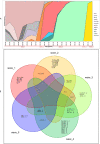
Fig. 1
Fig. 1. SARS-CoV-2 lineages circulating across the five COVID-19 waves in Kenya.
SARS-CoV-2 lineages circulating across the five COVID-19 waves in Kenya. Panel A shows monthly distribution of the major SARS-CoV-2 pango lineages and/or variant of concern/interest identified between May 2020 and January 2022. Red holizontal bars indicate the associated waves. Imported European lineages B.1 and B.1.1 dominated in waves one and two, although by wave two, there was an expansion of local lineages such as B.1.549, B.1.530 and A.23.1. Wave three was dominated by the Alpha VOC which progressively displaced the variants identified in the previous two waves. Wave four was the longest (lasting 5 months), and was dominated by the Delta VOC. Wave five, caused by the hyper transmissible Omicron variant, emerged stealthily, quickly spread and replaced Delta and the remnant Alpha variants, to become the dominant VOC. Panel B is a Venn diagram showing the relationships between the 40 Pangolin lineages across the five COVID-19 waves. Each wave had characteristic dominant lineages; some shared across the waves, but in general, the shared lineages were fewer. Red stars denote local lineages. Greek symbols denotes VOC or VOI (α = Alpha, β = Beta, δ = Delta, η = Eta, ο = Omicron).
Fig. 2
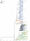
Fig. 2. Time-scaled phylogenetic tree of Kenyan genomes against selected global genomes.
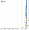
The tree was constructed with 316 genomes sampled from GISAID and 969 genomes from this study. The study samples were resolved into non-VOC/VOI lineages (grey branches), Delta (blue branches), Omicron (red branches), Alpha (green branches), Beta (black branches), Eta (teal branches) and the A.23.1 lineage (purple branches). No Kenyan samples branched with either the gamma variant (orange branches) or the Mu variant (crimson branches). Yellow tips represent global genomes, while blue tips represent the study samples. The different colors on branches represent the Pango lineages.
Fig. 3
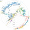
Fig. 3. Phylogenetic tree of the B.1.1.7 lineage from our samples and those sub-sampled globally.
The tree was constructed with 546 genomes, 381 from Kenya (182 from our study) and 165 sub-sampled from the globe. Genomes from other parts of the world include 44 genomes sampled from the earliest reported Alpha variants. The tree is rooted against the Wuhan/WHO1/2019 and Wuhan/Hu-1/2019 reference genomes (Genbank accession no. LR757998 and MN908947, respectively). The genomes from the study are shown in dark blue circular tips while other Kenyan genomes are shown in light blue circular tips. Genomes from other parts of the globe are shown in yellow circular tips. Kenyan samples branched from different parts of the tree, indicating multiple independent seeding events. Black arrows show samples from one of the first reported outbreak of the B.1.1.7 lineage in Kenya. Encircled red dots indicate earlier two independent introductions (18 January 2021) of the alpha variant outside the Nanyuki outbreak.
Fig. 4
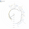
Fig. 4. Phylogenetic tree of the B.1.351 lineage from Kenyan samples and those sub-sampled globally.
The tree was constructed from 82 genomes - 27 from Kenya (12 from this study), and 55 sub-sampled genomes from around the world. The global sub-sample included 29 genomes sampled from the earliest reported beta variants. The tree is rooted against the Wuhan/WHO1/2019 and Wuhan/Hu-1/2019 reference genome (Genbank accession no. LR757998 and MN908947). The genomes from this study are shown in dark blue circular tips, while other Kenyan genomes are shown in light blue circular tips. Genomes from other parts of the globe are shown in yellow circular tips. South African genomes occurred in the basal parts of the tree, while majority of the Kenyan genomes were in the more derived parts of the tree.
Fig. 5
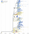
Fig. 5. Phylogenetic tree of the B.1.617.2 lineage from our study samples and those from around the world.
The tree was constructed with 836 genomes, 605 from Kenya (396 from this study), and 231 sub-sampled from the globe (including 29 early B.1.617.2 lineages all from India (Red branches). Genomes from this study are shown in dark blue circular tips, while other Kenyan genomes are shown in light blue circular tips. Kenyan samples branched with AY.16 pango lineage (grey branches) and AY.46 lineage (Black branches). The AY.16 lineage was the majority, followed by the AY.46 lineage. Samples encircled in red (AY.16) and black (AY.61) represent the earliest reported Delta introductions in, both from Nairobi.
Fig. 6
Fig. 6. Phylogenetic tree of the omicron variant from our study samples and those from across the world.
The tree was constructed with 1041 genomes, 827 from Kenya (188 from our study) and 214 sub-sampled from the globe that included eight early omicron genomes mostly from South Africa. The tree was rooted against the Wuhan/WHO1/2019 and Wuhan/Hu-1/2019 SARS-CoV-2 reference genome genbank accession no.s LR757998 and MN908947, respectively. Genomes from the study are shown in dark blue circular tips while other Kenyan genomes are shown in light blue circular tips. The earliest Kenyan samples (27th November 2021) are shown in light blue tips with red borders. Other than one sample Kenya/ILRI_COVM01771/2021 - GISAID identifier EPI_ISL_9093518 that branched with lineage BA.2, all other samples branched with the BA.1 lineage. A, B and C represent lineage diversification within the BA.1 cluster. Most Kenyan samples branched within the A cluster.

References

    1. Wu F, et al. A new coronavirus associated with human respiratory disease in China. Nature. 2020;579:265–269. doi: 10.1038/s41586-020-2008-3. - DOI - PMC - PubMed
    1. Andersen KG, Rambaut A, Lipkin WI, Holmes EC, Garry RF. The proximal origin of SARS-CoV-2. Nat. Med. 2020;26:450–452. doi: 10.1038/s41591-020-0820-9. - DOI - PMC - PubMed
    1. Li X, et al. Transmission dynamics and evolutionary history of 2019-nCoV. J. Med. Virol. 2020;92:501–511. doi: 10.1002/jmv.25701. - DOI - PMC - PubMed
    1. Wan, Y., Shang, J., Graham, R., Baric, R. S. & Li, F. Receptor Recognition by the Novel Coronavirus from Wuhan: an Analysis Based on Decade-Long Structural Studies of SARS Coronavirus. J. Virol. 94, e00127-20 (2020). - PMC - PubMed
    1. Wei X, Li X, Cui J. Evolutionary perspectives on novel coronaviruses identified in pneumonia cases in China. Natl. Sci. Rev. 2020;7:239–242. doi: 10.1093/nsr/nwaa009. - DOI - PMC - PubMed