. 2022 Aug 1;18(13):5001-5018.
doi: 10.7150/ijbs.70149. eCollection 2022.

ALKBH5/MAP3K8 axis regulates PD-L1+ macrophage infiltration and promotes hepatocellular carcinoma progression

Affiliations

ALKBH5/MAP3K8 axis regulates PD-L1+ macrophage infiltration and promotes hepatocellular carcinoma progression

Yu You et al. Int J Biol Sci. .

Abstract

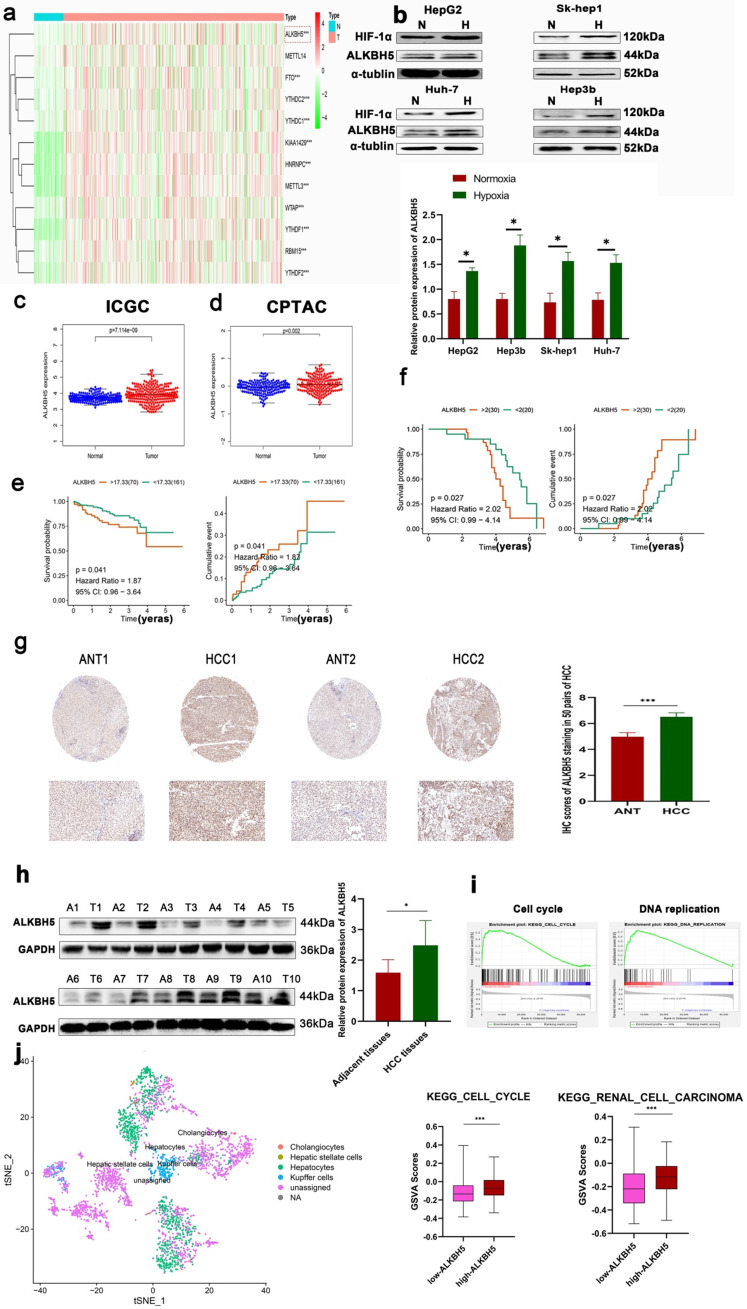
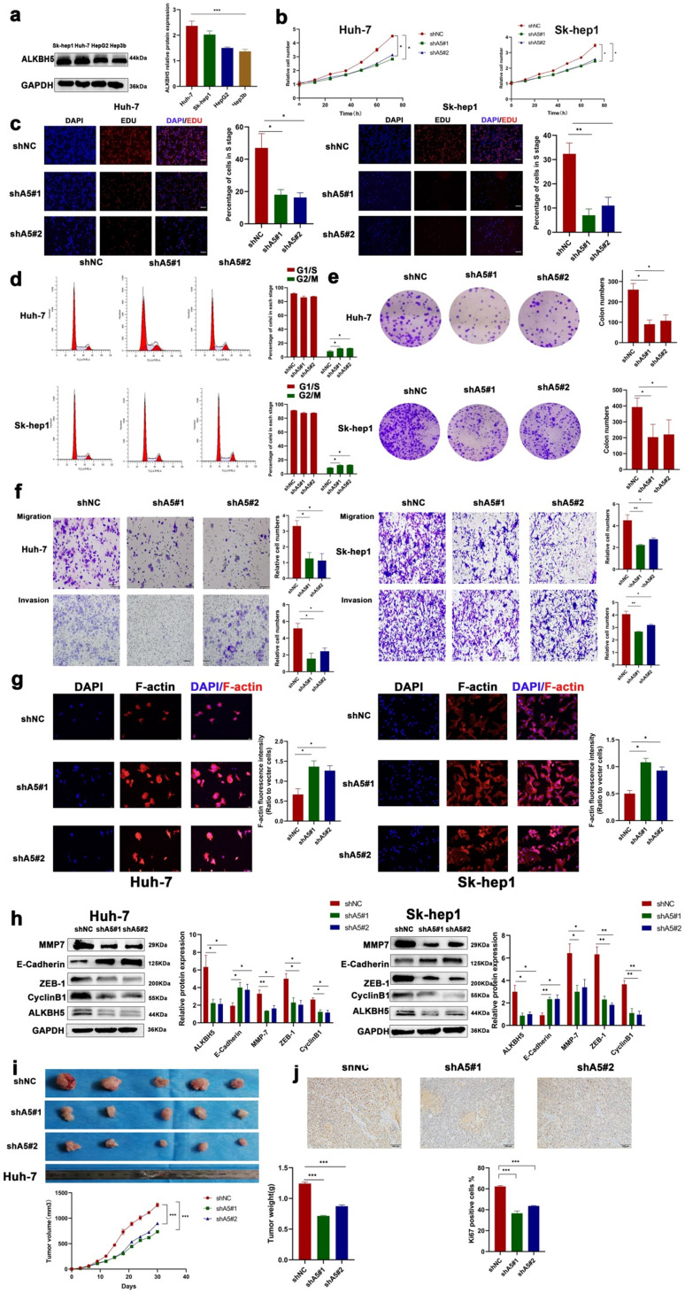
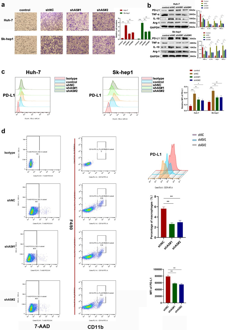
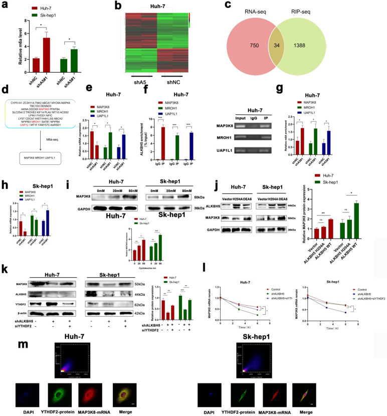
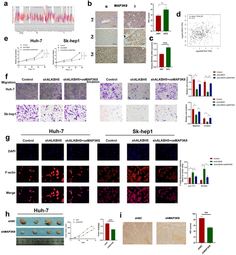
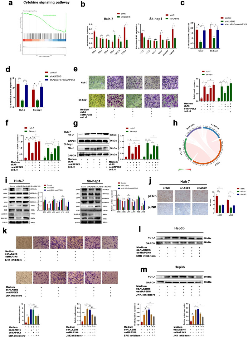
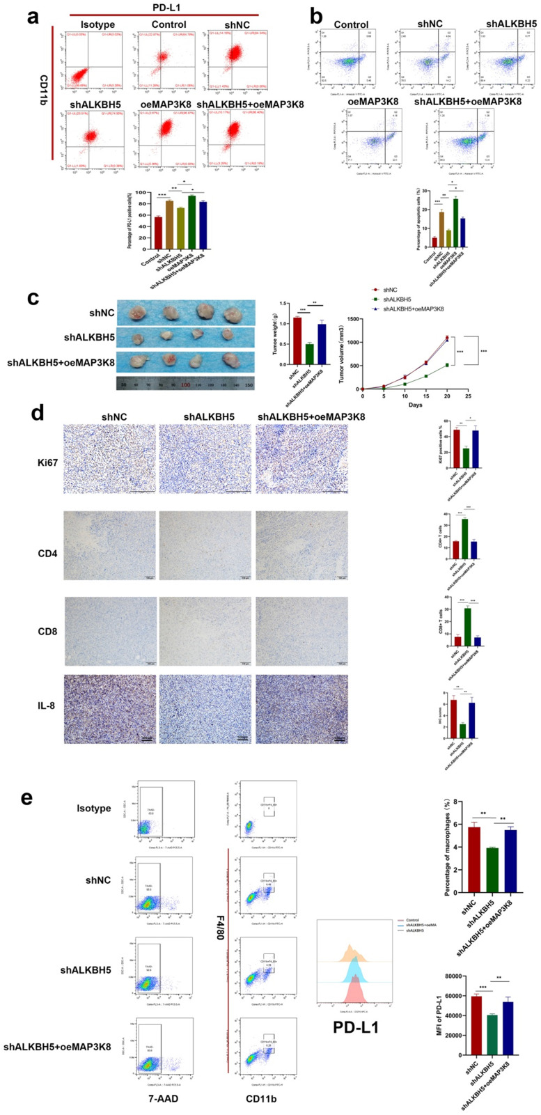
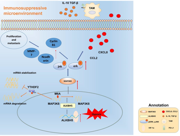
Hepatocellular carcinoma is one of the most common malignant tumors.M6A is a novel epigenetic modification that have been emerged as vital regulators for the progression of HCC. However, the regulatory role, clinical significance and the details of the modification, such as the impact on the local tumor environment, remain largely unclear. Our study showed that ALKBH5 was highly expressed in HCC and high ALKBH5 expression predicted a worse prognosis of HCC patients. Prediction of ALKBH5 function by tissue samples and single cell sequencing Gene Set Variation Analysis. Primary CD3 + T lymphocytes and bone marrow-derived macrophages were used to evaluate the effect of ALKBH5 on immune microenvironment. The results indicated that ALKBH5 promote HCC cell proliferation, metastasis and PD-L1+macrophage recruitment. Mechanistically the results showed that ALKBH5 regulates MAP3K8 expression in a m6A dependent manner which mediates the proliferation and metastasis of HCC cells. ALKBH5 also promotes the activation of JNK and ERK pathways through upregulating MAP3K8, thus regulating the expression of IL-8 and promoting macrophage recruitment. Taken together, these data show that ALKBH5 promotes HCC growth, metastasis and macrophage recruitment through ALKBH5/MAP3K8 axis and it may serve as a potential diagnostic marker and target for treatment of HCC patients.

Keywords: Hypoxia; Immune microenvironment; M6A; PD-L1; Tumor-associated macrophages.


Conflict of interest statement

Competing Interests: The authors have declared that no competing interest exists.

Figures

Figure 1
Figure 1
ALKBH5 is upregulated in patients with HCC and closely related to poor prognosis. (a) Analysis of differentially expressed m6A-related genes in HCC based on the TCGA database. (b) Western blot analysis in HepG2, HUH-7, Sk-hep1 and Hep3b cells with or without hypoxia, n=3. (c) Analysis of differentially expressed ALKBH5 in HCC based on the ICGC database. (d) Analysis of differentially expressed ALKBH5 in HCC based on the CPTAC database. (e) Survival analysis of ALKBH5 in HCC based on the ICGC database. (f) Survival analysis of ALKBH5 in HCC based on the 50 pairs IHC tissues. (g) IHC assay for ALKBH5 in HCC tissue and adjacent normal tissue n=50. (h) Western blot analysis of ALKBH5 in HCC tissue and paracancerous tissue n=10. (i) GSEA of ALKBH5 in HCC based on TCGA-LIHC dataset the TCGA database. (j) GSVA analysis of ALKBH5 in independent liver tumor cells (Total hepatocytes were regards as HCC cells) based on the single-cell sequencing dataset GSE149614. *P < 0.05; **P < 0.01; ***P < 0.001. Comparisons between the HCC and adjacent tissues were analyzed by paired t test, and comparisons between the other two groups were analyzed by nonpaired t test. Comparisons among multiple groups were analyzed by one-way ANOVA. Cell experiments were repeated in triplicate. All data are presented as the means ± SEM.
Figure 2
Figure 2
ALKBH5 promotes the proliferation, migration and invasion of HCC. (a) Western blot analysis of ALKBH5 protein expression in four HCC cell lines (HepG2, Sk-hep1, HUH-7 and Hep3b n=3. Notes: Proteintech (16837-1-ap) for ALKBH5 antibody (b) CCK8 assay for HCC cells with or without silencing ALKBH5 n=3. (c) EDU probe for HCC cells with or without silencing ALKBH5 n=3. (d) Cell cycle probe showed that cell cycle arrest induced by ALKBH5 silencing in hepatocellular carcinoma cells n=3. (e) Colony formation assay for HCC cells with or without silencing ALKBH5 n=3. (f) Cell migration and invasion assay for HCC cells n=3. (g) F-actin probe for HCC cells observed n=3. (h) Western blot was used to analyze the changes of metastasis and proliferation related markers (ZEB-1, MMP-7, CyclinB1 and E-Cadherin) with or without silencing ALKBH5 n=3. (i) Subcutaneous tumor in nude mice. Measurement of tumor volume and weight n=5. (j) IHC for Ki67 of subcutaneous tumor in nude mice n=5. *P < 0.05; **P < 0.01; ***P < 0.001. All data are presented as the means ± SEM. Student's t-test for independent samples and unequal variances was used to assess statistical significance. Comparisons among multiple groups were analyzed by one-way ANOVA. Comparisons at different time points were analyzed by repeated-measures ANOVA. Cell experiments were independently repeated three times.
Figure 3
Figure 3
ALKBH5 promotes the recruitment of PD-L1+ macrophages. (a) Transwell assays were used to measure the ability of tumor cells with or without silencing ALKBH5 to recruit macrophages. (b) RT-qPCR was used to analyze the expression of PD-L1 and polarization markers (Arg-1, IL-10 and TNF-α) in macrophages co-culturing with HCC cells. (c) Western blot assay of PD-L1 and polarization markers (Arg-1, IL-10 and TNF-α) expression in macrophages co-culturing with HCC cells. (c) Flow probe of PD-L1 expression in macrophages co-culturing with HCC cells. (d) Evaluation of PD-L1 positive macrophages in mouse subcutaneous tumors by flow cytometry, F4 / 80 and CD11b double positive labeled macrophages. All data are presented as the means ± SEM. Student's t-test for independent samples and unequal variances was used to assess statistical significance. Comparisons among multiple groups were analyzed by one-way ANOVA. Cell experiments were independently repeated three times.
Figure 4
Figure 4
ALKBH5 regulates MAP3K8 in an m6A -dependent manner. (a) The overall level of m6A changed after intervention of ALKBH5 expression by m6A colorimetry. (b) RNA-seq for HUH-7 cells with or without ALKBH5 knockdown n=2. (c) Intersection of the mRNA-seq and RIP-seq datasets (ALKBH5 binding target genes) (GSE144963). (d) Intersection of the mRNA-seq and m6A-seq datasets (Genes with increased m6A modification after silencing ALKBH5) (GSE87515). (e) RT-qPCR analysis of HUH-7 cells with or without silencing ALKBH5. (f) RIP-qPCR analysis of HUH-7 with or without silencing ALKBH5 in order to detect the combination of ALKBH5 with three genes (MAP3K8, MROH1, and UAP1L1). (g) M6a-IP-qPCR analysis of HUH-7 with or without silencing ALKBH5. (h) RT-qPCR analysis of Sk-hep1 with or without silencing ALKBH5. (i) Western blot analysis of the effect of cycloleucine on MAP3K8 expression. (j) Western blot analysis of the effect of mut-ALKBH5 H204A on MAP3K8 expression. (k) Western blot analysis of the effect of silencing YTHDF2 on MAP3K8 expression. (l) RT-qPCR analysis of the effect of silencing YTHDF2 on the half-life of MAP3K8 mRNA. (m) Colocalization of the YTHDF2 protein and MAP3K8 mRNA was observed by IF-FISH. *P < 0.05; **P < 0.01; ***P < 0.001. All data are presented as the means ± SEM. Student's t-test for independent samples and unequal variances was used to assess statistical significance. Comparisons among multiple groups were analyzed by one-way ANOVA. Comparisons at different time points were analyzed by repeated-measures ANOVA. Cell experiments were independently repeated three times.
Figure 5
Figure 5
MAP3K8 mediates ALKBH5 to promote the proliferation, migration and invasion of hepatocellular carcinoma. (a) Timer database analysis showed that MAP3K8 was highly expressed in HCC. (b) Immunohistochemical showed that MAP3K8 was highly expressed in HCC, n=50. (c) RT-qPCR analysis showed that MAP3K8 was highly expressed in HCC. n=70 (d) Correlation analysis between ALKBH5 and MAP3K8 based on the TCGA-LIHC dataset. (e) CCK-8 analysis of the role of MAP3K8 on the proliferation of hepatoma cells. (f) Transwell assays were used to analyze the effect of MAP3K8 on the migration and invasion of hepatoma cells. (g) Effect of MAP3K8 on the cytoskeleton of HCC detected by the F-actin probe. (h) Subcutaneous tumor in nude mice with or without MAP3K8 knockdown in HUH-7 cells. Measurement of tumor volume and weight n=4. (i) IHC for Ki67 of subcutaneous tumor in nude mice n=4. *P < 0.05; **P < 0.01; ***P < 0.001. All data are presented as the means ± SEM. Comparisons between the HCC and adjacent tissues were analyzed by paired t test, and comparisons between the other two groups were analyzed by non-paired t test. Comparisons among multiple groups were analyzed by one-way ANOVA. Cell experiments were independently repeated three times.
Figure 6
Figure 6
MAP3K8 mediates the recruitment of PD-L1+ macrophages by ALKBH5. (a) GSEA of ALKBH5 in HCC based on the above own mRNA-seq data. (b) Effect of ALKBH5 on macrophage chemokines measured by RT-qPCR. (c) Effect of MAP3K8 on IL-8 measured by RT-qPCR. (d) Effect of MAP3K8 on IL-8 measured by ELISA. (e) The effect of MAP3K8 and IL-8 on the recruitment of macrophages was detected by Transwell assay. (f) RT-qPCR of PD-L1 expression in macrophages. (g) Western blot of PD-L1 in macrophage. (h) Correlation analysis of the ALKBH5/MAP3K8/IL-8/CD68 axis based on the ICGC database. (i) Western blot analysis of the effect of ALKBH5 and MAP3K8 on ERK/JNK/p38 pathway. (j) IHC for p-JNK and p-ERK in subcutaneous tumor. (l) Effects of ERK inhibitors (LY3214996) and JNK inhibitors (JNK-IN-8) on the ability of ALKBH5 to interfere with the recruitment of macrophages in hepatoma cells (Hep3b). (l) Effects of ERK inhibitors (LY3214996) on the ability of ALKBH5 to induce PD-L1 expression of macrophage in hepatoma cells (Hep3b). (m) Effects of JNK inhibitors (JNK-IN-8) on the ability of ALKBH5 to induce PD-L1 expression of macrophage in hepatoma cells (Hep3b). *P < 0.05; **P < 0.01; ***P < 0.001. All data are presented as the means ± SEM. Student's t-test for independent samples and unequal variances was used to assess statistical significance. Comparisons among multiple groups were analyzed by one-way ANOVA. Cell experiments were independently repeated three times.
Figure 7
Figure 7
ALKBH5 promotes immunosuppressive microenvironment. (a) Effect of MAP3K8 and ALKBH5 on the expression of PD-L1 in macrophages (BMDM) induced by H22 cells by flow cytometry. (b) The apoptosis of CD3+T lymphocytes induced by the above treated macrophages (BMDM) was detected by flow cytometry. (c) H22 cells stably interfering with ALKBH5 and MAP3K8 were injected subcutaneously into mice, n=4. (d) The infiltration of CD4+ and CD8+ T lymphocytes and Ki67 positive percentage was analyzed by IHC n=4. (e) The ratio of PD-L1 + macrophages were analyzed by flow cytometry. TAMs were double positive for CD11b and F4 / 80 n=4. *P < 0.05; **P < 0.01; ***P < 0.001. All data are presented as the means ± SEM. Student's t-test for independent samples and unequal variances was used to assess statistical significance. Comparisons among multiple groups were analyzed by one-way ANOVA. Cell experiments were independently repeated three times.
Figure 8
Figure 8
Mechanism diagram. ALKBH5 regulates MAP3K8 expression in a m6A dependent manner, promotes the proliferation and metastasis of hepatoma cells, and promotes the expression of IL-8 by activating ERK / JNK pathway, thereby recruiting PD-L1 + macrophages to participate in shaping the immunosuppressive microenvironment.

References

    1. Huang DQ, El-Serag HB, Loomba R. Global epidemiology of NAFLD-related HCC: trends, predictions, risk factors and prevention. Nat Rev Gastroenterol Hepatol. 2021;18:223–38. - PMC - PubMed
    1. Singal AG, Lampertico P, Nahon P. Epidemiology and surveillance for hepatocellular carcinoma: New trends. J Hepatol. 2020;72:250–61. - PMC - PubMed
    1. Bray F, Ferlay J, Soerjomataram I. et al. Global cancer statistics 2018: GLOBOCAN estimates of incidence and mortality worldwide for 36 cancers in 185 countries. CA Cancer J Clin. 2018;68:394–424. - PubMed
    1. Li J, Zhu Y. Recent Advances in Liver Cancer Stem Cells: Non-coding RNAs, Oncogenes and Oncoproteins. Front Cell Dev Biol. 2020;8:548335. - PMC - PubMed
    1. Chen M, Wong CM. The emerging roles of N6-methyladenosine (m6A) deregulation in liver carcinogenesis. Mol Cancer. 2020;19:44. - PMC - PubMed

Publication types

MeSH terms