Review
. 2022 Aug 10:16:2545-2559.
doi: 10.2147/OPTH.S372011. eCollection 2022.

Open Globe Injuries: Review of Evaluation, Management, and Surgical Pearls

Affiliations
Review

Open Globe Injuries: Review of Evaluation, Management, and Surgical Pearls

Yujia Zhou et al. Clin Ophthalmol. .

Abstract

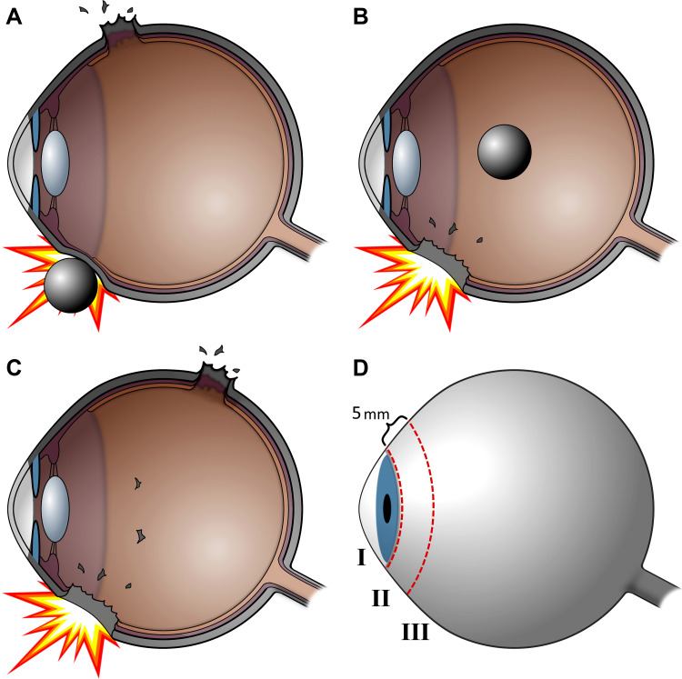
Ocular trauma may either be closed globe or open globe. Open globe injuries are full-thickness defects of the eyewall and are often differentiated by the mechanisms of injury from which they are caused: sharp or blunt trauma. They are ocular emergencies and can lead to substantial visual morbidity. Without timely intervention, damage is irreversible and leads to permanent vision loss. The goals of evaluation are to identify the mechanism of injury, characterize the extent of injury, and gather relevant history. If an open globe is suspected, ophthalmologic consultation should be requested. Once an open globe is diagnosed, preparations for surgery should be made immediately and steps should be taken to avoid further injury. Intraocular infection risk is relatively high, requiring immediate empiric systemic antibiotics. Emergent surgical exploration and primary closure is indicated whenever possible. After initial closure, secondary surgery and revision may be needed to improve vision outcomes, followed by extensive follow-up. In this review, best practices for evaluation and management are reviewed, with particular focus on the surgical approach and techniques.

Keywords: ocular trauma; open globe surgery; open globe surgical pearls; ophthalmic surgery; repair of open globe.


Conflict of interest statement

The authors report no conflicts of interest in this work.

Figures

Figure 1
Figure 1
Open Globe Classification. Cross sectional views are shown with various mechanisms of open globe injury. (A) In ruptured globes, the wound can occur elsewhere from the impact site. (B) A penetrating injury is an entry wound without an exit wound and may have a retained intraocular foreign body. (C) A perforating injury is a pair of entry and exit wounds caused by the same object. (D) Zone I injuries are limited to the cornea and limbus, while zone II injuries extend 5 mm posterior to the limbus and zone III injuries extend beyond 5 mm posterior to the limbus.
Figure 2
Figure 2
Penlight Open Globe Injury Findings. Selected findings by penlight displayed here, although examination may also be done by slit lamp. (A) Obvious left eye penetrating open globe with a retained fishing hook. (B) Penetrating injury of the left eye with aqueous humor leaking in a pool of fluorescein stain indicating a positive Seidel sign. (C) Right eye penetrating open globe injury with prolapsed iris. (D) Left eye penetrating open globe injury with a bead of extruded vitreous. (E) Right eye penetrating open globe injury with displaced lens extruding from a corneal laceration. (F) Dense 360-degree subconjunctival hemorrhage in the setting of ground-level fall suggesting occult open globe injury. (G) Obvious left eye globe rupture with significant volume loss causing a deformed anterior chamber. (H) Obvious right eye ruptured globe with significant volume loss causing disruption of the scleral contour. (I) Right eye peaked pupil pointing to an occult open globe injury of the inferotemporal sclera. (J) Left eye globe rupture with displaced lens and missing temporal section of iris.
Figure 3
Figure 3
Computed Tomography of Open Globe Injuries. Open globe injury wounds are represented by dotted dark red lines and sutures by solid black lines, numbered in order of placement. (A) CT axial image of a right eye penetrating globe injury with deflated globe colloquially known as a “mushroom sign”, in addition to intraconal air. (B) CT sagittal image of a left eye penetrating globe injury with vitreous hyper-attenuation in a dependent distribution indicating layered vitreous hemorrhage. (C) CT axial image of a left eye penetrating globe injury with a retained metallic intraocular foreign body, later determined to be a steel ball bearing. (D) CT axial image of a left eye penetrating open globe injury with shallow left anterior chamber, and distances labelled in yellow (right eye, 3.3 mm) and white (left eye, 2.5 mm).
Figure 4
Figure 4
Suture Order. Open globe injury wounds are represented by dotted dark red lines and sutures by solid black lines, numbered in order of placement. If the wound is not “visco-tight”, sutures marked by an asterisk may be placed first to facilitate an initial “visco-tight” seal. (A) Place sutures to align the limbus first in corneoscleral wounds. (B) Place corneal sutures from the limbus to the central visual axis, using shorter, deeper, and more closely spaced sutures towards the center of the cornea. (C) For lacerations traversing the diameter of the cornea, place sutures in a centripetal alternating order. (D) Use sequential bisecting sutures for linear scleral wounds, first in the middle of the wound, then between remaining open segments until the wound is closed. (E) Close linear segments of complex and stellate wounds first, then close intersections with a mattress or purse-string suture. (F) For wounds extending posterior to the equator, place sutures in an anterior to posterior order until surgical exposure is inadequate.
Figure 5
Figure 5
Suture Technique. Wounds are represented by dotted dark red lines and sutures by solid black lines. Cross sections represent sclera with the left side oriented towards the anterior globe. (A) Sutures should be placed perpendicular to the wound. (B) The length of a suture is approximately equal to the length of wound it seals, and these zones of sealing (gray) must overlap. (C) Tighten sutures to fully appose wound edges. (D) Sutures should enter and exit the wound perpendicular to the eye wall, equidistant to the wound on both sides. (E) Wounds that open at an oblique angle require a longer suture favoring the overriding side of the wound. (F) Only corneal sutures near the central visual axis may be full thickness; sutures should be partial thickness otherwise. (G) Suture knots should be rotated 90° and buried away from the visual axis if possible. (H) Oversew preserved pericardium or scleral patches to replace lost tissue.
Figure 6
Figure 6
Postoperative Right Eye Photographs. (A) A Patient’s right eye with nasal coloboma, corneal scarring, and corneal sutures after a repair of a stellate wound created by penetrating open globe injury. Wounds should be watertight and free of vitreous at case conclusion. (B) The same patient’s right eye after penetrating keratoplasty as secondary repair of corneal open globe injury.

References

    1. Kuhn F, Morris R, Witherspoon CD, Heimann K, Jeffers JB, Treister G. A standardized classification of ocular trauma. Ophthalmology. 1996;103(2):240–243. doi:10.1016/S0161-6420(96)30710-0 - DOI - PubMed
    1. Kolb H. Gross Anatomy of the Eye. Salt Lake City (UT): University of Utah Health Sciences Center; 1995. - PubMed
    1. Navon SE. Management of the ruptured globe. Int Ophthalmol Clin. 1995;35(1):71–91. doi:10.1097/00004397-199503510-00009 - DOI - PubMed
    1. Duke-Elder SMPA. System of Ophthalmology Vol. 14, Part 1. London: KimptonSt. Louis: Mosby; 1972.
    1. Kuhn F, Pelayes DE. Management of the ruptured eye. Eur Ophth Rev. 2009;3(1):48–50. doi:10.17925/EOR.2009.03.01.48 - DOI

LinkOut - more resources