Structure and Interaction of Ceramide-Containing Liposomes with Gold Nanoparticles as Characterized by SERS and Cryo-EM
- PMID: 35983312
- PMCID: PMC9377338
- DOI: 10.1021/acs.jpcc.2c01930
Structure and Interaction of Ceramide-Containing Liposomes with Gold Nanoparticles as Characterized by SERS and Cryo-EM
Abstract
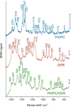
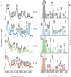
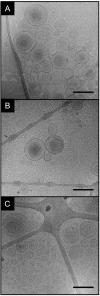
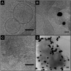
Due to the great potential of surface-enhanced Raman scattering (SERS) as local vibrational probe of lipid-nanostructure interaction in lipid bilayers, it is important to characterize these interactions in detail. The interpretation of SERS data of lipids in living cells requires an understanding of how the molecules interact with gold nanostructures and how intermolecular interactions influence the proximity and contact between lipids and nanoparticles. Ceramide, a sphingolipid that acts as important structural component and regulator of biological function, therefore of interest to probing, lacks a phosphocholine head group that is common to many lipids used in liposome models. SERS spectra of liposomes of a mixture of ceramide, phosphatidic acid, and phosphatidylcholine, as well as of pure ceramide and of the phospholipid mixture are reported. Distinct groups of SERS spectra represent varied contributions of the choline, sphingosine, and phosphate head groups and the structures of the acyl chains. Spectral bands related to the state of order of the membrane and moreover to the amide function of the sphingosine head groups indicate that the gold nanoparticles interact with molecules involved in different intermolecular relations. While cryogenic electron microscopy shows the formation of bilayer liposomes in all preparations, pure ceramide was found to also form supramolecular, concentric stacked and densely packed lamellar, nonliposomal structures. That the formation of such supramolecular assemblies supports the intermolecular interactions of ceramide is indicated by the SERS data. The unique spectral features that are assigned to the ceramide-containing lipid model systems here enable an identification of these molecules in biological systems and allow us to obtain information on their structure and interaction by SERS.
© 2022 The Authors. Published by American Chemical Society.
Conflict of interest statement
The authors declare no competing financial interest.
Figures


Similar articles
-
SERS and Cryo-EM Directly Reveal Different Liposome Structures during Interaction with Gold Nanoparticles.J Phys Chem Lett. 2018 Dec 6;9(23):6767-6772. doi: 10.1021/acs.jpclett.8b03191. Epub 2018 Nov 15. J Phys Chem Lett. 2018. PMID: 30421928
-
Molecular Structure and Interactions of Lipids in the Outer Membrane of Living Cells Based on Surface-Enhanced Raman Scattering and Liposome Models.Anal Chem. 2021 Jul 27;93(29):10106-10113. doi: 10.1021/acs.analchem.1c00964. Epub 2021 Jul 15. Anal Chem. 2021. PMID: 34264630
-
Impact of the ceramide subspecies on the nanostructure of stratum corneum lipids using neutron scattering and molecular dynamics simulations. Part I: impact of CER[NS].Chem Phys Lipids. 2018 Aug;214:58-68. doi: 10.1016/j.chemphyslip.2018.05.006. Epub 2018 May 30. Chem Phys Lipids. 2018. PMID: 29859142
-
Recent developments on gold nanostructures for surface enhanced Raman spectroscopy: Particle shape, substrates and analytical applications. A review.Anal Chim Acta. 2021 Jul 11;1168:338474. doi: 10.1016/j.aca.2021.338474. Epub 2021 Apr 5. Anal Chim Acta. 2021. PMID: 34051992 Review.
-
Novel optical nanosensors for probing and imaging live cells.Nanomedicine. 2010 Apr;6(2):214-26. doi: 10.1016/j.nano.2009.07.009. Epub 2009 Aug 20. Nanomedicine. 2010. PMID: 19699322 Review.
Cited by
-
Design Principles Of Inorganic-Protein Hybrid Materials for Biomedicine.Exploration (Beijing). 2025 Mar 6;5(3):20240182. doi: 10.1002/EXP.20240182. eCollection 2025 Jun. Exploration (Beijing). 2025. PMID: 40585774 Free PMC article. Review.
-
Gold Nanoparticle Adsorption and Uptake are Directed by Particle Capping Agent.Small Sci. 2025 May 19;5(7):2500060. doi: 10.1002/smsc.202500060. eCollection 2025 Jul. Small Sci. 2025. PMID: 40666033 Free PMC article.
-
Tunable Nanomaterials of Intracellular Crystallization for In Situ Biolabeling and Biomedical Imaging.Chem Biomed Imaging. 2023 Mar 30;1(9):767-784. doi: 10.1021/cbmi.3c00021. eCollection 2023 Dec 25. Chem Biomed Imaging. 2023. PMID: 39473839 Free PMC article. Review.
-
In Situ Raman Study of Neurodegenerated Human Neuroblastoma Cells Exposed to Outer-Membrane Vesicles Isolated from Porphyromonas gingivalis.Int J Mol Sci. 2023 Aug 28;24(17):13351. doi: 10.3390/ijms241713351. Int J Mol Sci. 2023. PMID: 37686157 Free PMC article.
References
-
- Michel R.; Plostica T.; Abezgauz L.; Danino D.; Gradzielski M. Control of the Stability and Structure of Liposomes by Means of Nanoparticles. Soft Matter 2013, 9 (16), 4167–4177. 10.1039/c3sm27875a. - DOI
LinkOut - more resources
Full Text Sources
Miscellaneous
