

Disseminated Superficial Actinic Porokeratosis (DSAP): A Case Report Highlighting the Clinical, Dermatoscopic, and Pathology Features of the Condition
- PMID: 35983404
- PMCID: PMC9376211
- DOI: 10.7759/cureus.26923
Disseminated Superficial Actinic Porokeratosis (DSAP): A Case Report Highlighting the Clinical, Dermatoscopic, and Pathology Features of the Condition
Abstract
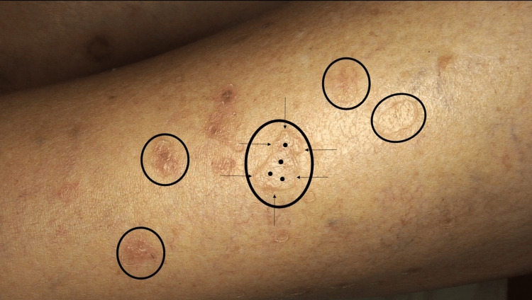
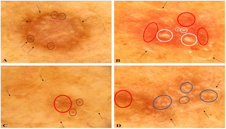
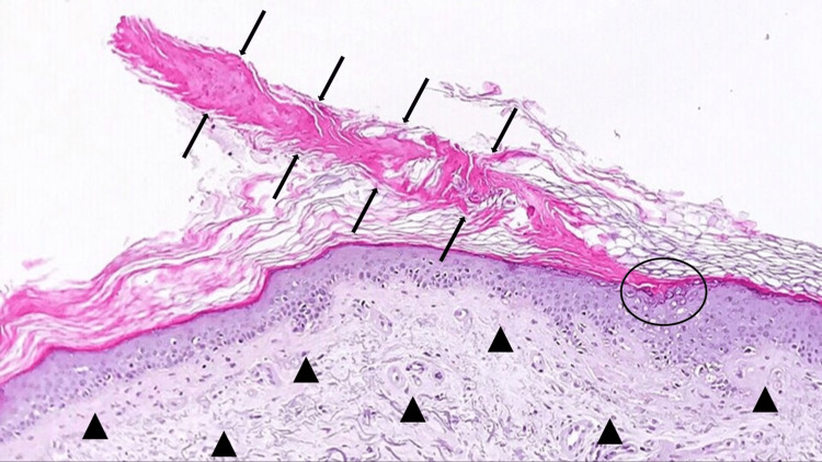
Porokeratosis describes a heterogenic group of keratinization disorders in which lesions are papules and plaques that demonstrate central atrophy surrounded by a hyperkeratotic margin. Clinical variants include not only porokeratosis of Mibelli, but also disseminated superficial, disseminated actinic superficial, linear, punctate, and palmaris et plantaris disseminata. Porokeratosis has a risk of malignant transformation. A woman with disseminated superficial actinic porokeratosis (DSAP) whose lesions presented as pruritic plaques and papules is described. The diagnosis was suspected clinically, supported by dermoscopy findings, and confirmed histologically. The condition-associated pruritus was managed symptomatically; her skin lesions will be monitored clinically. Clinical manifestations, dermatoscopic features, pathology findings, and treatment options for DSAP are summarized.
Keywords: cornoid lamella; dermatoscopy; disseminated superficial actinic porokeratosis (dsap); genitogluteal; linear; mibelli; porokeratosis.
Copyright © 2022, Waqar et al.
Conflict of interest statement
The authors have declared financial relationships, which are detailed in the next section.
Figures
References
-
- Porokeratosis: an enigma beginning to unravel. Das A, Vasudevan B, Talwar A. Indian J Dermatol Venereol Leprol. 2022;88:291–299. - PubMed
-
- Porokeratosis: a review of its pathophysiology, clinical manifestations, diagnosis, and treatment. (Article in Spanish) Vargas-Mora P, Morgado-Carrasco D, Fustà-Novell X. Actas Dermosifiliogr (Engl Ed) 2020;111:545–560. - PubMed
-
- Genitogluteal porokeratosis in an HIV-positive man: a case report and review of the literature on genital porokeratosis. Bari O, Vazirnia A, Cohen PR, Romero LS. Dermatol Online J. 2018;24 - PubMed
-
- Disseminated superficial actinic porokeratosis co-existing with linear and verrucous porokeratosis in an elderly woman: update on the genetics and clinical expression of porokeratosis. Murase J, Gilliam AC. J Am Acad Dermatol. 2010;63:886–891. - PubMed
-
- Clinical and dermoscopic features of pigmented disseminated superficial actinic porokeratosis: case report and literature review. Sotoodian B, Mahmood MN, Salopek TG. J Cutan Med Surg. 2018;22:229–231. - PubMed
Publication types
LinkOut - more resources
Full Text Sources