Review
. 2022 Oct 3;51(19):8216-8257.
doi: 10.1039/d2cs00387b.

Discovery of small molecule ligands for the von Hippel-Lindau (VHL) E3 ligase and their use as inhibitors and PROTAC degraders

Affiliations
Review

Discovery of small molecule ligands for the von Hippel-Lindau (VHL) E3 ligase and their use as inhibitors and PROTAC degraders

Claudia J Diehl et al. Chem Soc Rev. .

Abstract

The von Hippel-Lindau (VHL) Cullin RING E3 ligase is an essential enzyme in the ubiquitin-proteasome system that recruits substrates such as the hypoxia inducible factor for ubiquitination and subsequent proteasomal degradation. The ubiquitin-proteasome pathway can be hijacked toward non-native neo-substrate proteins using proteolysis targeting chimeras (PROTACs), bifunctional molecules designed to simultaneously bind to an E3 ligase and a target protein to induce target ubiquitination and degradation. The availability of high-quality small-molecule ligands with good binding affinity for E3 ligases is fundamental for PROTAC development. Lack of good E3 ligase ligands as starting points to develop PROTAC degraders was initially a stumbling block to the development of the field. Herein, the journey towards the design of small-molecule ligands binding to VHL is presented. We cover the structure-based design of VHL ligands, their application as inhibitors in their own right, and their implementation into rationally designed, potent PROTAC degraders of various target proteins. We highlight the key findings and learnings that have provided strong foundations for the remarkable development of targeted protein degradation, and that offer a blueprint for designing new ligands for E3 ligases beyond VHL.


Conflict of interest statement

The Ciulli laboratory receives or has received sponsored research support from Almirall, Amgen, Amphista Therapeutics, Boehringer Ingelheim, Eisai, Merck KGaA, Nurix Therapeutics, Ono Pharmaceutical and Tocris-BioTechne. A. C. is a scientific founder, advisor, and shareholder of Amphista Therapeutics, a company that is developing targeted protein degradation therapeutic platforms. C. D. reports no competing interest.

Figures

Fig. 1
Fig. 1. Schematic overview of the UPS, where E3 can consist of a single protein or a multi-subunit protein complex (a) and mechanism of action of PROTAC molecules inducing targeted protein degradation (b).
Fig. 2
Fig. 2. Crystal structure of VCB (PDB 1VCB) (a), cocrystal structure of HIF-1α peptide bound to VCB (PDB 1LM8) (b), and structure of the full length VCB-Cul2-Rbx1 multidomain complex (PDB 5N4W) (c).
Fig. 3
Fig. 3. Initial structure-guided design of VHL inhibitors. Cocrystal structure of hydroxy-HIF-1α (green) bound to VHL (PDB 1LM8) (a), nomenclature of VHL inhibitor subsections (b) and first reported binders (c).
Fig. 4
Fig. 4. Cocrystal structure of 5 bound to VCB (PDB 3ZRC).
Fig. 5
Fig. 5. Structure-guided optimisation of the RHS (a) and LHS (b) in Hyp-based inhibitors.
Fig. 6
Fig. 6. Cocrystal structure of inhibitor 12 bound to VCB (PDB 4B9K), derived from combinatorial optimisation of LHS and RHS.
Fig. 7
Fig. 7. Development of second-generation VHL inhibitors: RHS optimisation starting from benchmarking compound 13 and cocrystal structure of 13 with VCB (PDB 4W9C) (a); disclosure of VHL inhibitor VH032 in the course of LHS optimisation and cocrystal structure of VH032 bound to VCB (PDB 4W9H) (b).
Fig. 8
Fig. 8. Development of second generation VHL inhibitors: structure–activity relationships at the LHS pocket (a), cocrystal structure of inhibitor 20 bound to VCB (PDB 5NVW) (b), and cocrystal structure of VH101 bound to VCB (PDB 5NVX) (c).
Fig. 9
Fig. 9. Development of 2nd generation VHL inhibitors: further LHS optimisation towards reduced cytotoxicity (a) and cocrystal structure of inhibitor VH298 (24) bound to VCB (PDB 5LLI) (b).
Fig. 10
Fig. 10. 19F spy molecules for binding in Hyp site of VHL (a) and BODIPY-FL-PEG4-VH032 fluorescent probe (b).
Fig. 11
Fig. 11. Pyrrolidine's conformational preference in 4-functionalised prolines (a), synthesised F-Hyp diastereomers (b), F-Hyp containing derivatives of VH032 (c) and cocrystal structure of a F-Hyp containing HIF-1α 19-mer peptide bound to VCB (PDB 6GFX) (d).
Fig. 12
Fig. 12. VHL inhibitors containing thioamide bioisosteres and superimposition of cocrystal structures of 35 (green), 36 (yellow) and 37 (cyan) with VCB (PDB 5NVY, 6FMJ, 6FMK).
Fig. 13
Fig. 13. VHL inhibitors with benzylic modifications.
Fig. 14
Fig. 14. VHL inhibitors exploiting the subpocket between His110 and Tyr112.
Fig. 15
Fig. 15. Alternative VHL inhibitor derived from virtual screening approach.
Fig. 16
Fig. 16. Exit vectors for linker attachment used in VHL-recruiting PROTAC design.
Fig. 17
Fig. 17. Development of first VHL targeting PROTAC degrader. Structure of MZ1 (49) (a), synthetic route to access such degraders (b), and ternary cocrystal structure of MZ1 bound to VCB and Brd4BD2 (c), PROTAC–protein interactions (d) and de novo PPI between Brd4BD2 and VHL (e) (PDB 5T35).
Fig. 18
Fig. 18. First ERRα (50) and RIPK2 (51) targeting VHL-recruiting PROTACs.
Fig. 19
Fig. 19. Synthetic strategy to access phenolic linked HaloPROTACs (a), and structures of HaloPROTAC3 (52) (b) and optimised HaloPROTAC-E (53) (c).
Fig. 20
Fig. 20. Development of further BET targeting PROTACs. Brd2/Brd3/Brd4 pan-selective degrader ARV-771 (54) (a), and Brd3/Brd4 selective degrader MZP-54 (55) (b).
Fig. 21
Fig. 21. PROTAC 3i (56), chemical probe selectively degrading TBK1.
Fig. 22
Fig. 22. E3 ligase targeting PROTACs. Homo-PROTAC CM11 (57) inducing self-degradation of VHL (a), and CRBN-VHL Hetero-PROTACs inducing the selective degradation of CRBN (b).
Fig. 23
Fig. 23. Bcl-xL targeting PROTACs DT2216 (63) (a) and 64 (b), and ternary cocrystal structure of 64 bound to Bcl-xL and VCB (PDB 6ZHC) highlighting PROTAC–protein interactions (c) and de novo PPIs (d).
Fig. 24
Fig. 24. First FAK targeting PROTAC 65 (a), degrader BI-0319 (66) featuring improved selectivity (b), and structure and ternary cocrystal structure of GSK215 (67) bound to FAK and VCB (PDB 7PI4) (c).
Fig. 25
Fig. 25. Ternary cocrystal structures of WDR5/MS33/VCB (PDB 7JTO) (a) and WDR5/MS67/VCB (PDB 7JTP) (b).
Fig. 26
Fig. 26. Ternary cocrystal structure of WDR5/Homer/VCB (PDB 7Q2J).
Fig. 27
Fig. 27. EGFR targeting Gefitinib-based PROTAC 3 (71).
Fig. 28
Fig. 28. SGK3-selective VHL-recruiting degrader SGK3-PROTAC1 (72).
Fig. 29
Fig. 29. Design of LHS thioether conjugation vector from ternary crystal structure of VCB, MZ1 and Brd4BD2 (PDB 5T35) and structure of the optimised Brd4BD2 selective degrader AT1 (73).
Fig. 30
Fig. 30. LRRK2 degraders XL01126 (74) and XL01134 (75).
Fig. 31
Fig. 31. Development of AR degrader ARD-69 (80) featuring a benzylic exit vector between the VHL ligand and the linker.
Fig. 32
Fig. 32. Development of AR degraders build from low-affinity VHL binding ligands featuring the linker exit vector in benzylic position.
Fig. 33
Fig. 33. Synthetic routes to attach alkyl linkers (a), branched ether-containing linkers (b) and branched alkyl linkers (c) to the benzylic position of VHL binders.
Fig. 34
Fig. 34. Initial benzylic tethered SMARAC2/4 targeting PROTAC 83 (a) and SMARCA2-selective PROTAC 84 including visualisation of ionic de novo PPIs (yellow dotted lines) in the ternary crystal structure of degrader 84 bound to SMARCA2BD and VCB (PDB 7Z76).
Fig. 35
Fig. 35. Further optimised orally bioavailable SMARCA2 degraders 85 and ACBI2 (86) featuring benzylic linker attachment points.
Fig. 36
Fig. 36. Development of Brd7/9 selective VHL-recruiting degraders 87 and VZ185 (88) featuring phenolic linker vectors.
Fig. 37
Fig. 37. Structure-based design of phenolic tethered SMARCA2/4 degraders: cocrystal structure of the initial degrader 89 bound to VCB and SMARCA2BD (PDB 6HAY), and structures of degraders 90 and ACBI1 (91) derived from structure-guided design.
Fig. 38
Fig. 38. Isoform selectivity of p38 degraders driven by differing linker tethering vectors of the VHL-recruiting ligand.
Fig. 39
Fig. 39. Optimised CDK6 degraders 94 and 95 featuring phenolic tethering vector of the VHL-recruiting ligand.
Fig. 40
Fig. 40. ERα targeting VHL-recruiting PROTACs derived from DNA-encoded library screening.
Fig. 41
Fig. 41. Amide-to-ester conversion BET protein targeting PROTACs recruiting VHL leading to improved pharmacokinetics and cellular potency.
Fig. 42
Fig. 42. Synthetic strategy to access macroPROTAC-1 (100) and cocrystal structure of macroPROTAC-1 bound to VCB and Brd4BD2 (PDB 6SIS).
Fig. 43
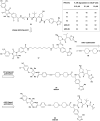
Fig. 43. Trivalent PROTACs. Trivalent degrader SIM1 (101) targeting both BDs of Brd4 via two incorporated BET ligands (a), and trivalent degrader DP-V-4 (102) simultaneously targeting EGFR and PARP proteins (b).
Fig. 44
Fig. 44. Reversible switching between an active, degrading trans-isomer and an inactive cis-isomer of photoPROTAC1 (103) upon irradiation with 415 nm viz. 530 nm.
Fig. 45
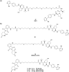
Fig. 45. Photocaged PROTACs. Photocleavable DEACM-caged ERRα degrader 104 (a), and photocleavable DMNB-caged BET degrader 105 (b).
Fig. 46
Fig. 46. Folate-caged BET degrader 106.
Fig. 47
Fig. 47. Enzymatic activated PROTACs. Quinone-caged HaloPROTAC 107 activated by NOQ1-mediated reduction (a) and generation of ROS via NOQ1-mediated reduction of β-lapachone (110) activating ROS-responsive HaloPROTAC 108 and BET degrader 109 (b).
Fig. 48
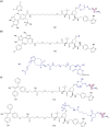
Fig. 48. Degrader-antibody conjugates as tissue-selective delivery systems. BET degrader-antibody conjugates 112 and 113 attached using disulfide bonding (a) or strain-promoted azide–alkyne cycloaddition (b) and ERα targeting degrader-conjugates featuring disulfide (114) and pyrophosphate (115) linkages to the antibody (c).
Fig. 49
Fig. 49. VHL-recruiting degraders used in TAG degradation platforms: FKBP12F36V fusion protein targeting degrader dTAGV-1 (116) (a) and Brd4BD2 L387A (BromoTag) targeting degrader AGB1 (117) (b).
Fig. 50
Fig. 50. Oligonucleotide-containing PROTACs. Schematic peptide-based RNA-PROTAC 118 (a), mode of action of TRAFTACs as TF degradation technology (b) and schematic DNA-nucleotide containing TF degrader 119 (c).
None
Claudia J. Diehl
None
Alessio Ciulli

References

    1. Myung J. Kim K. B. Crews C. M. Med. Res. Rev. 2001;21:245–273. doi: 10.1002/med.1009. - DOI - PMC - PubMed
    1. Fang S. Weissman A. M. Cell. Mol. Life Sci. 2004;61:1546–1561. doi: 10.1007/s00018-004-4129-5. - DOI - PMC - PubMed
    1. Goldberg A. L. Biochem. Soc. Trans. 2007;35:12–17. doi: 10.1042/BST0350012. - DOI - PubMed
    1. Shang F. Taylor A. Free Radical. Biol. Med. 2011;51:5–16. doi: 10.1016/j.freeradbiomed.2011.03.031. - DOI - PMC - PubMed
    1. Bulatov E. Ciulli A. Biochem. J. 2015;467:365–386. doi: 10.1042/BJ20141450. - DOI - PMC - PubMed

MeSH terms