. 2022 Oct;195(3):263-274.
doi: 10.1007/s10549-022-06698-x. Epub 2022 Aug 19.

BluePrint breast cancer molecular subtyping recognizes single and dual subtype tumors with implications for therapeutic guidance

Affiliations

BluePrint breast cancer molecular subtyping recognizes single and dual subtype tumors with implications for therapeutic guidance

Midas M Kuilman et al. Breast Cancer Res Treat. 2022 Oct.

Abstract

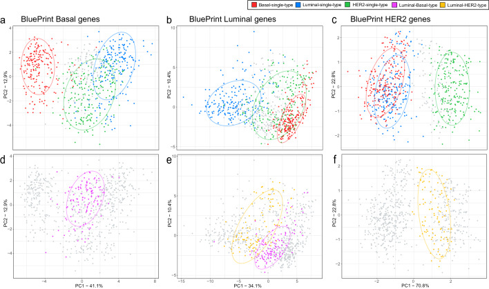
Purpose: BluePrint (BP) is an 80-gene molecular subtyping test that classifies early-stage breast cancer (EBC) into Basal, Luminal, and HER2 subtypes. In most cases, breast tumors have one dominant subtype, representative of a single activated pathway. However, some tumors show a statistically equal representation of more than one subtype, referred to as dual subtype. This study aims to identify and examine dual subtype tumors by BP to understand their biology and possible implications for treatment guidance.

Methods: The BP scores of over 15,000 tumor samples from EBC patients were analyzed, and the differences between the highest and the lowest scoring subtypes were calculated. Based upon the distribution of the differences between BP scores, a threshold was determined for each subtype to identify dual versus single subtypes.

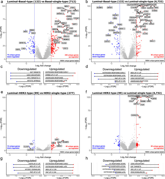
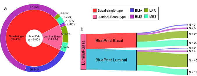
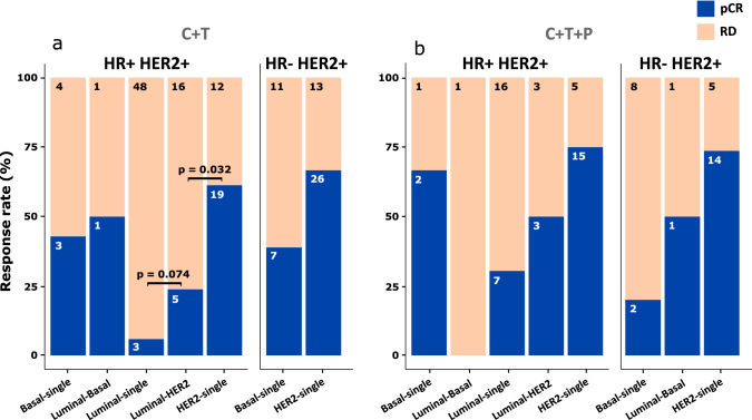
Results: Approximately 97% of samples had one single activated BluePrint molecular subtype, whereas ~ 3% of samples were classified as BP dual subtype. The most frequently occurring dual subtypes were the Luminal-Basal-type and Luminal-HER2-type. Luminal-Basal-type displays a distinct biology from the Luminal single type and Basal single type. Burstein's classification of the single and dual Basal samples showed that the Luminal-Basal-type is mostly classified as 'luminal androgen receptor' and 'mesenchymal' subtypes, supporting molecular evidence of AR activation in the Luminal-Basal-type tumors. Tumors classified as Luminal-HER2-type resemble features of both Luminal-single-type and HER2-single-type. However, patients with dual Luminal-HER2-type have a lower pathological complete response after receiving HER2-targeted therapies in addition to chemotherapy in comparison with patients with a HER2-single-type.

Conclusion: This study demonstrates that BP identifies tumors with two active functional pathways (dual subtype) with specific transcriptional characteristics and highlights the added value of distinguishing BP dual from single subtypes as evidenced by distinct treatment response rates.

Keywords: BluePrint; Breast cancer; Genomic testing; Molecular subtypes; Single and dual subtypes.


Conflict of interest statement

All authors (MMK, AE, AB, JCH, RB, DW, ACM, WMA, LM, and AMG) are non-commercial employees of Agendia, the commercial entity that markets the 80-gene signature as BluePrint. AMG is named inventor on the patent for the 80-gene signature used in this study. No writing assistance was utilized in the production of this manuscript.

Figures

Fig. 1
Fig. 1
Principle component analysis using the three BluePrint signature gene sets (Basal-type, N = 28, panels a and d; Luminal-type, N = 58, panels b and e; HER2-type, N = 4, panels c and f). The x-axis shows variance explained for the first principle component (PC) and the y-axis show the variance explained for the second PC of the correspondent BluePrint signature gene set. ac Clustering of Basal-single-type, Luminal-single-type, and HER2-single-type samples based on BluePrint signature genes. df Clustering of Luminal-Basal-type and Luminal-HER2-type based on BluePrint signature genes. ac shows coloring of single subtype samples (blue, Luminal-single-type; green, HER2-single-type; red, Basal-single-type) whereas the dual subtype samples are colored grey. df shows this in reverse where the dual subtypes are colored (yellow, Luminal-HER2-type; pink, Luminal-Basal-type) and the single subtypes are shown in grey. The ellipses reported in each subfigure illustrate the 80% confidence intervals of the single and dual subtypes
Fig. 2
Fig. 2
Differential gene expression analysis between BluePrint single and dual subtypes. The x-axis and y-axis report the Log2 fold change and the FDR-adjusted p-values (− Log10(FDR)), respectively. Number of tumor samples used for the analysis are shown in between brackets in titles. Significance thresholds of ≤ 0.05 FDR and a log2 fold change of ≥ 1 were used. Red and blue dots illustrate significant differentially expressed genes. The strongest differentially expressed genes are labeled (abs(logFC) ≥ 2 or −Log10 adj p-value ≥ 50). Differentially expressed genes are identified in the following comparisons: a Luminal-Basal-type versus Basal-single-type. b Luminal-Basal-type versus Luminal-single-type, e Luminal-HER2-type versus HER2-single-type, and f Luminal-HER2-type versus Luminal-single-type. Similarly, differentially expressed pathways are shown between c Luminal-Basal-type versus Basal-single-type. d Luminal-Basal-type versus Luminal-single-type, g Luminal-HER2-type versus HER2-single-type, and h Luminal-HER2-type versus Luminal-single-type. FDR = false discovery rate, UP = upregulated, DN = downregulated
Fig. 3
Fig. 3
a Sankey plot showing the further stratification of the standard BluePrint (BP) Basal, Luminal, and HER2 subtypes with full-genome microarray data available, into the BP single and dual subtypes. b Sankey plot illustrating the re-classification of clinical-based subtypes (based on hormone receptors (HR) and human epidermal growth factor receptor 2 (HER2) status) to BP-based single-type molecular subtypes (Basal-single-type, Luminal-single-type, HER2-single-type). c Further stratification of the same clinical-based subtypes as in (b) to the BP-based dual subtypes (Luminal-HER2-type, Luminal-Basal-type, HER2-Basal-type, and Luminal-HER2-Basal-type). d, e Boxplots reporting for each single and dual subtype category (x-axis), the level and spread of estrogen receptor and Ki67 positivity based on Immunohistochemistry assessment (y-axis). Significant differential positivity between ER and Ki67 was assumed at a p-value < 0.05 determined with a t-test between subtype categories. To note, for 4511 of the 9573 tumor samples with clinical annotation, HR and HER2 status were not available (Table S1)
Fig. 4
Fig. 4
BluePrint (BP) dual subtype classification compared with Burstein’s classification of triple-negative breast cancer tumors [12]. a The inner circle contains percentages of the BP Basal-single-type and BP Luminal-Basal-type. The outer circle illustrates the correspondent Burstein classification into Basal-like immuno-activated (BLIA), Basal-like immuno-suppressed (BLIS), Luminal androgen receptor (LAR), or Mesenchymal (MES). b) Samples with the Luminal-Basal-type were split based on standard BluePrint classification to illustrate their distribution over BLIA, BLIS, LAR, and MES subtypes. Significant differential classification of Burstein subtypes was assumed at a p-value ≤ 0.05 determined with a Chi-Square test of Independence between subtypes
Fig. 5
Fig. 5
Distribution of pathologically confirmed HER2+ patients of the NBRST trial [–20] based on the BluePrint single and dual subtype classification and their treatment response (N = 253). Patients are grouped based on their therapy regimen [chemotherapy (C) plus Trastuzumab (T) (panel a) or C + T and Pertuzumab (P) (panel b)], and their HR and HER2 status (HR+ HER2 + or HR- HER2+. The colored bars represents if a tumor did (pCR, blue) or did not [Residual Disease (RD), bisque] achieve pathological complete response (pCR). p-value determined with a chi-square test of independence between subtypes. Of the entire NBRST set (n = 289), 253 samples are showed due to low numerosity of HER2-Basal-type (n = 19) and Luminal-HER2-Basal (n = 17)

Similar articles

Cited by

References

    1. Krijgsman O, Roepman P, Zwart W, et al. A diagnostic gene profile for molecular subtyping of breast cancer associated with treatment response. Breast Cancer Res Treat. 2012;133:37–47. doi: 10.1007/s10549-011-1683-z. - DOI - PubMed
    1. Wallden B, Storhoff J, Nielsen T, et al. Development and verification of the PAM50-based Prosigna breast cancer gene signature assay. BMC Med Genomics. 2015;8:1–14. doi: 10.1186/s12920-015-0129-6. - DOI - PMC - PubMed
    1. Perou CM, Sørile T, Eisen MB, et al. Molecular portraits of human breast tumours. Nature. 2000;406:747–752. doi: 10.1038/35021093. - DOI - PubMed
    1. Sørlie T, Perou CM, Tibshirani R, et al. Gene expression patterns of breast carcinomas distinguish tumor subclasses with clinical implications. Proc Natl Acad Sci USA. 2001;98:10869–10874. doi: 10.1073/pnas.191367098. - DOI - PMC - PubMed
    1. Press O, Guzman R, Cervantes M, et al. Characterization of HER2 status by fluorescence in situ hybridization (FISH) and immunohistochemistry (IHC) New York: Springer; 2014. - PubMed