. 2022 Aug 19;17(8):e0273219.
doi: 10.1371/journal.pone.0273219. eCollection 2022.

Effect of preconditioning on propofol-induced neurotoxicity during the developmental period

Affiliations

Effect of preconditioning on propofol-induced neurotoxicity during the developmental period

Satoshi Shibuta et al. PLoS One. .

Abstract

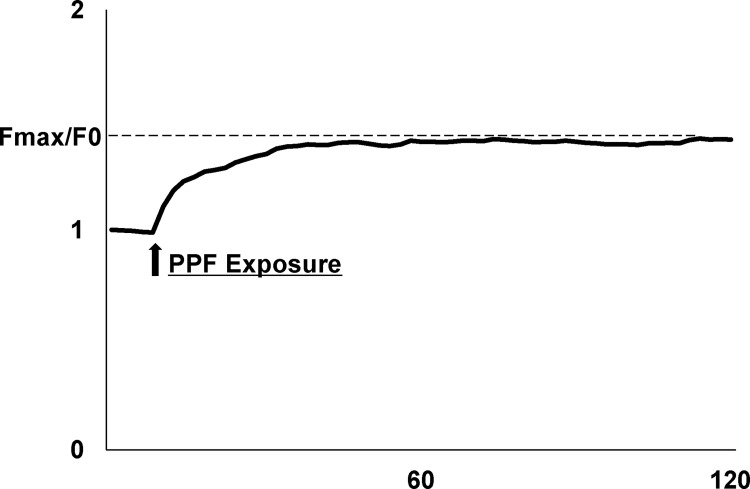
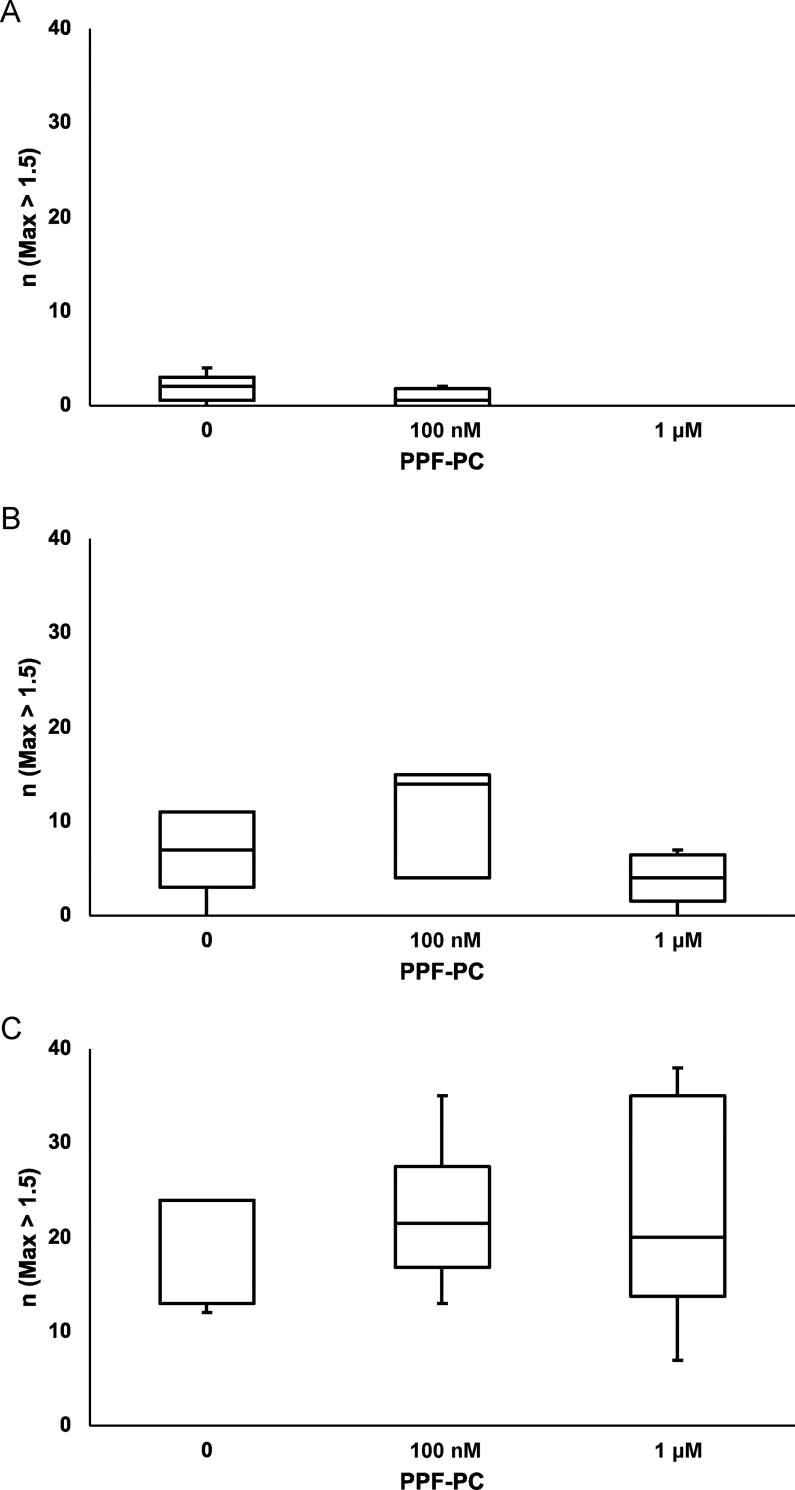
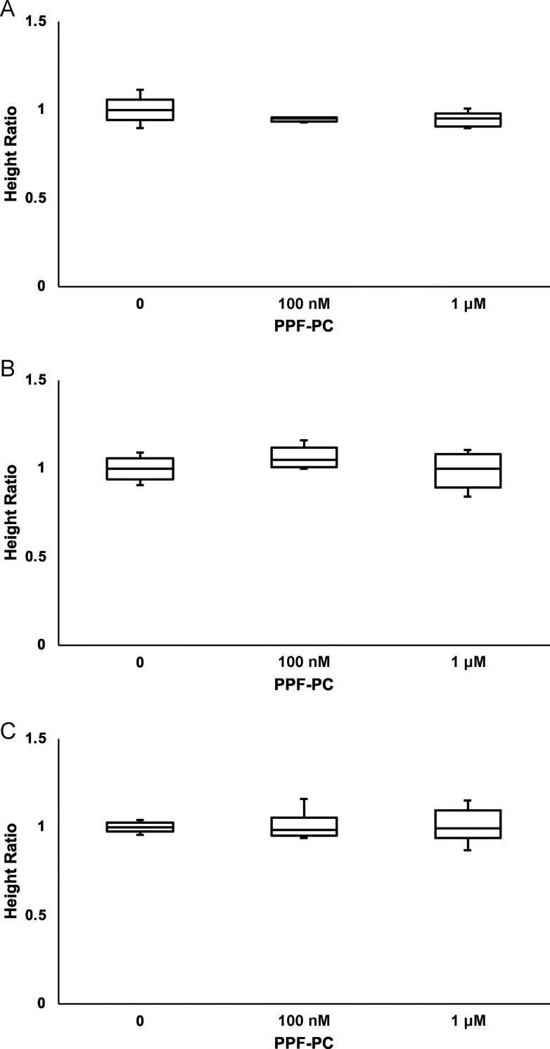
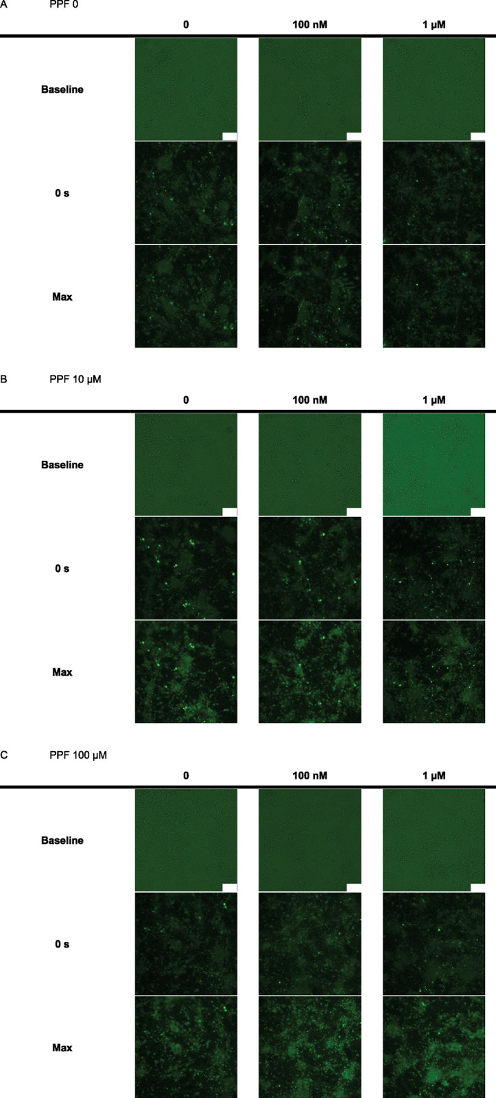
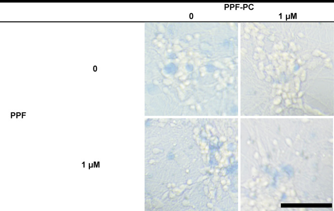
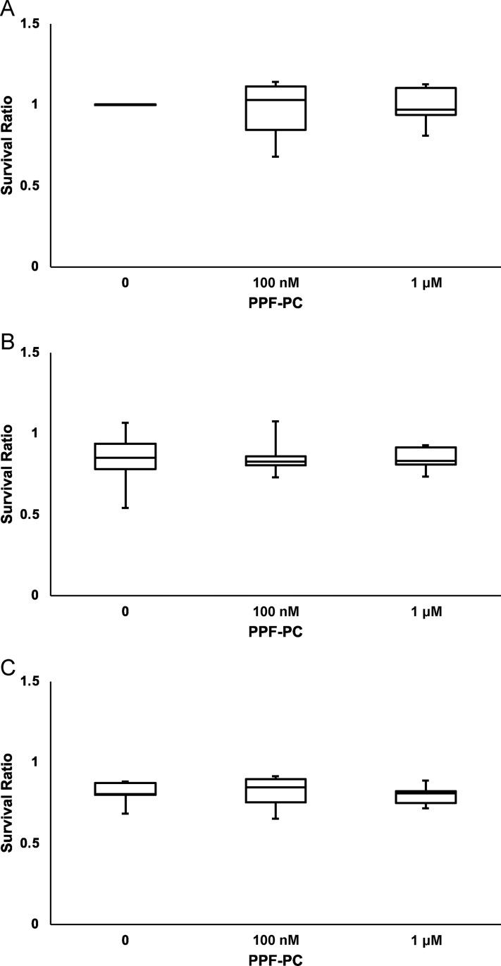
At therapeutic concentrations, propofol (PPF), an anesthetic agent, significantly elevates intracellular calcium concentration ([Ca2 +]i) and induces neural death during the developmental period. Preconditioning enables specialized tissues to tolerate major insults better compared with tissues that have already been exposed to sublethal insults. Here, we investigated whether the neurotoxicity induced by clinical concentrations of PPF could be alleviated by prior exposure to sublethal amounts of PPF. Cortical neurons from embryonic day (E) 17 Wistar rat fetuses were cultured in vitro, and on day in vitro (DIV) 2, the cells were preconditioned by exposure to PPF (PPF-PC) at either 100 nM or 1 μM for 24 h. For morphological observations, cells were exposed to clinical concentrations of PPF (10 μM or 100 μM) for 24 h and the survival ratio (SR) was calculated. Calcium imaging revealed significant PPF-induced [Ca2+]i elevation in cells on DIV 4 regardless of PPF-PC. Additionally, PPF-PC did not alleviate neural cell death induced by PPF under any condition. Our findings indicate that PPF-PC does not alleviate PPF-induced neurotoxicity during the developmental period.


Conflict of interest statement

The authors have declared that no competing interests exist.

Figures

Fig 1
Fig 1. Chemical structure of propofol (PPF).
Fig 2
Fig 2. The intensity of the fluorescence (F/F0) over time in a cultured primary neuron.
PPF application led to an increase in the temporal intracellular calcium concentration ([Ca2+]i). The arrow indicates PPF exposure and onset. To evaluate the PPF application effect, we observed Fmax/F0 as height calculated from the [Ca2 +]i response in the neuron.
Fig 3
Fig 3. The number of the neurons that had Fmax (i.e., height) > 1.5 in 50 neurons.
When neurons were exposed to vehicle (DMSO dissolved in NBS) on DIV 4, almost no neurons showed a significant height increase (exceeding F max > 1.5), regardless of exposure to PPF-PC on DIV 2 (A). Both 10 μM (B) and 100 μM (C) PPF significantly increased the number of neurons whose Fmax was > 1.5 compared to vehicle. However, PPF-PC did not affect the number of neurons whose F max was > 1.5. The differences between the means were calculated using analysis of variance (ANOVA), followed by the Tukey–Kramer honestly significant difference test as a post-hoc test.
Fig 4
Fig 4. Height ratios (HR) of neurons in response to vehicle or PPF.
HR of all groups were irrelevant to PPF-PC on DIV 2; (A) vehicle (DMSO dissolved in NBS). PPF exposure at 10 μM (B) and 100 (C) μM increased Fmax/F0, significantly. PPF-PC on DIV 2 did not affect Fmax/F0. The differences between the means were calculated using analysis of variance (ANOVA), followed by the Tukey–Kramer honestly significant difference test as a post-hoc test.
Fig 5
Fig 5. Images of the time course of neurons from all groups demonstrating the changes of [Ca2 +]i in response to PPF application on DIV 4.
In the vehicle groups (dimethyl sulfoxide [DMSO]), the fluorescence intensities of the neurons showed little change (A), whereas in all 10 μM (B) and 100 μM (C) PPF exposure groups, the fluorescence intensities of neurons on DIV 4 were significantly high, and were irrelevant to PPF-PC on DIV 2. 0s: shortly before DMSO of PPF application. Max; most nearly the moment when Fmax is recorded. Since each neuron showed its own Fmax (peak moment of intracellular calcium concentration), not all the neurons showed Fmax at the same time in these images. Scale Bar = 100 μm.
Fig 6
Fig 6. Transmitted light microphotographs of primary cultured cortical neurons exposed to vehicle (DMSO in PBS) or 100 μM PPF on DIV 3, taken 24 h after the exposure.
Regardless of PPF-PC on DIV 2, PPF exposure on DIV 3 significantly induced neuronal death compared to vehicle. Scale Bar = 100 μm.
Fig 7
Fig 7
Survival ratio (SR) of primary cultured cortical neurons exposed to vehicle (DMSO in PBS (A); PPF at (B) 10 or (C) 100 μM) for 24 h during DIV 3 and 4. Although exposure to PPF elicited a significant neuronal death, the decrease in SRs on DIV 3 and 4 were irrelevant to PPF-PC on DIV 2. The differences between the means were calculated using ANOVA, followed by the Tukey–Kramer honestly significant difference test as a post-hoc test.

Similar articles

Cited by

References

    1. Shibuta S, Kanemura S, Uchida O, Mashimo T. The influence of initial target effect-site concentrations of propofol on the similarity of effect-sites concentrations at loss and return of consciousness in elderly female patients with the Diprifusor system. J Anaesthesiol Clin Pharmacol. 2012;28: 194–199. doi: 10.4103/0970-9185.94851 - DOI - PMC - PubMed
    1. Loepke AW, Soriano SG. An assessment of the effects of general anesthetics on developing brain structure and neurocognitive function. Anesth Analg. 2008;106: 1681–1707. doi: 10.1213/ane.0b013e318167ad77 - DOI - PubMed
    1. Kahraman S, Zup SL, McCarthy MM, Fiskum G. GABAergic mechanism of propofol toxicity in immature neurons. J Neurosurg Anesthesiol. 2008;20: 233–240. doi: 10.1097/ANA.0b013e31817ec34d - DOI - PMC - PubMed
    1. Bercker S, Bert B, Bittigau P, Felderhoff-Müser U, Bührer C, Ikonomidou C, et al.. Neurodegeneration in newborn rats following propofol and sevoflurane anesthesia. Neurotox Res. 2009;16: 140–147. doi: 10.1007/s12640-009-9063-8 - DOI - PubMed
    1. Karen T, Schlager GW, Bendix I, Sifringer M, Herrmann R, Pantazis C, et al.. Effect of propofol in the immature rat brain on short- and long-term neurodevelopmental outcome. PLOS ONE. 2013;8: e64480. doi: 10.1371/journal.pone.0064480 - DOI - PMC - PubMed

Publication types