

Classical and Learned MR to Pseudo-CT Mappings for Accurate Transcranial Ultrasound Simulation
- PMID: 35984788
- PMCID: PMC7616982
- DOI: 10.1109/TUFFC.2022.3198522
Classical and Learned MR to Pseudo-CT Mappings for Accurate Transcranial Ultrasound Simulation
Abstract
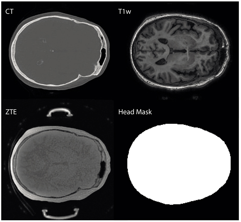
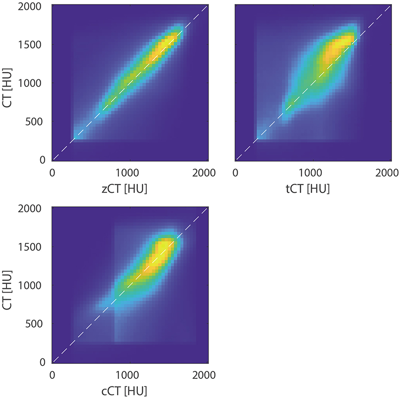
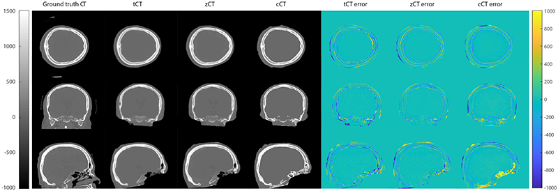
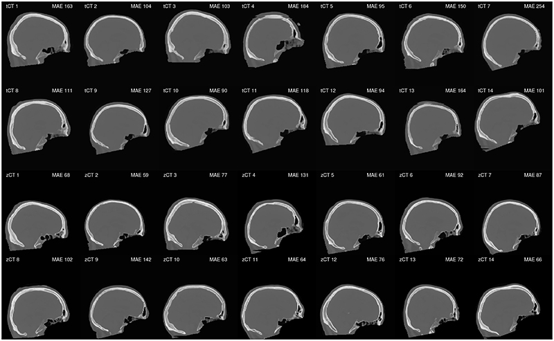
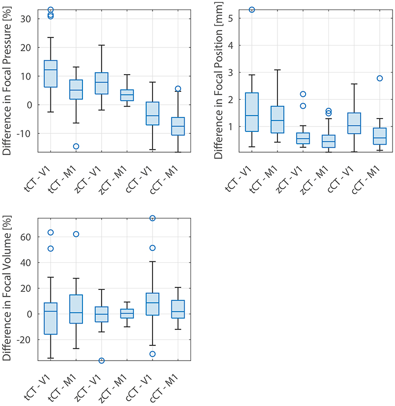
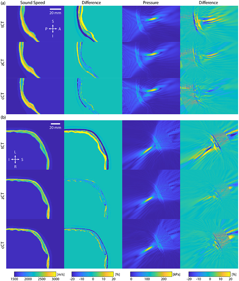
Model-based treatment planning for transcranial ultrasound therapy typically involves mapping the acoustic properties of the skull from an X-ray computed tomography (CT) image of the head. Here, three methods for generating pseudo-CT (pCT) images from magnetic resonance (MR) images were compared as an alternative to CT. A convolutional neural network (U-Net) was trained on paired MR-CT images to generate pCT T images from either T1-weighted or zero-echo time (ZTE) MR images (denoted tCT and zCT, respectively). A direct mapping from ZTE to pCT was also implemented (denoted cCT). When comparing the pCT and ground-truth CT images for the test set, the mean absolute error was 133, 83, and 145 Hounsfield units (HU) across the whole head, and 398, 222, and 336 HU within the skull for the tCT, zCT, and cCT images, respectively. Ultrasound simulations were also performed using the generated pCT images and compared to simulations based on CT. An annular array transducer was used targeting the visual or motor cortex. The mean differences in the simulated focal pressure, focal position, and focal volume were 9.9%, 1.5 mm, and 15.1% for simulations based on the tCT images; 5.7%, 0.6 mm, and 5.7% for the zCT; and 6.7%, 0.9 mm, and 12.1% for the cCT. The improved results for images mapped from ZTE highlight the advantage of using imaging sequences, which improves the contrast of the skull bone. Overall, these results demonstrate that acoustic simulations based on MR images can give comparable accuracy to those based on CT.
Figures
Similar articles
-
Deep learning approaches using 2D and 3D convolutional neural networks for generating male pelvic synthetic computed tomography from magnetic resonance imaging.Med Phys. 2019 Sep;46(9):3788-3798. doi: 10.1002/mp.13672. Epub 2019 Jul 26. Med Phys. 2019. PMID: 31220353
-
Zero TE-based pseudo-CT image conversion in the head and its application in PET/MR attenuation correction and MR-guided radiation therapy planning.Magn Reson Med. 2018 Oct;80(4):1440-1451. doi: 10.1002/mrm.27134. Epub 2018 Feb 18. Magn Reson Med. 2018. PMID: 29457287
-
Pseudo-CT generation from multi-parametric MRI using a novel multi-channel multi-path conditional generative adversarial network for nasopharyngeal carcinoma patients.Med Phys. 2020 Apr;47(4):1750-1762. doi: 10.1002/mp.14062. Epub 2020 Feb 21. Med Phys. 2020. PMID: 32012292
-
Transcranial MR Imaging-Guided Focused Ultrasound Interventions Using Deep Learning Synthesized CT.AJNR Am J Neuroradiol. 2020 Oct;41(10):1841-1848. doi: 10.3174/ajnr.A6758. Epub 2020 Sep 3. AJNR Am J Neuroradiol. 2020. PMID: 32883668 Free PMC article.
-
Zero-Echo-Time and Dixon Deep Pseudo-CT (ZeDD CT): Direct Generation of Pseudo-CT Images for Pelvic PET/MRI Attenuation Correction Using Deep Convolutional Neural Networks with Multiparametric MRI.J Nucl Med. 2018 May;59(5):852-858. doi: 10.2967/jnumed.117.198051. Epub 2017 Oct 30. J Nucl Med. 2018. PMID: 29084824 Free PMC article.
Cited by
-
Parameter optimisation for mitigating somatosensory confounds during transcranial ultrasonic stimulation.bioRxiv [Preprint]. 2025 Mar 19:2025.03.19.642045. doi: 10.1101/2025.03.19.642045. bioRxiv. 2025. Update in: Brain Stimul. 2025 Jul-Aug;18(4):1224-1236. doi: 10.1016/j.brs.2025.06.009. PMID: 40166137 Free PMC article. Updated. Preprint.
-
ENHANCING TRANSCRANIAL FOCUSED ULTRASOUND TREATMENT PLANNING WITH SYNTHETIC CT FROM ULTRA-SHORT ECHO TIME (UTE) MRI: A MULTI-TASK DEEP LEARNING APPROACH.Proc IEEE Int Symp Biomed Imaging. 2024 May;2024:10.1109/isbi56570.2024.10635176. doi: 10.1109/isbi56570.2024.10635176. Epub 2024 Aug 22. Proc IEEE Int Symp Biomed Imaging. 2024. PMID: 39844940 Free PMC article.
-
Evaluation of synthetically generated computed tomography for use in transcranial focused ultrasound procedures.J Med Imaging (Bellingham). 2023 Sep;10(5):055001. doi: 10.1117/1.JMI.10.5.055001. Epub 2023 Sep 22. J Med Imaging (Bellingham). 2023. PMID: 37744953 Free PMC article.
-
Robust deep learning estimation of cortical bone porosity from MR T1-weighted images for individualized transcranial focused ultrasound planning.medRxiv [Preprint]. 2024 Jul 18:2024.07.18.24310644. doi: 10.1101/2024.07.18.24310644. medRxiv. 2024. PMID: 39072036 Free PMC article. Preprint.
-
Generating Patient-Specific Acoustic Simulations for Transcranial Focused Ultrasound Procedures Based on Optical Tracking Information.IEEE Open J Ultrason Ferroelectr Freq Control. 2023;3:146-156. doi: 10.1109/ojuffc.2023.3318560. Epub 2023 Sep 25. IEEE Open J Ultrason Ferroelectr Freq Control. 2023. PMID: 38222464 Free PMC article.
References
-
- Elias WJ, Lipsman N, Ondo WG, Ghanouni P, Kim YG, Lee W, Schwartz M, Hynynen K, Lozano AM, Shah BB, et al. A randomized trial of focused ultrasound thalamotomy for essential tremor. New England Journal of Medicine. 2016;375(8):730–739. - PubMed
-
- Legon W, Sato TF, Opitz A, Mueller J, Barbour A, Williams A, Tyler WJ. Transcranial focused ultrasound modulates the activity of primary somatosensory cortex in humans. Nature Neuroscience. 2014;17(2):322–329. - PubMed
-
- Gasca-Salas C, Fernández-Rodríguez B, Pineda-Pardo JA, Rodriguez-Rojas R, Obeso I, Hernandez-Fernandez F, Del Alamo M, Mata D, Guida P, Ordas-Bandera C, et al. Blood-brain barrier opening with focused ultrasound in Parkinson’s disease dementia. Nature communications. 2021;12(1):1–7. doi: 10.1038/s41467-021-21022-9. - DOI - PMC - PubMed
-
- Sun J, Hynynen K. Focusing of therapeutic ultrasound through a human skull: a numerical study. The Journal of the Acoustical Society of America. 1998;104(3):1705–1715. - PubMed
