. 2022 Aug 19;17(8):e0272277.
doi: 10.1371/journal.pone.0272277. eCollection 2022.

Dysregulation of the hippocampal neuronal network by LGI1 auto-antibodies

Affiliations

Dysregulation of the hippocampal neuronal network by LGI1 auto-antibodies

Elodie Fels et al. PLoS One. .

Abstract

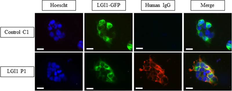
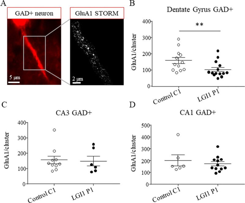
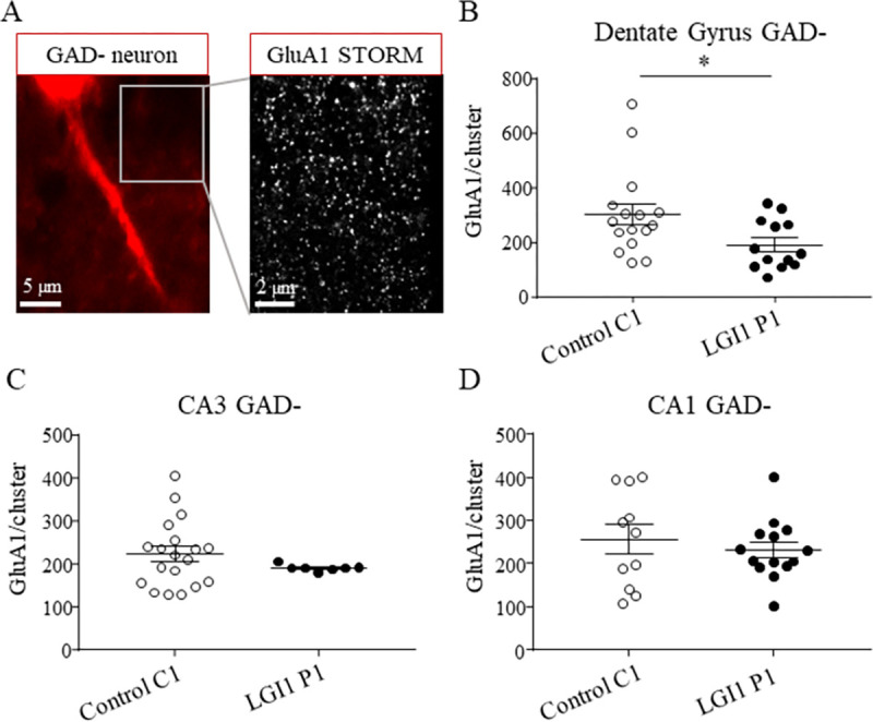
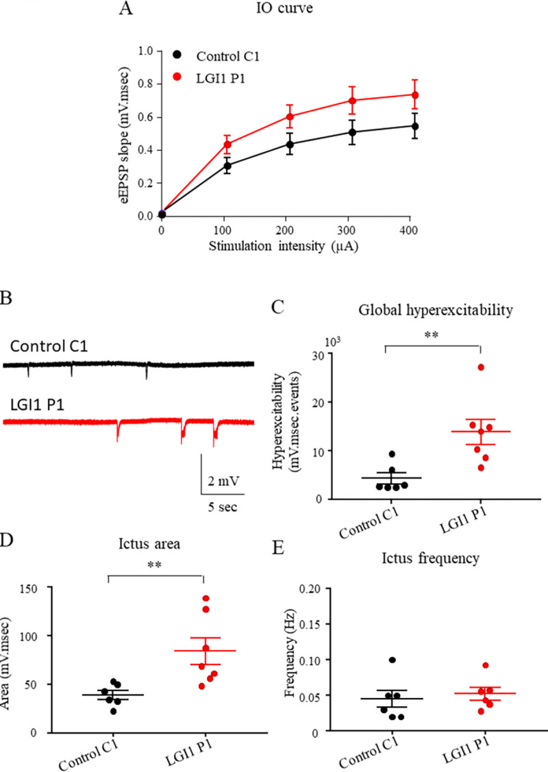
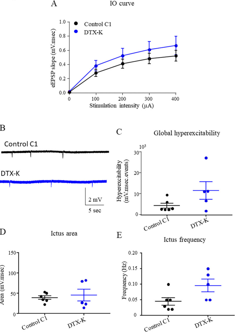
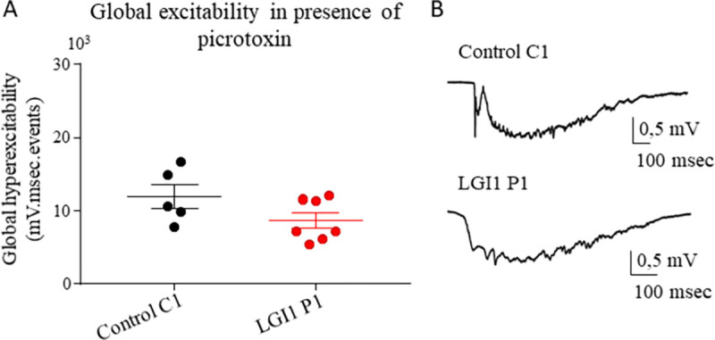
LGI1 is a neuronal secreted protein highly expressed in the hippocampus. Epileptic seizures and LGI1 hypo-functions have been found in both ADLTE, a genetic epileptogenic syndrome and LGI1 limbic encephalitis (LE), an autoimmune disease. Studies, based mainly on transgenic mouse models, investigated the function of LGI1 in the CNS and strangely showed that LGI1 loss of function, led to a decreased AMPA-receptors (AMPA-R) expression. Our project intends at better understanding how an altered function of LGI1 leads to epileptic seizures. To reach our goal, we infused mice with LGI1 IgG purified from the serum of patients diagnozed with LGI1 LE. Super resolution imaging revealed that LGI1 IgG reduced AMPA-R expression at the surface of inhibitory and excitatory neurons only in the dentate gyrus of the hippocampus. Complementary electrophysiological approaches indicated that despite reduced AMPA-R expression, LGI1 IgG increased the global hyperexcitability in the hippocampal neuronal network. Decreased AMPA-R expression at inhibitory neurons and the lack of LGI1 IgG effect in presence of GABA antagonist on excitability, led us to conclude that LGI1 function might be essential for the proper functioning of the overall network and orchestrate the imbalance between inhibition and excitation. Our work suggests that LGI1 IgG reduced the inhibitory network activity more significantly than the excitatory network shedding lights on the essential role of the inhibitory network to trigger epileptic seizures in patients with LGI1 LE.


Conflict of interest statement

The authors have declared that no competing interests exist.

Figures

Fig 1
Fig 1. Purified IgG from LGI1 LE patient contain LGI1 Abs.
HEK293T cells were transfected with LGI1-GFP and ADAM22 plasmids. Purified IgG from LGI1 LE patient are colocalized with LGI1-GFP signal at the cell surface of HEK293T transfected cells while no signal was found in cells treated with purified IgG from healthy subject. Scale bar = 20μm.
Fig 2
Fig 2. LGI1 Abs decrease the expression of AMPA-R inside clusters at the surface of interneurons in the hippocampus.
Epifluorescence images (100x objective) of an inhibitory GAD+ neuron expressing td-tomato (left image). Magnification of the corresponding super-resolution STORM image showing the expression of GluA1 subunits along the neuronal extension (right image) (A). The total density of surface GluA1 subunits decreased after LGI1 P1 IgG infusion compared to control C1 IgG infusion at the surface of interneurons in the DG (Mann-Whitney **p = 0.0091, nCtrl = 12 neurons, nLGI1 = 14 neurons) (B) but not in CA3 region (Mann-Whitney test p = 0.63, nCtrl = 11 neurons, nLGI1 = 6 neurons) (C) nor CA1 region of the hippocampus (Mann-Whitney t-test p = 0.68, nCtrl = 6 neurons, nLGI1 = 12 neurons) (D). Data are represented as means ± SEM (analysis realized from nCtrl = 4 mice, nLGI1 = 3 mice).
Fig 3
Fig 3. LGI1 Abs decrease the expression of AMPA-R inside clusters at the surface of excitatory neurons in the hippocampus.
Epifluorescence images (100x objective) of GAD- region representing excitatory neurons (left image). The magnification showing the super-resolution STORM image of the expression of GluA1 subunits in the GAD- region (right image) (A). The total density of surface GluA1 subunits decreased after LGI1 P1 IgG infusion compared to control C1 IgG infusion in GAD- area in the DG of the hippocampus (Unpaired t test *p = 0.031, nCtrl = 16 area, nLGI1 = 13 area) (B) but not in CA3 region (Mann-Whitney test p = 0.33, nCtrl = 19 area, nLGI1 = 9 area) (C) nor CA1 (Unpaired t-test p = 0.49, nCtrl = 11 area, nLGI1 = 14 area) (D). Data are represented as mean ± SEM (analysis realized from nCtrl = 4 mice, nLGI1 = 3 mice.
Fig 4
Fig 4. LGI1 abs increase induced neuronal hyperexcitability in the hippocampus.
LFP were recorded in CA1 region of acute hippocampal slices from mice infused for 7 days with control C1 IgG, LGI1 P1 IgG. Increasing stimulations intensity were delivered to Schaffer collaterals and the slope of eEPSP in CA1 area were plotted. No effect of the infusion of control C1 IgG, LGI1 P1 IgG on the IO curve was detected (two-way ANOVA test, p = 0.27; nCtrl = 12; nLGI1 = 14) (A). Examples of ictus recorded in slices infused with control C1 IgG, LGI1 P1 IgG (B). The global hyperexcitability of the neuronal network is increased after LGI1 P1 IgG infusion compared to control C1 IgG infusion (Kruskal-Wallis test *p = 0.02, Dunn’s post Hoc test LGI1-Ctrl **p = 0.0082) (C) as well as the area of the ictus (Kruskal-Wallis test *p = 0.02, Dunn’s post Hoc test LGI1-Ctrl **p = 0.12) (D) while frequency is unchanged (Kruskal-Wallis test *p = 0.40, Dunn’s post Hoc test LGI1-Ctrl **p>0.99) (E). Data are represented as mean ± SEM (nCtrl = 6 mice, nLGI1 = 7 mice).
Fig 5
Fig 5. DTX-K infusion does not increase neuronal hyperexcitability in the hippocampus.
LFP were recorded in CA1 region of acute hippocampal slices from mice infused for 7 days with control C1 IgG or DTX-K. Increasing stimulations intensity were delivered to Schaffer collaterals and the slope of eEPSP in CA1 area were plotted. No effect of the infusion of control C1 IgG vs DTX-K on the IO curve was detected (two-way ANOVA test, p = 065; nCtrl = 12; nDTX-K = 8) (A). Examples of ictus recorded in slices infused with control C1 IgG or DTX-K (B). The global hyperexcitability of the neuronal network is not increased significantly after DTX-K infusion compared to control C1 IgG infusion (Dunn’s post Hoc test DTX-K- Ctrl p = 0.11) (C) as well as the area of the ictus (Fisher LSD post Hoc test, Ctrl-DTX-K p = 0.73) (D) while frequency is not significantly increased (Kruskal-Wallis test, p = 0.09) (E). Data are represented as mean ± SEM (nCtrl = 6 mice, nDTX-K = 5 mice).
Fig 6
Fig 6. LGI1 abs have no effect on the global hyperexcitability when the inhibitory network was blocked.
(A) LFP in presence of 4-AP (40μM) and picrotoxin (100μM) were recorded in CA1 area of acute hippocampal slices from mice infused for 7 days with control C1 or LGI1 P1 IgG. No difference was found in the global hyperexcitability between control C1 IgG and LGI1 P1 IgG infused neuronal network when recordings were performed in presence of picrotoxine (Mann-Whitney test p = 0.20, nCtrl = 6 mice, nLGI1 = 7 mice). (B) Examples of ictus recorded in presence of 4-AP and picrotoxine. Data are represented as mean ± SEM (nCtrl = 6 mice, nLGI1 = 7 mice).

Similar articles

Cited by

References

    1. Kalachikov S, Evgrafov O, Ross B, Winawer M, Barker-Cummings C, Boneschi FM, et al.. Mutations in LGI1 cause autosomal-dominant partial epilepsy with auditory features. Nat Genet. 2002. Mar;30(3):335–41. doi: 10.1038/ng832 - DOI - PMC - PubMed
    1. Senechal KR, Thaller C, Noebels JL. ADPEAF mutations reduce levels of secreted LGI1, a putative tumor suppressor protein linked to epilepsy. Hum Mol Genet. 2005. Jun 15;14(12):1613–20. doi: 10.1093/hmg/ddi169 - DOI - PubMed
    1. Fukata Y, Lovero KL, Iwanaga T, Watanabe A, Yokoi N, Tabuchi K, et al.. Disruption of LGI1–linked synaptic complex causes abnormal synaptic transmission and epilepsy. Proc Natl Acad Sci. 2010. Feb 23;107(8):3799–804. doi: 10.1073/pnas.0914537107 - DOI - PMC - PubMed
    1. Zhou Y-D, Lee S, Jin Z, Wright M, Smith SEP, Anderson MP. Arrested maturation of excitatory synapses in autosomal dominant lateral temporal lobe epilepsy. Nat Med. 2009. Oct;15(10):1208–14. doi: 10.1038/nm.2019 - DOI - PMC - PubMed
    1. Irani SR, Alexander S, Waters P, Kleopa KA, Pettingill P, Zuliani L, et al.. Antibodies to Kv1 potassium channel-complex proteins leucine-rich, glioma inactivated 1 protein and contactin-associated protein-2 in limbic encephalitis, Morvan’s syndrome and acquired neuromyotonia. Brain. 2010. Sep;133(9):2734–48. doi: 10.1093/brain/awq213 - DOI - PMC - PubMed

MeSH terms

Substances