. 2022 Aug 19;8(33):eabo3153.
doi: 10.1126/sciadv.abo3153. Epub 2022 Aug 19.

Intermediates in SARS-CoV-2 spike-mediated cell entry

Affiliations

Intermediates in SARS-CoV-2 spike-mediated cell entry

Tara C Marcink et al. Sci Adv. .

Abstract

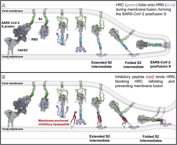
SARS-CoV-2 cell entry is completed after viral spike (S) protein-mediated membrane fusion between viral and host cell membranes. Stable prefusion and postfusion S structures have been resolved by cryo-electron microscopy and cryo-electron tomography, but the refolding intermediates on the fusion pathway are transient and have not been examined. We used an antiviral lipopeptide entry inhibitor to arrest S protein refolding and thereby capture intermediates as S proteins interact with hACE2 and fusion-activating proteases on cell-derived target membranes. Cryo-electron tomography imaged both extended and partially folded intermediate states of S2, as well as a novel late-stage conformation on the pathway to membrane fusion. The intermediates now identified in this dynamic S protein-directed fusion provide mechanistic insights that may guide the design of CoV entry inhibitors.


Figures

Fig. 1.
Fig. 1.. VLP and tEV as surrogates for SARS-CoV-2 virus-cell fusion.
(A) (ia) Plasmids encoding the SARS-CoV-2 spike (S), envelope (E), membrane (M), and N-terminal HiBiT-tagged nucleoprotein (HiBiT-N) were cotransfected, and (ib) plasmids encoding hACE2 were separately transfected into HEK293T cells. (ii) Supernatant fluids were harvested after 2 days. VLPs and tEVs were purified by size exclusion chromatography (SEC). (iii) Purified VLPs and tEVs were mixed and used for (iv) fusion assays and (v) cryo-ET. (B) Schematic of spike activation using exogenous protease with resultant fusion measured by complementation between tEV LgBiT and VLP HiBiT from the VLP (fluorescent yellow). (C) Monomeric and dimeric SARS-CoV-2 inhibitory peptides were assessed in viral fusion assays using VLPs and tEVs. Relative luminescence units (RLU) were plotted 30 min after the temperature was raised to 37°C.
Fig. 2.
Fig. 2.. Visualization of VLPs and tEVs by cryo-electron tomography.
(A) VLPs (yellow) and tEVs (red) show particle sizes ranging from 50 to 500 nm with a mean of 200 nm for VLPs (n = 50) and 175 nm for tEVs (n = 50). (B and C) Contrast-inverted tomogram slice of two different-sized VLPs. Insets show prefusion spikes without (top insets) and with (bottom insets) prefusion spike atomic model (PDB ID: 5x58) fit into the raw tomogram densities. (D and E) Contrast-inverted tomogram slices of tEVs at 4°C (D) and 37°C (E) with densities attributable to hACE2 on the surface of tEVs (red arrowheads). (F) X and Y projections from the final subtomogram reconstruction of hACE2. (G and H) Side view and top view without (G) and with (H) hACE2 model (PDB ID: 6M1D) (11) fit into the subtomogram averaged density map. (I) Fourier shell correlation (FSC) plots of the hACE2 density map with and without a mask. (J) Distribution of VLP membrane to tEV membrane distance measurements where spike intermediates were identified. Intermediate spikes whose VLP membrane to tEV membrane distance measured less than 15 nm (n = 116) were in a partially refolded state, while above 15 nm (n = 76), these spikes were in an extended state. Spike intermediates range from 3 to 29 nm (n = 192). Scale bars, (B to E) 10 nm and (F) 5 nm.
Fig. 3.
Fig. 3.. Interaction between hACE2 and prefusion spike.
(A) Schematic derived from a coarse-grained molecular mechanics (CG-MM) simulation guided by the tomographic densities of prefusion spike (S1 + S2) bound to hACE2. (B, F, H, and K) Contrast-inverted slices through tomograms of VLPs containing spike and tEVs containing hACE2. (C, D, G, I, J, L, and M) Enlarged views of tomograms with densities attributed to S (yellow arrows) interacting with densities attributed to hACE2 (shades of red arrowheads). (E) Isosurface representation of a tomogram from (B) to (D) with densities attributed to four hACE2 dimers [shades of red arrowheads corresponding to arrowheads in (C) and (D)] and two S proteins with their RBDs oriented upward (yellow arrowhead). Ribbon models fitted into the density map are displayed in purple for hACE2 (PDB ID: 7L7F) and orange for S (PDB ID: 6X2B). Full set of tomogram z slices and the isosurface representation are displayed as movie S1 for (B) to (E). Scale bars, (B, F, H, and K) 50 nm and (C to E, G, I, J, L, and M) 10 nm.
Fig. 4.
Fig. 4.. Extended intermediate state of the spike protein.
(A) Schematic derived from a CG-MM simulation guided by the tomographic densities of extended intermediate states of S2. (B and D) Contrast-inverted slices from tomograms of VLPs bearing S and tEVs bearing hACE2 showing densities (green arrowheads) that appear to be spikes in their extended state. (C and E) Enlarged views of tomograms showing densities (green arrowheads) attributed to S proteins in an extended state. (F and G) Distance plot of S intermediates measured between the green arrows of (B) and (D), respectively. a.u., arbitrary units. (H to J) Additional enlarged views from tomogram slices showing extended intermediate S (green arrowheads) spanning VLP and tEV membranes. Densities matching hACE2 are found in close proximity to the region of S insertion (red arrowheads). Scale bars, (B and D) 50 nm and (C, E, and H to J) 10 nm.
Fig. 5.
Fig. 5.. Partially folded intermediate state of the spike protein.
(A) Schematic derived from a CG-MM simulation guided by the tomographic densities of partially folded intermediate states of S2 spanning the viral and host membranes. (B, E, H, L, and N) Contrast-inverted slices from tomograms of VLPs bearing S and tEVs bearing hACE2, showing densities attributable to the partially folded intermediate state of S. (C, F, I to K, M, and O) Enlarged regions from tomogram slices showing densities attributable to S partially folded intermediates (purple arrowheads) linking VLP and tEV membranes. (D and G) Isosurface representations of (C) and (F) with the postfusion S (PDB ID: 6M3W) fitted into the map density. Movie from z slices of tomogram displayed in (K) and (M) is shown as movies S2 and S3, respectively. Scale bars, (B, E, H, L, and N) 50 nm and (C, D, F, G, I to K, M, and O) 10 nm.
Fig. 6.
Fig. 6.. Proposed model of fusion inhibition by [SARSHRC-PEG4]2-chol.
(A) Model of interaction between SARS-CoV-2 prefusion S on the viral envelope and hACE2 on the host cell membrane. After detachment of S1, S2 unfolds and inserts into the host membrane. Refolding of S2 to a postfusion state leads to membrane fusion. (B) Model of antiviral lipopeptide mechanism with the lipopeptide anchored into the host membrane and binding the HRN region of S2, perturbing the final conformational changes of S2 intermediates and preventing membrane fusion.

Similar articles

Cited by

References

    1. Walls A. C., Park Y.-J., Tortorici M. A., Wall A., McGuire A. T., Veesler D., Structure, function, and antigenicity of the SARS-CoV-2 spike glycoprotein. Cell 181, 281–292.e6 (2020). - PMC - PubMed
    1. Qing E., Kicmal T., Kumar B., Hawkins G. M., Timm E., Perlman S., Gallagher T., Dynamics of SARS-CoV-2 spike proteins in cell entry: Control elements in the amino-terminal domains. MBio 12, e01590 (2021). - PMC - PubMed
    1. Outlaw V. K., Bovier F. T., Mears M. C., Cajimat M. N., Zhu Y., Lin M. J., Addetia A., Lieberman N. A. P., Peddu V., Xie X., Shi P. Y., Greninger A. L., Gellman S. H., Bente D. A., Moscona A., Porotto M., Inhibition of coronavirus entry in vitro and ex vivo by a lipid-conjugated peptide derived from the SARS-CoV-2 spike glycoprotein HRC domain. MBio 11, e01935-20 (2020). - PMC - PubMed
    1. Bosch B. J., van der Zee R., de Haan C. A., Rottier P. J., The coronavirus spike protein is a class I virus fusion protein: Structural and functional characterization of the fusion core complex. J. Virol. 77, 8801–8811 (2003). - PMC - PubMed
    1. de Vries R. D., Schmitz K. S., Bovier F. T., Predella C., Khao J., Noack D., Haagmans B. L., Herfst S., Stearns K. N., Drew-Bear J., Biswas S., Rockx B., McGill G., Dorrello N. V., Gellman S. H., Alabi C. A., de Swart R. L., Moscona A., Porotto M., Intranasal fusion inhibitory lipopeptide prevents direct-contact SARS-CoV-2 transmission in ferrets. Science 371, 1379–1382 (2021). - PMC - PubMed

Substances