

Base-Stacking Heterogeneity in RNA Resolved by Fluorescence-Detected Circular Dichroism Spectroscopy
- PMID: 35984918
- PMCID: PMC9442794
- DOI: 10.1021/acs.jpclett.2c01778
Base-Stacking Heterogeneity in RNA Resolved by Fluorescence-Detected Circular Dichroism Spectroscopy
Abstract
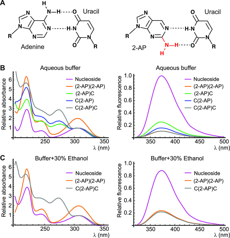
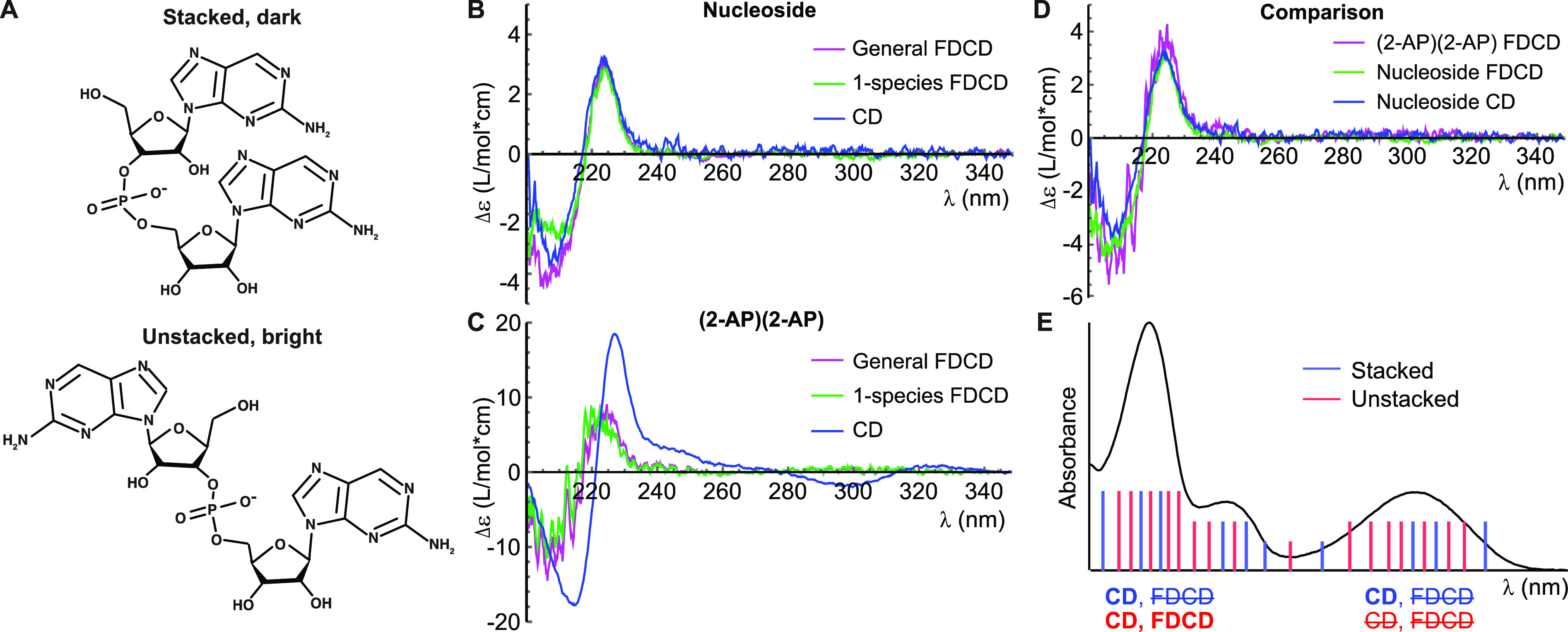
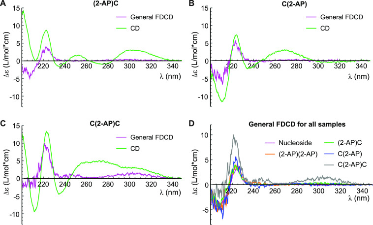
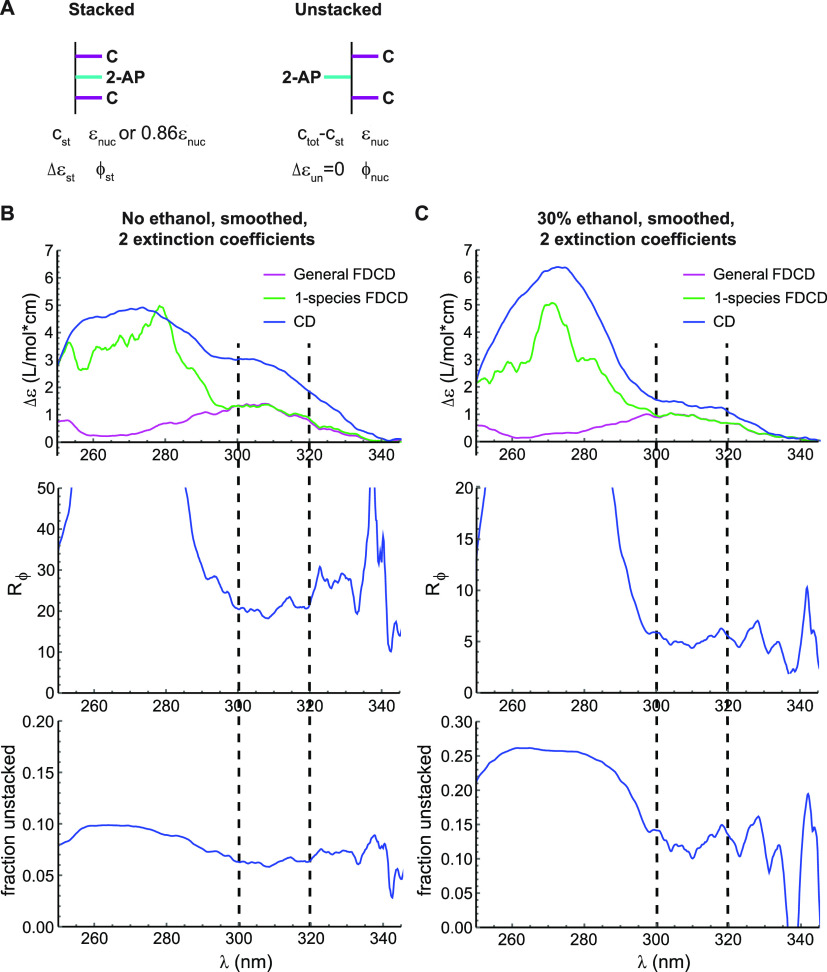
RNA plays a critical role in many biological processes, and the structures it adopts are intimately linked to those functions. Among many factors that contribute to RNA folding, van der Waals interactions between adjacent nucleobases stabilize structures in which the bases are stacked on top of one another. Here, we utilize fluorescence-detected circular dichroism spectroscopy (FDCD) to investigate base-stacking heterogeneity in RNA labeled with the fluorescent adenine analogue 2-aminopurine (2-AP). Comparison of standard (transmission-detected) CD and FDCD spectra reveals that in dinucleotides, 2-AP fluorescence is emitted almost exclusively by unstacked molecules. In a trinucleotide, some fluorescence is emitted by a population of stacked and highly quenched molecules, but more than half originates from a minor ∼10% population of unstacked molecules. The combination of FDCD and standard CD measurements reveals the prevalence of stacked and unstacked conformational subpopulations as well as their relative fluorescence quantum yields.
Conflict of interest statement
The authors declare no competing financial interest.
Figures
Similar articles
-
Low-energy circular dichroism of 2-aminopurine dinucleotide as a probe of local conformation of DNA and RNA.Proc Natl Acad Sci U S A. 2004 Mar 9;101(10):3426-31. doi: 10.1073/pnas.0400591101. Epub 2004 Mar 1. Proc Natl Acad Sci U S A. 2004. PMID: 14993592 Free PMC article.
-
Site-specific variations in RNA folding thermodynamics visualized by 2-aminopurine fluorescence.Biochemistry. 2007 Dec 11;46(49):13948-60. doi: 10.1021/bi7011977. Epub 2007 Nov 13. Biochemistry. 2007. PMID: 17997580 Free PMC article.
-
Influence of base stacking and hydrogen bonding on the fluorescence of 2-aminopurine and pyrrolocytosine in nucleic acids.Biochemistry. 2006 Aug 1;45(30):9145-55. doi: 10.1021/bi060479t. Biochemistry. 2006. PMID: 16866360
-
Probing RNA conformational dynamics and heterogeneity using femtosecond time-resolved fluorescence spectroscopy.Methods. 2009 Oct;49(2):128-35. doi: 10.1016/j.ymeth.2009.04.001. Epub 2009 Apr 9. Methods. 2009. PMID: 19362148 Review.
-
2-Aminopurine as a fluorescent probe of DNA conformation and the DNA-enzyme interface.Q Rev Biophys. 2015 May;48(2):244-79. doi: 10.1017/S0033583514000158. Epub 2015 Apr 17. Q Rev Biophys. 2015. PMID: 25881643 Review.
Cited by
-
Probing and perturbing riboswitch folding using a fluorescent base analogue.Photochem Photobiol. 2024 Mar-Apr;100(2):419-433. doi: 10.1111/php.13896. Epub 2023 Dec 14. Photochem Photobiol. 2024. PMID: 38098287 Free PMC article.
-
Impacts of sequence and structure on pyrrolocytosine fluorescence in RNA.Nucleic Acids Res. 2025 Apr 10;53(7):gkaf262. doi: 10.1093/nar/gkaf262. Nucleic Acids Res. 2025. PMID: 40207631 Free PMC article.
-
Towards control of excitonic coupling in DNA-templated Cy5 aggregates: the principal role of chemical substituent hydrophobicity and steric interactions.Nanoscale. 2023 Feb 16;15(7):3284-3299. doi: 10.1039/d2nr05544a. Nanoscale. 2023. PMID: 36723027 Free PMC article.
References
-
- Jhunjhunwala A.; Ali Z.; Bhattacharya S.; Halder A.; Mitra A.; Sharma P. On the Nature of Nucleobase Stacking in RNA: A Comprehensive Survey of Its Structural Variability and a Systematic Classification of Associated Interactions. J. Chem. Inf. Model. 2021, 61, 1470–1480. 10.1021/acs.jcim.0c01225. - DOI - PubMed
MeSH terms
Substances
Grants and funding
LinkOut - more resources
Full Text Sources
