. 2022 Dec 26;62(24):6715-6726.
doi: 10.1021/acs.jcim.2c00507. Epub 2022 Aug 19.

DHFR Mutants Modulate Their Synchronized Dynamics with the Substrate by Shifting Hydrogen Bond Occupancies

Affiliations

DHFR Mutants Modulate Their Synchronized Dynamics with the Substrate by Shifting Hydrogen Bond Occupancies

Ebru Cetin et al. J Chem Inf Model. .

Abstract

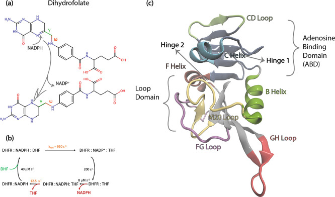
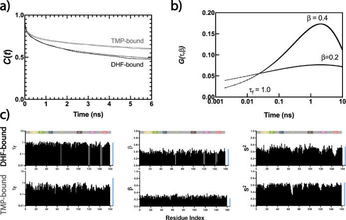
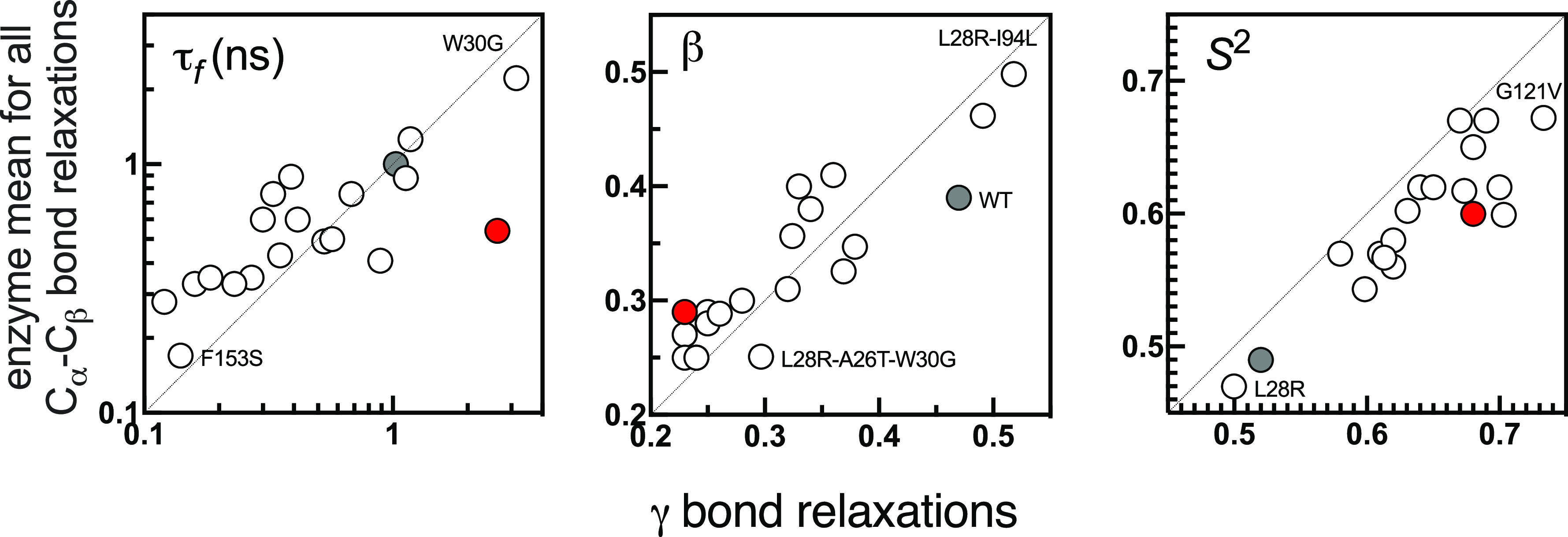
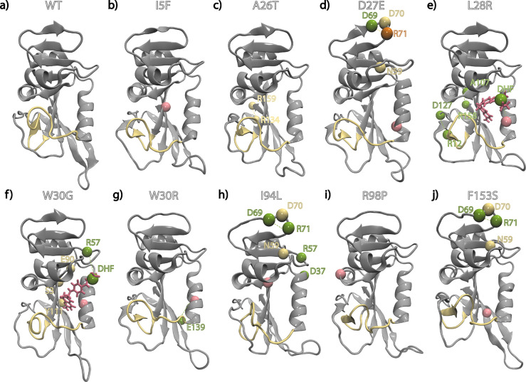
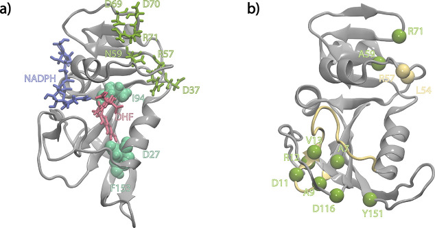
Antibiotic resistance is a global health problem in which mutations occurring in functional proteins render drugs ineffective. The working mechanisms of the arising mutants are seldom apparent; a methodology to decipher these mechanisms systematically would render devising therapies to control the arising mutational pathways possible. Here we utilize Cα-Cβ bond vector relaxations obtained from moderate length MD trajectories to determine conduits for functionality of the resistance conferring mutants of Escherichia coli dihydrofolate reductase. We find that the whole enzyme is synchronized to the motions of the substrate, irrespective of the mutation introducing gain-of-function or loss-of function. The total coordination of the motions suggests changes in the hydrogen bond dynamics with respect to the wild type as a possible route to determine and classify the mode-of-action of individual mutants. As a result, nine trimethoprim-resistant point mutations arising frequently in evolution experiments are categorized. One group of mutants that display the largest occurrence (L28R, W30G) work directly by modifying the dihydrofolate binding region. Conversely, W30R works indirectly by the formation of the E139-R30 salt bridge which releases energy resulting from tight binding by distorting the binding cavity. A third group (D27E, F153S, I94L) arising as single, resistance invoking mutants in evolution experiment trajectories allosterically and dynamically affects a hydrogen bonding motif formed at residues 59-69-71 which in turn modifies the binding site dynamics. The final group (I5F, A26T, R98P) consists of those mutants that have properties most similar to the wild type; these only appear after one of the other mutants is fixed on the protein structure and therefore display clear epistasis. Thus, we show that the binding event is governed by the entire enzyme dynamics while the binding site residues play gating roles. The adjustments made in the total enzyme in response to point mutations are what make quantifying and pinpointing their effect a hard problem. Here, we show that hydrogen bond dynamics recorded on sub-μs time scales provide the necessary fingerprints to decipher the various mechanisms at play.


Conflict of interest statement

The authors declare no competing financial interest.

Figures

Figure 1
Figure 1
(a) Chemical structure of dihydrofolate; the γ bond is shown in green and the neighboring ω bond in red. The glutamate tail is to the right of the γ bond, and to its left is the pterin ring. Hydride transfer positions indicated by arrows. (b) Catalytic cycle of DHFR. Hydride transfer rate and the rate-limiting step are in orange. (c) DHFR structure (PDB code 1RX2); the colors for the loops and helices are used consistently throughout the paper.
Figure 2
Figure 2
(a) Cα–Cβ bond vector relaxations averaged over all residues, displayed for WT DHFR in DHF-bound (black) and TMP-bound (gray) forms; lines fitted by eq 4 are shown by the dashed lines. (b) Distribution of relaxation times for β = 0.4 and β = 0.2; the weighted average τf = 1 ns in both cases. (c) Residue-by-residue curve fit values for fast relaxations in WT DHFR for the DHF-bound and TMP-bound form.
Figure 3
Figure 3
Comparison of enzyme mean (over 159 residues) versus that of γ bond relaxations for all of the systems studied. WT is shown by the gray filled circle. Mutants with extreme values are labeled. y = x line shown to guide the eye in each case. Best-fitting lines (not shown) have R2 values of 0.81, 0.85, and 0.89, respectively, each with p value <0.0001. The red dot is the data for the γ bond of WT-TMP showing how the τf value deviates significantly from that of the enzyme mean, completely destroying the concerted motions.
Figure 4
Figure 4
Changes in hydrogen bonding profiles of the mutants with respect to the WT where the M20 loop is displayed in yellow ribbon representation. Residues for which there is significant change in hydrogen bond occupancies are shown as spheres. Rose, mutated residue; green, residue with formed/increased occupancies; yellow, residue with lost/decreased occupancies; tangerine, residue with shifting occupancy from one partner to another.
Figure 5
Figure 5
Stabilization of the cryptic site. (a) I94L, D27E, and F153S mutants (green) disrupt the hydrogen bond occupancies in the loop domain residues shown in olive. Positions of DHF and NADPH are shown to guide the eye. (b) Residues whose hydrogen bond occupancies are disrupted are displayed for the N59A mutant. Color coding is the same as in Figure 4.

Similar articles

Cited by

References

    1. Toprak E.; Veres A.; Michel J.-B.; Chait R.; Hartl D. L.; Kishony R. Evolutionary paths to antibiotic resistance under dynamically sustained drug selection. Nat. Genet. 2012, 44 (1), 101–105. 10.1038/ng.1034. - DOI - PMC - PubMed
    1. Yeh P. J.; Hegreness M. J.; Aiden A. P.; Kishony R. Drug interactions and the evolution of antibiotic resistance. Nature Reviews Microbiology 2009, 7, 460–466. 10.1038/nrmicro2133. - DOI - PMC - PubMed
    1. Palmer A. C.; Toprak E.; Baym M.; Kim S.; Veres A.; Bershtein S.; Kishony R. Delayed commitment to evolutionary fate in antibiotic resistance fitness landscapes. Nat. Commun. 2015, 6 (1), 7385.10.1038/ncomms8385. - DOI - PMC - PubMed
    1. Ayhan D. H.; Tamer Y. T.; Akbar M.; Bailey S. M.; Wong M.; Daly S. M.; Greenberg D. E.; Toprak E. Sequence-Specific Targeting of Bacterial Resistance Genes Increases Antibiotic Efficacy. PLOS Biology 2016, 14 (9), e100255210.1371/journal.pbio.1002552. - DOI - PMC - PubMed
    1. Thompson S.; Zhang Y.; Ingle C.; Reynolds K. A.; Kortemme T. Altered expression of a quality control protease in E. coli reshapes the in vivo mutational landscape of a model enzyme. eLife 2020, 9, e5347610.7554/eLife.53476. - DOI - PMC - PubMed

Publication types

Substances