Review
. 2022 Sep 21;110(18):2916-2928.
doi: 10.1016/j.neuron.2022.07.012. Epub 2022 Aug 18.

Neuroblastoma: When differentiation goes awry

Affiliations
Review

Neuroblastoma: When differentiation goes awry

Maged Zeineldin et al. Neuron. .

Abstract

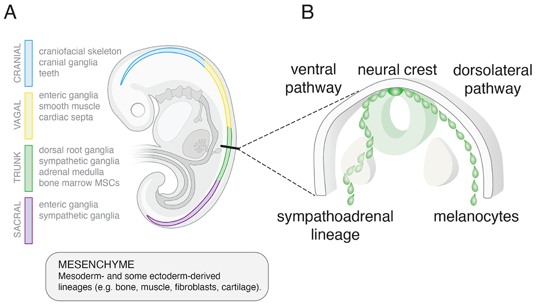
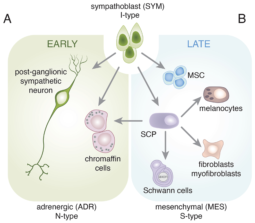
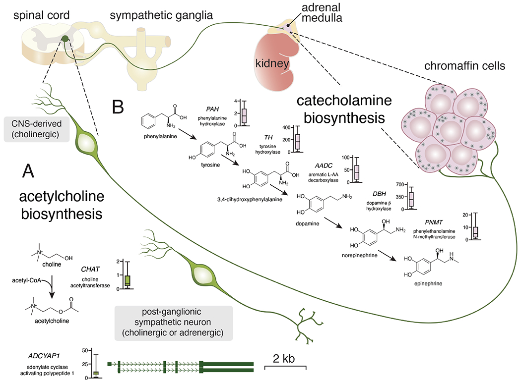
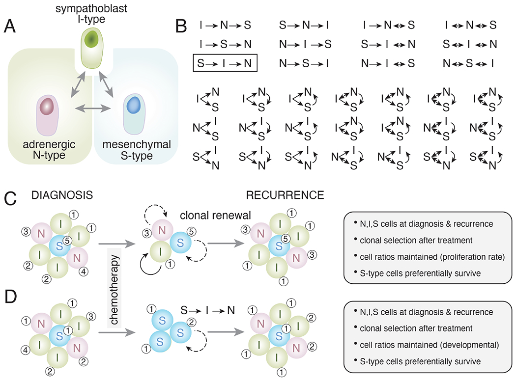
Neuroblastoma is a leading cause of cancer-related death in children. Accumulated data suggest that differentiation arrest of the neural-crest-derived sympathoadrenal lineage contributes to neuroblastoma formation. The developmental arrest of these cell types explains many biological features of the disease, including its cellular heterogeneity, mutational spectrum, spontaneous regression, and response to drugs that induce tumor cell differentiation. In this review, we provide evidence that supports the notion that arrested neural-crest-derived progenitor cells give rise to neuroblastoma and discuss how this concept could be exploited for clinical management of the disease.


Conflict of interest statement

Declaration of interests The authors declare no competing interests.

Figures

Figure 1.
Figure 1.. Extrinsic cues regulate neural crest lineage specification.
A) Drawing of a human embryo showing the neural crest (4-color). The cellular lineages derived from the neural crest vary based on their rostral-caudal location as shown (cranial (blue), vagal (yellow), trunk (green), sacral (purple)). Transplantation studies have shown that lineage specification is regulated primarily by the extrinsic cues rather than cell-autonomous factors. Trunk neural crest cells transplanted into the cranial region will produce cranial lineages and vice versa. Neural crest cells are multipotent and can give rise to both neuronal and non-neuronal (mesenchymal) cell types. This is particularly relevant for neuroblastomas that arise from the sympathoadrenal lineage of the trunk neural crest because they have both neuronal (N-type) and mesenchymal (S-type) cell populations. B) Drawing of a cross section of the trunk neural crest showing the two paths of migration. Both paths occur bilaterally but for simplicity the ventral pathway is shown on the left side of the embryo and the dorsolateral pathway is shown on the right. Extrinsic cues in these different regions of the embryo contribute to specification of the sympathoadrenal lineage and melanocytes. Thus, not only is the position along the rostral caudal axis important for neural crest derived cell lineage specification but the path of migration (ventral versus dorsolateral) is also important.
Figure 2.
Figure 2.. Sympathoblast cell fate specification.
Schematic drawing of major cell fate specification events in the sympathoadrenal lineage and their derivatives. This is not intended to be comprehensive but rather to highlight the multipotency of the sympathoblasts. The neural crest is a transient population and after cells have migrated out of the trunk neural crest along the ventral pathway, a subset of cells commits to become sympathoblasts (I-type). Shortly after the neural crest cells have dispersed and the sympathoblasts have been specified, they form two cell types with neuronal (N-type) features. The post-ganglionic sympathetic neurons may be cholinergic or adrenergic. The chromaffin cells lack traditional dendrites and axons but synthesize catecholamines (epinephrine and norepinephrine) and release those neurotransmitters into the circulation from the adrenal medulla. These are some of the first developmental events of the sympathoadrenal lineage and help to establish the patterning of the sympathetic ganglia and developing adrenal gland. B) Later during development, the sympathoblasts produce Schwann cell precursors (SCPs) and mesenchymal stem cells (MSCs). The SCPs can produce mature Schwann cells that associate with the axons of the sympathetic neurons. They can also produce mesenchymal cell types such as fibroblasts and myofibroblasts. The MSCs migrate to the bone marrow where they contribute to the hematopoietic stem cell niche. Importantly, SCPs have been shown to be the major source of chromaffin cells in the late stages of adrenal development. This resembles the interconversion of N-type and S-type neuroblastoma cells. Therefore, while individual cell types (e.g., SCPs) are multipotent, the sympathoblasts are the most immature cell population in this lineage and may resemble the I-type neuroblastoma cells. This is not intended to be comprehensive, and each arrow may include several distinct stages of cell fate specification and differentiation. For example, bridge cells are thought to be an intermediate state from SCP to chromaffin cells during the later stages of adrenal medulla development.
Figure 3.
Figure 3.. Expression of sympathoadrenal neurotransmitters and neuropeptides in neuroblastoma.
Simplified drawing of the neuronal connections in the sympathoadrenal lineage. Central nervous system (CNS) derived pre-ganglionic neurons synapse directly with chromaffin cells of the adrenal medulla where they release acetylcholine. Once stimulated, the chromaffin cells release epinephrine or norepinephrine into the circulation. Some chromaffin cells release epinephrine and some release norepinephrine from their dense core vesicles. A) Acetylcholine biosynthesis is regulated by choline acetyltransferase and the expression is relatively low (box plot) in the 191 representative neuroblastoma tumors. Acetylcholine is used for acute stimulation of the chromaffin cells. The neuropeptide encoded by ADCYAP1 is used for more chronic stimulation. While expression of ADCYAP1 is low in most neuroblastomas, a subset has very high levels of expression suggesting inter-tumor heterogeneity. B) Drawing of the biosynthetic pathway for catecholamines with relevant gene and abbreviation for each enzyme. Next to each gene name is a boxplot of RNA expression (FPKM) from 191 neuroblastomas tumors (www.stjude.org/pecan). The chromaffin cells of the adrenal medulla secrete catecholamines as do a subset of post-ganglionic sympathetic neurons that are derived from sympathoblasts.
Figure 4.
Figure 4.. Neuroblastoma differentiation.
A) Drawing of one hypothetical simplified relationship between immature (I-type) neuronal (N-type) and mesenchymal (S-type) neuroblastoma cells. B) With just these 3 cell types, there are 34 possible relationships in terms of the ability of cells to produce other cell phenotypes and to interconvert. C,D) These 34 different possible relationships are relevant when considering clonal selection and clonal evolution after treatment with chemotherapy. Broad spectrum systemic chemotherapy primarily kills the proliferating cells. In both examples (C,D), the tumor composition is equal with primarily I-type (sympathoblast-like) cells and some with more differentiated neuronal features (N-type) and a small subset of mesenchymal cells (S-type). Chemotherapy eliminates most dividing cells, and it has been shown previously that S-type cells are more likely to survive treatment. This may be due to intrinsic drug resistance or a lower rate of proliferation. In the model in (C), each cell population is clonal and repopulates the same cell phenotype. Some cells may divide more rapidly (solid arrows) than others (dashed arrows). The numbers (1-5) indicate hypothetical genetic clonal relationships. In the model in (D), the S-type cells that survive treatment can produce cells with the other phenotypes (I-type, N-type) according to one of the 34 possible models (box in (B)). The S-type cells produce I-type cells, and they produce N-type cells thereby reconstituting the cellular heterogeneity of the original tumor. Importantly, the two models give very similar results (gray boxes to right), and the major difference is in the cellular mechanism. Specifically, in both models, N-type, I-type, and S-type cells are present at diagnosis and recurrence. Clonal selection occurs in both models as there are fewer clones in the recurrent tumor than the tumor at diagnosis. The approximate cell rations are maintained in both models either by their proliferation rate, developmental trajectory, or both. In both models, the S-type cells preferentially survive and therapeutic intervention that promotes the differentiation of I-type cells to N-type cells would slow recurrence but may not be curative. Therefore, these two models are consistent with our current understanding of neuroblastoma and the major difference is in the cellular mechanisms that lead to recurrence. These examples are illustrative only and future research should focus on detailed in vivo clonal analysis to discern which of the 34 possible combinations of lineage relationships are relevant in neuroblastoma.

Similar articles

Cited by

References

    1. Abbasi MR, Rifatbegovic F, Brunner C, Mann G, Ziegler A, Pötschger U, Crazzolara R, Ussowicz M, Benesch M, Ebetsberger-Dachs G, et al. (2017). Impact of Disseminated Neuroblastoma Cells on the Identification of the Relapse-Seeding Clone. Clin Cancer Res 23, 4224–4232. - PMC - PubMed
    1. Alleboina S, Aljouda N, Miller M, and Freeman KW (2021). Therapeutically targeting oncogenic CRCs facilitates induced differentiation of NB by RA and the BET bromodomain inhibitor. Mol Ther Oncolytics 23, 181–191. - PMC - PubMed
    1. Anderson DJ (1993). Molecular control of cell fate in the neural crest: the sympathoadrenal lineage. Annu Rev Neurosci 16, 129–158. - PubMed
    1. Bailey P (1926). A classification of the tumors of the glioma group on a histogenetic basis with a correlated study of prognosis (Philadelphia: J.B. Lippincott Company; ).
    1. Bechmann N, Berger I, Bornstein SR, and Steenblock C (2021). Adrenal medulla development and medullary-cortical interactions. Mol Cell Endocrinol 528, 111258. - PubMed

Publication types