. 2022 Oct 11;55(10):1924-1939.e5.
doi: 10.1016/j.immuni.2022.08.003. Epub 2022 Aug 12.

The pre-exposure SARS-CoV-2-specific T cell repertoire determines the quality of the immune response to vaccination

Affiliations

The pre-exposure SARS-CoV-2-specific T cell repertoire determines the quality of the immune response to vaccination

Carina Saggau et al. Immunity. .

Abstract

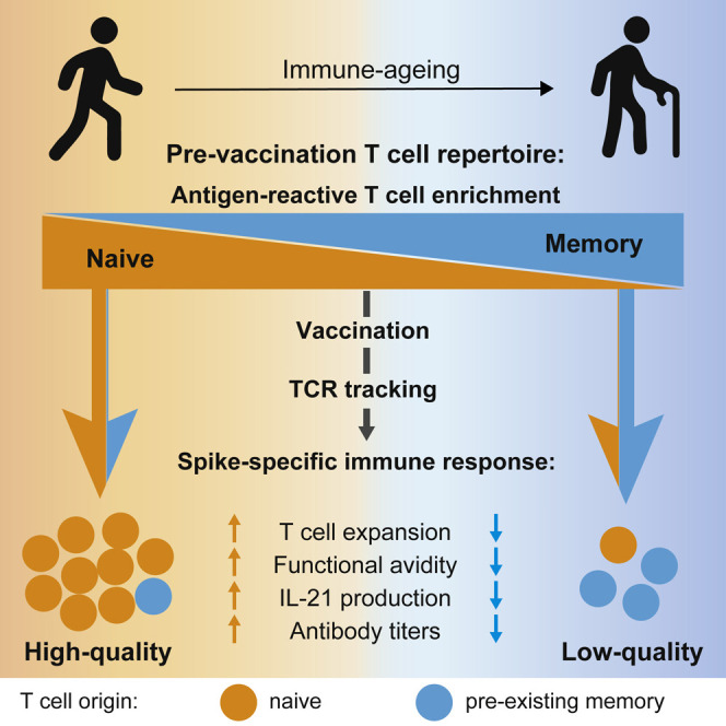
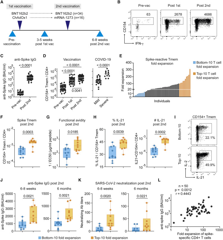
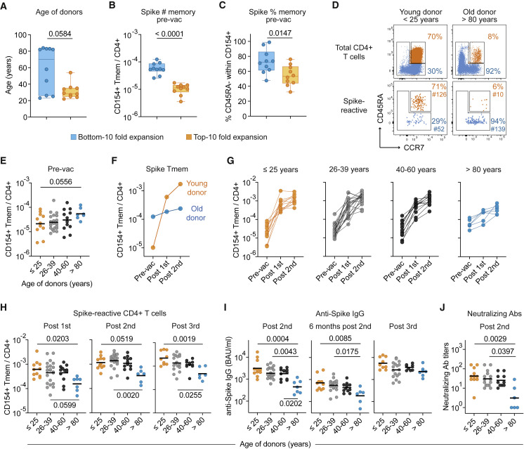
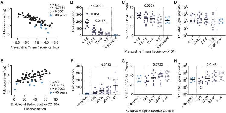
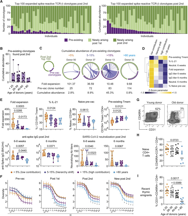
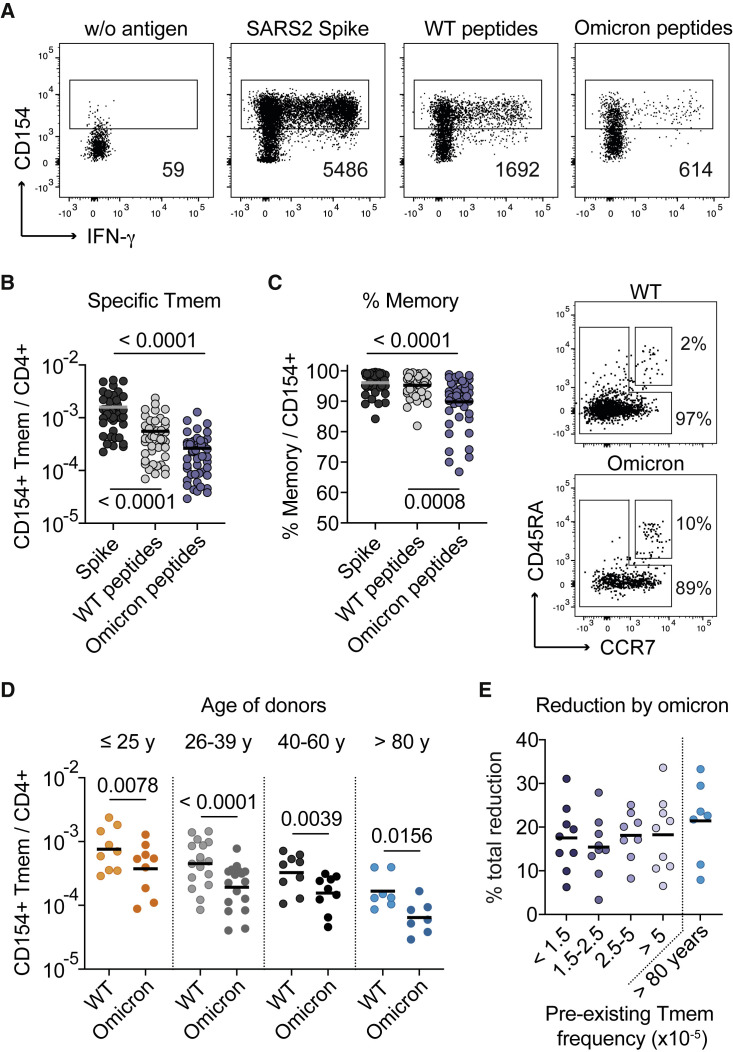
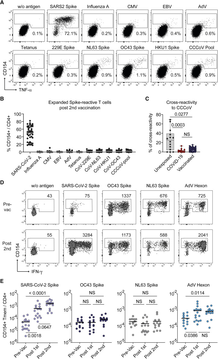
SARS-CoV-2 infection and vaccination generates enormous host-response heterogeneity and an age-dependent loss of immune-response quality. How the pre-exposure T cell repertoire contributes to this heterogeneity is poorly understood. We combined analysis of SARS-CoV-2-specific CD4+ T cells pre- and post-vaccination with longitudinal T cell receptor tracking. We identified strong pre-exposure T cell variability that correlated with subsequent immune-response quality and age. High-quality responses, defined by strong expansion of high-avidity spike-specific T cells, high interleukin-21 production, and specific immunoglobulin G, depended on an intact naive repertoire and exclusion of pre-existing memory T cells. In the elderly, T cell expansion from both compartments was severely compromised. Our results reveal that an intrinsic defect of the CD4+ T cell repertoire causes the age-dependent decline of immune-response quality against SARS-CoV-2 and highlight the need for alternative strategies to induce high-quality T cell responses against newly arising pathogens in the elderly.

Keywords: CD154; CD40L; COVID-19; SARS-CoV-2; TCR tracking; antigen-reactive T cell enrichment; antigen-specific CD4+ T cells; vaccination.


Conflict of interest statement

Declaration of interests E.R. is employee of Evotec SE. O.K. is employee of Peptides & elephants GmbH. O.C. reports grants or contracts from MSD, Pfizer; consulting fees from Biocon, Molecular Partners, Noxxon, PSI; honoraria for lectures from Abbott, Pfizer; participation on a data safety monitoring board or advisory board from Jannsen, PSI. P.K. reports grants or contracts from BMBF, B-FAST, and NAPKON of the Network University Medicine and the State of North Rhine-Westphalia; consulting fees from Ambu GmbH, Gilead Sciences, Noxxon N.V., Pfizer Pharma; honoraria for lectures from Ambu GmbH, Gilead Sciences, MSD Sharp & Dohme GmbH, Pfizer Pharma GmbH, Scilink Comunicación Científica SC; participation on an advisory board from Ambu GmbH, Gilead Sciences, Pfizer Pharma.

Figures

None
Graphical abstract
Figure 1
Figure 1
High- and low-quality CD4+ T cell responses develop upon SARS-CoV-2 vaccination (A) Overview of the study design. Blood was drawn at the indicated time points and analyzed for SARS-CoV-2-specific CD4+ T cells and IgG. 34 individuals of our study received two doses of the BioNTech/Pfizer BNT162b2 mRNA vaccine, while 16 individuals received a heterologous immunization with AstraZeneca ChAdOx1 followed by Moderna mRNA-1273. (B) Representative dot plot examples for the ex vivo detection of SARS-CoV-2 spike-reactive CD4+ T cells by ARTE pre- and post-vaccination. Absolute cell counts after magnetic CD154+ enrichment from 1x10e7 PBMCs are indicated. (C) Serum anti-spike IgG antibody concentrations pre- and post-second vaccination. (D) Frequencies of SARS-CoV-2 spike-reactive Tmems pre- and post-vaccination (left) in comparison to convalescent COVID-19 patients with mild (non-hospitalized) or severe disease (based on WHO criteria WHO4–7) (right). (E) Vaccination-induced expansion of spike-reactive CD4+ Tmems was calculated for each donor based on the frequency pre-vaccination and after the second vaccination. Individuals with the lowest (“bottom 10”) and highest (“top 10”) expansion rate are indicated in blue or orange, respectively. (F) Bottom 10 and top 10 expanders were determined as described in (E) and compared for spike-reactive Tmem frequencies. (G) Spike-reactive CD154+ Tmems were FACS-purified, expanded, and re-stimulated with decreasing antigen concentration in the presence of autologous antigen-presenting cells. Reactive cells were determined by re-expression of CD154 and TNF-α, and EC50 values were calculated from dose-response curves. (H) Ex vivo IL-21 production of spike-reactive T cells, within CD154+ Tmems (left) or within total CD4+ T cells (right). (I) Dot plot example of spike-specific IL-21 and IL-2 production of a bottom 10 or top 10 donor. (J and K) Anti-spike IgG concentrations (J) and SARS-CoV-2 serum neutralization titers (K) at 6–8 weeks and 6 months post-second vaccination. (L) Spearman correlation of the fold expansion of spike-reactive CD4+ T cells and anti-spike IgG concentrations measured 6–8 weeks post-second vaccination. Each symbol in (C, D, F–H, J–L) represents one donor; horizontal lines indicate geometric mean in (C, D). Box-and-whisker plots display quartiles and range in (F–H, J, K). Statistical differences: Friedman test with Dunn’s post hoc test in (D, left); two-tailed Mann-Whitney test in (D, right) and (C, F–H, J, K). See also Figures S1–S3.
Figure 2
Figure 2
The spike-specific immune response declines with age (A–C) Bottom 10 and top 10 expanders were determined as described in Figure 1E and analyzed for (A) age, (B) frequencies of SARS-CoV-2 spike-reactive Tmem pre-vaccination, and (C) the proportion of CD45RA Tmem cells within spike-reactive cells pre-vaccination. (D) Representative CD45RA and CCR7 staining of total CD4+ T cells (upper plots) and spike-reactive CD154+ cells pre-vaccination of a young (<25 years) and old (>80 years) individual. Percentages of naive and memory cells and absolute cell counts of enriched spike-reactive T cells are indicated. (E) Frequencies of spike-reactive memory cells pre-vaccination in different age groups. (F) Vaccination-induced expansion of spike-reactive memory cells in the two representative donors from (D). (G) Vaccination-induced expansion of spike-reactive memory cells in different age groups. (≤25, n = 11; 26–39, n = 21; 40–60, n = 12; >80, n = 6). (H) Frequencies of spike-reactive CD4+ Tmems post-first, post-second, or post-third vaccination in different age groups. (I) Serum anti-spike IgG concentrations 6–8 weeks or 6 months post-second vaccination or post-third vaccination in different age groups. (J) SARS-CoV-2 serum neutralization titers 6–8 weeks post-second vaccination. Each symbol in (A–C) and (E–J) represents one donor; box-and-whisker plots display quartiles and range in (A–C). Horizontal lines indicate geometric mean in (E, H, I, J). Statistical differences: two-tailed Mann-Whitney test in (A–C); Kruskal-Wallis test with Dunn’s post hoc test in (E, H, I, J). See also Figure S3.
Figure 3
Figure 3
Pre-existing memory cells expand poorly upon vaccination (A) Spearman correlation between the frequencies of spike-reactive Tmems pre-vaccination (x axis) and the fold expansion post-second vaccination (y axis). (B–D) Donors were grouped by their pre-vaccination frequencies of spike-reactive Tmems (<1.5, n = 8; 1.5–2.5, n = 14; 2.5–5, n = 10; >5, n = 11). Donors >80 years of age (n = 6) are depicted as separate group. For each bin, the (B) fold expansion post-second vaccination, (C) percentage of IL-21 production within CD154+ Tmems, and (D) functional avidity of expanded spike-reactive T cells is indicated. (E) Spearman correlation between the proportion of spike-reactive naive cells pre-vaccination (x axis) and the fold expansion post-second vaccination (y axis). (F–H) Donors were grouped by their pre-vaccination proportion of spike-reactive naive cells (<20, n = 9; 20–30, n = 11; 30–45, n = 12; >45, n = 11). Donors >80 years of age (n = 6) are depicted as separate group. For each bin, the (F) fold expansion post-second vaccination, (G) percentage of IL-21 production within CD154+ Tmems, and (H) functional avidity of expanded spike-reactive T cells is indicated. Each symbol in (A–H) represents one donor; horizontal lines indicate mean in (B, C, F, G) and geometric mean in (D, H). Statistical differences: Kruskal-Wallis test with Dunn’s post hoc test in (B–D, F–H). To determine differences between the younger age groups, donors >80 years of age were excluded from statistical analysis. See also Figure S3.
Figure 4
Figure 4
TCR sequencing post-vaccination identifies primarily newly arising clonotypes (A–C) Spike-reactive memory T cells were ex vivo sorted pre- and post-vaccination and analyzed by bulk TCR sequencing. Analysis of the top 100 expanded TCR-β spike-reactive clonotypes post-second vaccination is shown. (A) The number of clonotypes (left graph) and their cumulative relative abundance (right graph) derived from the pre-existing, post-first or post-second vaccination repertoire is indicated. Each bar represents one donor (n = 37). (B) Cumulative abundance of pre-existing or newly arising clonotypes found in the top 100 TCR repertoire post-second vaccination for different age groups (>25, n = 9; 26–39, n = 14; 40–60, n = 9, >80, n = 5). (C) Each circos plot represents the TCR-β sequences of the detected clonotypes pre-vaccination and the top 100 clonotypes post-second vaccination of one donor. Connecting lines show TCR-β sequences that are present at both time points. The fold expansion, the absolute number of clonotypes detected pre-vaccination, and the cumulative abundance of pre-existing clonotypes in the post-vaccine repertoire are indicated for each representative donor. (D) Donors were grouped according to the cumulative abundance of pre-existing clonotypes in the top 100 post-vaccination repertoire (<5%, n = 11; 5–15%, n = 11; >15%, n = 9; donors >80 years of age, n = 5). Mean values for the indicated parameters were calculated, Z score normalized for each parameter and are shown as heatmap. (E) Donors were grouped as described in (D) and the fold expansion, IL-21 production within CD154+ Tmems, the proportion of spike-reactive naive T cells pre-vaccination, the frequencies of pre-existing Tmems, and anti-spike IgG concentrations or SARS-CoV-2 serum neutralization titers post-second vaccination are indicated. (F) Rényi diversity profiles were calculated for mean values of the different pre-existing memory bins (<5%, n = 11; 5–15%, n = 11; >15%, n = 9; >80y, n = 5). By varying the scaling parameter α, different diversity indices are calculated. At values of 0, 1, 2, and infinite richness, Shannon diversity, Simpson diversity, and Berger-Parker index are calculated (see STAR Methods). Thus, the sample with the highest value at α = 0 has the highest richness, i.e., highest number of clonotypes, but the lower value at α = infinite indicates a higher proportion of the most abundant TCRs, i.e., lower population diversity. A sample with a profile that is overall higher than the profiles of other samples is therefore more diverse. Diversity of the total spike-specific TCR-β repertoire pre-vaccination, post-first, and post-second vaccination as well as of the newly arising clonotypes post-second vaccination are shown. (G) Representative CD45RA and CD31 staining of a young and old individual. (H) Proportion of naive (CD45RA+CCR7+) within total CD4+ T cells in different age groups. (I) Proportion of RTE (CD45RA+CD31+) within total CD4+ T cells in different age groups. Each symbol in (B, E, H, I) represents one donor. Box-and-whisker plots display quartiles and range in (B); truncated violin plots with quartiles and range are shown in (E). Symbols and error bars indicate mean and SEM in (F). Horizontal lines indicate mean in (H, I). Statistical differences: Kruskal-Wallis test with Dunn’s post hoc test in (E, F, H, I). See also Figures S4 and S5.
Figure 5
Figure 5
T cell reactivity against SARS-CoV-2 Omicron varies between individuals (A) Representative dot plot examples showing antigen-reactive T cells against the full-length spike, a pool of 80 peptides affected by the Omicron B.1.1.529 BA.1 mutations (Omicron peptides), and the corresponding wild-type peptides from one donor. Absolute cell counts after magnetic CD154+ enrichment from 1x10e7 PBMCs are indicated. (B) Frequencies of reactive CD4+ memory T cells against the indicated antigens analyzed post-third vaccination (n = 45). (C) Proportion of CD45RA memory T cells within reactive CD154+CD4+ T cells (n = 45) and representative CD45RA and CCR7 staining for the wild-type and Omicron peptide-reactive T cells. (D) Frequencies of reactive CD4+ Tmems against the wild-type and Omicron peptides analyzed post-third vaccination in different age groups (<25, n = 9; 26–39, n = 17; 40–60, n = 9; >80, n = 7). (E) Donors were grouped by their pre-vaccination frequencies of spike-reactive Tmems as described in Figure 3, and the percentage of total reduction in spike-specific T cells by the Omicron peptides is depicted. Each symbol in (B–E) represents one donor; horizontal lines indicate mean in (B, C, E) and geometric mean in (D). Statistical differences: Kruskal-Wallis test with Dunn’s post hoc test in (B–D).
Figure 6
Figure 6
CCCoV cross-reactive T cells barely contribute to the SARS-CoV-2 vaccine response (A–C) SARS-CoV-2 spike-reactive CD154+ Tmems were expanded post-second vaccination and restimulated in presence of autologous antigen-presenting cells with different antigens, including individual spike proteins from CCCoVs or a pool thereof. (A) Representative dot plots for re-stimulation. Percentage of CD154+TNFα+ cells within CD4+ is indicated. (B) Summarized reactivity of the expanded cell lines against different antigens (n = 46). (C) Percentage of cross-reactivity of SARS-CoV-2 spike-reactive cells to the pool of CCCoV spike proteins in unexposed donors (n = 14), COVID-19 patients (n = 18), and SARS-CoV-2 vaccinees (n = 46). (D and E) CD4+ T cells specific for SARS-CoV-2 spike, OC43 spike, NL63 spike, and AdV hexon were analyzed in 16 individuals receiving a heterologous vaccination with the adenovirus-vector-based ChAdOx1 vaccine, followed by Moderna mRNA-1273. (D) Representative dot plot examples for ex vivo detection of antigen-reactive CD4+ T cells by ARTE pre- and post-vaccination. Absolute cell counts after magnetic CD154+ enrichment from 1x10e7 PBMCs are indicated. (E) Frequencies of Tmems reactive for the indicated antigens pre- and post-SARS-CoV-2 vaccination (n = 16). Each symbol in (B, C, E) represents one donor; horizontal lines indicate mean in (B, E). Box-and-whisker plots display quartiles and range in (C). Statistical differences: Kruskal-Wallis test with Dunn’s post hoc test in (C, E). See also Figure S6.

Similar articles

Cited by

References

    1. Allen J.C., Toapanta F.R., Chen W., Tennant S.M. Understanding immunosenescence and its impact on vaccination of older adults. Vaccine. 2020;38:8264–8272. doi: 10.1016/j.vaccine.2020.11.002. - DOI - PMC - PubMed
    1. Anderson E.J., Rouphael N.G., Widge A.T., Jackson L.A., Roberts P.C., Makhene M., Chappell J.D., Denison M.R., Stevens L.J., Pruijssers A.J., et al. Safety and immunogenicity of SARS-CoV-2 mRNA-1273 vaccine in older adults. N. Engl. J. Med. 2020;383:2427–2438. doi: 10.1056/NEJMoa2028436. - DOI - PMC - PubMed
    1. Bacher P., Heinrich F., Stervbo U., Nienen M., Vahldieck M., Iwert C., Vogt K., Kollet J., Babel N., Sawitzki B., et al. Regulatory T cell specificity directs tolerance versus allergy against aeroantigens in humans. Cell. 2016;167:1067–1078.e16. doi: 10.1016/j.cell.2016.09.050. - DOI - PubMed
    1. Bacher P., Hohnstein T., Beerbaum E., Röcker M., Blango M.G., Kaufmann S., Röhmel J., Eschenhagen P., Grehn C., Seidel K., et al. Human anti-fungal Th17 Immunity and pathology rely on cross-reactivity against candida albicans. Cell. 2019;176:1340–1355.e15. doi: 10.1016/j.cell.2019.01.041. - DOI - PubMed
    1. Bacher P., Rosati E., Esser D., Martini G.R., Saggau C., Schiminsky E., Dargvainiene J., Schröder I., Wieters I., Khodamoradi Y., et al. Low-Avidity CD4(+) T Cell responses to SARS-CoV-2 in unexposed individuals and humans with severe COVID-19. Immunity. 2020;53:1258–1271.e5. doi: 10.1016/j.immuni.2020.11.016. - DOI - PMC - PubMed

Publication types