. 2022 Sep 14;30(9):1219-1230.e7.
doi: 10.1016/j.chom.2022.07.014. Epub 2022 Aug 18.

Structure of trimeric pre-fusion rabies virus glycoprotein in complex with two protective antibodies

Affiliations

Structure of trimeric pre-fusion rabies virus glycoprotein in complex with two protective antibodies

Weng M Ng et al. Cell Host Microbe. .

Abstract

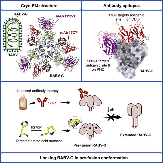
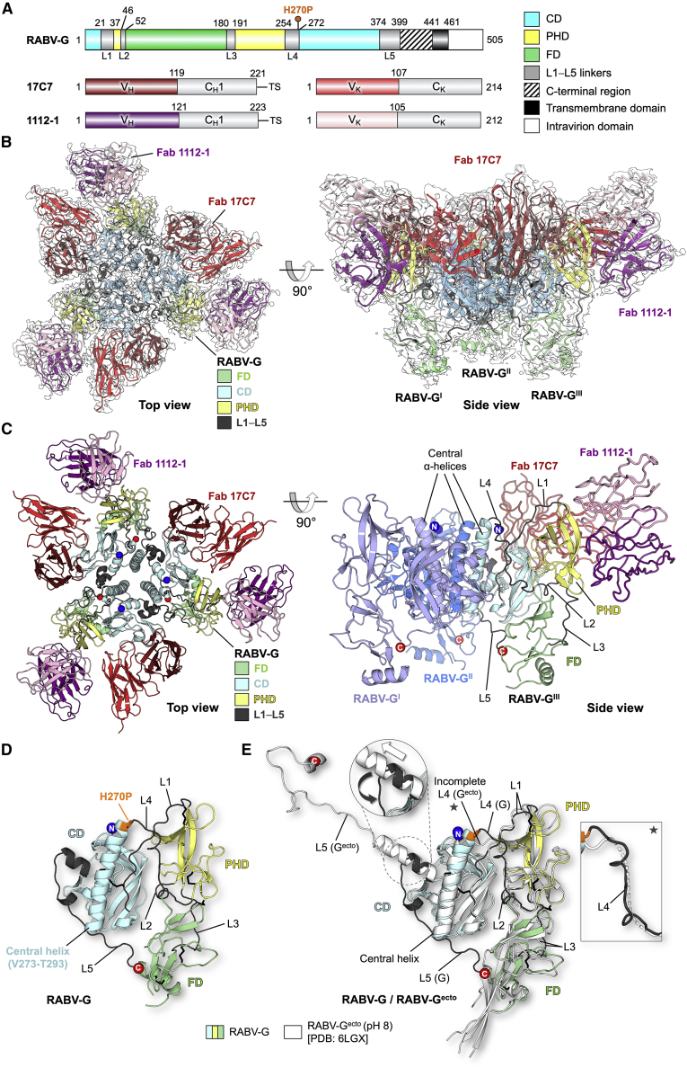
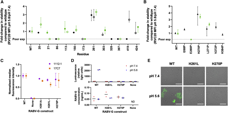
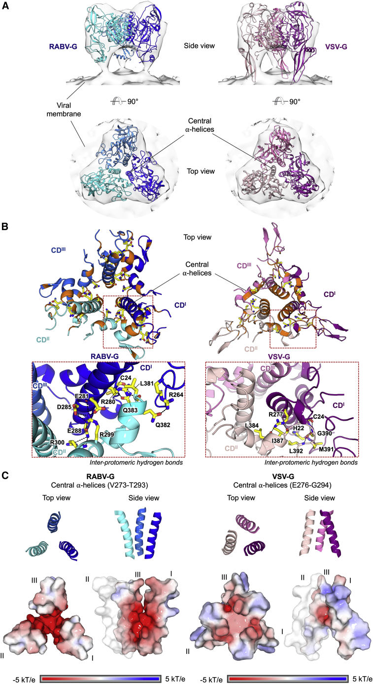
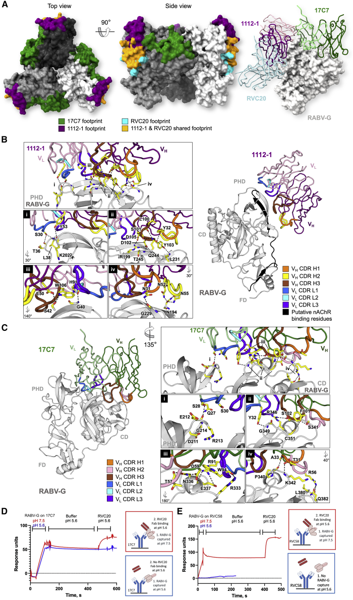
Rabies virus (RABV) causes lethal encephalitis and is responsible for approximately 60,000 deaths per year. As the sole virion-surface protein, the rabies virus glycoprotein (RABV-G) mediates host-cell entry. RABV-G's pre-fusion trimeric conformation displays epitopes bound by protective neutralizing antibodies that can be induced by vaccination or passively administered for post-exposure prophylaxis. We report a 2.8-Å structure of a RABV-G trimer in the pre-fusion conformation, in complex with two neutralizing and protective monoclonal antibodies, 17C7 and 1112-1, that recognize distinct epitopes. One of these antibodies is a licensed prophylactic (17C7, Rabishield), which we show locks the protein in pre-fusion conformation. Targeted mutations can similarly stabilize RABV-G in the pre-fusion conformation, a key step toward structure-guided vaccine design. These data reveal the higher-order architecture of a key therapeutic target and the structural basis of neutralization by antibodies binding two key antigenic sites, and this will facilitate the development of improved vaccines and prophylactic antibodies.

Keywords: antibody neutralization; glycoprotein; rabies virus; structure; viral fusion.


Conflict of interest statement

Declaration of interests S.F. and A.D.D. are named inventors on a patent invention relating to stabilization of RABV-G by the H270P mutation.

Figures

None
Graphical abstract
Figure 1
Figure 1
Structure of pre-fusion RABV-G trimer in complex with Fabs 17C7 and 1112-1 (A) Schematic of RABV-G domain boundaries. Linear map of RABV-G protein sequence is drawn to scale using DOG software (Ren et al., 2009), with domains colored as indicated in the legend, showing “palindromic” architecture typical of class III fusion proteins (CD, central domain; PHD, pleckstrin homology domain; FD, fusion domain). The H270P point mutation in our protein construct is indicated with a pin above the map. C-terminal region (shaded) is unresolved in our structure. (Bottom) Linear maps of Fabs 17C7 and 1112-1 heavy and kappa light chains, colored and labeled accordingly. VH, VK, CH1, and CK denote the antibody variable heavy, variable kappa light, constant 1 heavy, and constant kappa light-chain domains, respectively. TS, TwinStrep tag. (B) 2.8-Å cryo-EM map with the resulting structure of trimeric RABV-G shown in top and side view orientations. Single copies of 17C7 and 1112-1 were observed to bind to each protomer of RABV-G. The atomic model is fitted into the corresponding cryo-EM map (white) and colored according to domain with the variable regions of Fab 17C7 and Fab 1112-1 colored red and purple (darker shade for heavy chain, lighter shade for light chain), respectively. The constant regions of the Fabs were disordered in the reconstruction and therefore were not built. (C) Atomic model of the RABV-G-Fab 17C7-Fab 1112-1 complex. (Top view) The protein molecules are displayed in cartoon representation and colored accordingly as labeled. N- and C-termini are shown as blue and red spheres, respectively. (Side view) Only one copy of each Fab is shown in ribbon representation. Two RABV-G protomers are colored blue and light purple for visual clarity. The remaining copy is colored as shown in top view. (D) Conformational features revealed by atomic model of RABV-G. A single protomer of RABV-G is shown in cartoon representation. PHD is colored yellow, CD in blue, and FD in green, whereas the inter-domain linkers (L1−L5) are colored in dark gray. N- and C-termini are shown as blue and red spheres, respectively. The point mutation H270P is colored orange and shown in stick representation. (E) Structure superimposition of our trimeric RABV-G with a previously reported monomeric RABV-G ectodomain structure obtained at pH 8.0. (RABV-Gecto, white cartoon, PDB: 6LGX) (Yang et al., 2020). When domains were aligned separately, the PHD and CD aligned more closely than the FD (calculated root-mean-square deviations 1.0-Å over 72 equiv C⍺ atoms for PHD, and 0.6-Å/128 C⍺ for CD, and 2.4-Å/88 C⍺ for FD). Differences in the L4 and L5 linkers between our trimeric RABV-G structure and the previous RABV-Gecto structure are highlighted in the inset. For further information, see Figures S1–S3 and Tables S1 and S2.
Figure 2
Figure 2
Targeted mutations at two sites in L4 stabilize pre-fusion RABV-G Wild-type (WT) and mutant RABV-G constructs were expressed on transiently transfected Expi293 cells, and reactivity with site I (RVC20), site II (1112-1), and site III (17C7) IgG was assessed by flow cytometry. Cell-surface expression levels of all constructs are shown in Table S3. (A) Effect of histidine substitutions. All histidine residues in the RABV-G ectodomain were mutated to alanine and to leucine, with exceptions detailed in methods. Pre-fusion protein stability was measured by calculating median fluorescence intensity (MFI) of the pre-fusion-specific mAb RVC20 (Hellert et al., 2020; De Benedictis et al., 2016) after binding at pH 5.8 as a proportion of that after binding to the same construct at pH 7.4: this proportion was 0.11 for untagged WT RABV-G, and 0.12 for WT RABV-G expressed as a fusion protein. Results are expressed as fold change in this proportion compared with WT protein. Filled and open symbols denote introduction of alanine and leucine, respectively. Black and green symbols denote untagged constructs and those expressed as GFP fusion proteins, respectively. Points represent median and error bars represent range of four technical replicates across two experiments (a transfection with each of two independent DNA preparations on each of 2 days). “Poor exp” denotes constructs with cell-surface expression <33% of the level of WT RABV-G, as assessed by RVC20 binding at pH 7.4 (Table S3). (B) Effect of potentially helix-breaking substitutions with proline. Residues in L4/L5 regions expected to form helices in post-fusion protein were substituted with proline. Colors, replication (points and error bars), and the definition of poor cell-surface expression are as for (A). (C) H261A/L and H270P retain site II and site III antigenicity, as evidenced by 1112-1 and 17C7 binding. Untagged constructs were used. MFI is expressed as a proportion of that observed with WT RABV-G with each antibody. “None” denotes MFI on cells transfected with an irrelevant antigen. The replication strategy and meaning of points and error bars were as for (A). (D and E) H261L and H270P mutations abolish RABV-G-mediated cell-cell fusion. Acid-triggered cell-cell fusion was monitored in a dual-reporter luminescence and fluorescence assay. (D) shows luciferase activity (upper graph) and expression level measured by ELISA (lower graph) for samples from the same experiment. Points each represent median of three or more technical replicates (as described in STAR Methods section) using a single DNA preparation. Line indicates median of biological replicates using independent DNA preparations. ND indicates not detectable. (E) shows GFP activity, imaged by fluorescence microscopy. Scale bars, 100 μm.
Figure 3
Figure 3
Structural comparison of pre-fusion RABV-G and VSV-G reveals contrasting modes of inter-protomeric interactions at the trimerization core (A) Fitting of pre-fusion RABV-G (blue) and VSV-G (pink; PDB: 5I2S) into a subtomographic average map of pre-fusion VSV-G (semi-transparent gray; EMD-9331) (Si et al., 2018). RABV-G and VSV-G structures are shown in cartoon representation and colored in different shades of blue and pink, respectively. Maps are shown as transparent surfaces. (B) Zoom-in views of the G trimerization core involved in inter-protomeric interactions. Residues involved in hydrogen bonding (black dashed lines) are shown as sticks, with the carbon, nitrogen, oxygen, and sulfur constituents colored yellow, blue, red, and dark yellow, respectively. Residues involved in non-polar interactions are colored orange. The zoom-in panels of the CD α-helices shows the inter-protomeric hydrogen bonds. This analysis demonstrates that the trimerization core of RABV-G is largely mediated by polar interactions between charged residues, including a network of hydrogen bonds formed by negatively charged Glu281, Asp285, and Glu288 on one protomer and positively charged Arg299 and Arg300 on the adjacent protomer. Hydrophobic interactions are formed at the periphery and bottom of the central α-helices. In contrast, VSV-G displays the reverse pattern of inter-protomeric interactions, where the core is largely maintained by hydrophobic interactions with hydrogen bonds formed at the periphery. (C) Electrostatic potential of the central α-helices. The central α-helices of RABV-G (left) and VSV-G (right) are shown as cartoons (top) and surfaces (bottom). The surfaces are colored according to the electrostatic potential in the range of ±5 kT/e, as calculated by adaptive Poisson-Boltzmann solver (APBS). Both RABV-G and VSV-G display negatively charged trimerization cores. This characteristic is especially prominent in RABV-G, where the carboxyl groups of Glu274, Glu275, Glu281, Glu282, Asp285, and Glu288 side chains line the central α-helices.
Figure 4
Figure 4
Structural basis for antibody-mediated RABV neutralization: multiple sites of vulnerability on the membrane-distal crown of the RABV-G trimer (A) Visualization of structurally characterized anti-RABV antibody epitopes. (Left) Footprints exhibited by RVC20 (site I-targeting antibody, PDB: 6TOU; cyan), 1112-1 (site II; purple), and 17C7 (site III; green) plotted on trimeric RABV-G (white, gray, and dark gray surface). MAbs 1112-1 and RVC20 are shown to target distinct yet overlapping epitopes (orange surface). For clarity, the variable regions of the Fab fragments of mAbs RVC20, 1112-1, and 17C7 bound to a single RABV-G protomer are shown as ribbons (right). MAbs (B) 1112-1 and (C) 17C7 target the PHD and CD domains of RABV-G, respectively. Detailed interactions between complementarity-determining regions (CDRs) of each antibody with RABV-G (gray cartoon) are highlighted in the boxed panels. Residues involved in the antibody-antigen interactions are shown as yellow sticks, with the CDR loops colored as indicated. In our structure, one molecule of each Fab binds to each protomer of the RABV-G trimer. (B) The 1112-1 epitope encompasses three β strands and five loops on the PHD, which include residues 175-203 (highlighted black) that have been previously implicated in nicotinic acetylcholine receptor (nAChR) recognition by RABV (Lentz, 1990). RABV-G residues Asn194, Arg199, Gln244, and Thr245 form extensive hydrogen bond networks with 1112-1 CDR residues including Asn52, Asn55, Ser101, Asp102, Tyr103, and Asp105. Detailed interactions are provided in Figure S8. (C) 17C7 has been observed to bind mostly to the CD in addition to a small contact with the PHD. Notably, 17C7 engages residues Asn336 and Arg346. Mutations at these locations have been shown to confer resistance to 17C7 neutralization (Sloan et al., 2007; Wang et al., 2011). The 17C7 light chain also forms hydrogen bonds with Lys330 and Arg333, which have been reported to play roles in recognition of the receptor p75NTR (Tuffereau et al., 1998) and in neuroinvasion by RABV (Coulon et al., 1998). Detailed interactions are provided in Figure S8. (D and E) Locking of RABV-G in pre-fusion conformation by 17C7 and RVC58. SPR traces demonstrating that when RABV-G is captured by site-III-binding mAbs 17C7 or RVC58, respectively, at pH 7.4, followed by incubation at pH 5.6, binding of the pre-fusion conformation-specific RVC20 Fab remains possible. RVC20 and RVC58 are specific for the pre-fusion conformation: RVC58 fails to capture RABV-G at pH 5.6, and RVC20 does not bind at pH 5.6, unless RABV-G had previously been captured by 17C7 in pre-fusion conformation. Figure S9E demonstrates similar results obtained with a different assay format, in which the initial interaction between the site-III-binding antibody and RABV-G occurs in solution, and the conformation-specific RVC20 Fab is immobilized on the chip surface. See Figures S5–S9 and Table S1 for more related information.

References

    1. Adams P.D., Grosse-Kunstleve R.W., Hung L.W., Ioerger T.R., Mccoy A.J., Moriarty N.W., Read R.J., Sacchettini J.C., Sauter N.K., Terwilliger T.C. PHENIX: building new software for automated crystallographic structure determination. Acta Crystallogr. D Biol. Crystallogr. 2002;58:1948–1954. - PubMed
    1. Albertini A.A.V., Baquero E., Ferlin A., Gaudin Y. Molecular and cellular aspects of rhabdovirus entry. Viruses. 2012;4:117–139. - PMC - PubMed
    1. Aldrich C., Leroux-Roels I., Huang K.B., Bica M.A., Loeliger E., Schoenborn-Kellenberger O., Walz L., Leroux-Roels G., Von Sonnenburg F., Oostvogels L. Proof-of-concept of a low-dose unmodified mRNA-based rabies vaccine formulated with lipid nanoparticles in human volunteers: a phase 1 trial. Vaccine. 2021;39:1310–1318. - PMC - PubMed
    1. Balazs A.B., Bloom J.D., Hong C.M., Rao D.S., Baltimore D. Broad protection against influenza infection by vectored immunoprophylaxis in mice. Nat. Biotechnol. 2013;31:647–652. - PMC - PubMed
    1. Baquero E., Albertini A.A., Raux H., Abou-Hamdan A., Boeri-Erba E., Ouldali M., Buonocore L., Rose J.K., Lepault J., Bressanelli S., Gaudin Y. Structural intermediates in the fusion-associated transition of Vesiculovirus glycoprotein. EMBO J. 2017;36:679–692. - PMC - PubMed