. 2022 Sep-Oct;19(5):614-623.
doi: 10.21873/cgp.20346.

Nuclear Respiratory Factor 1 Overexpression Inhibits Proliferation and Migration of PC3 Prostate Cancer Cells

Affiliations

Nuclear Respiratory Factor 1 Overexpression Inhibits Proliferation and Migration of PC3 Prostate Cancer Cells

Chun-Hsien Wu et al. Cancer Genomics Proteomics. 2022 Sep-Oct.

Abstract

Background/aim: The role of nuclear respiratory factor 1 (NRF1) on the prostate cancer progression is controversial. We aimed to investigate the effect of NRF1 overexpression on the metastasis potential of PC3 prostate cancer cells and the associated molecular mechanisms.

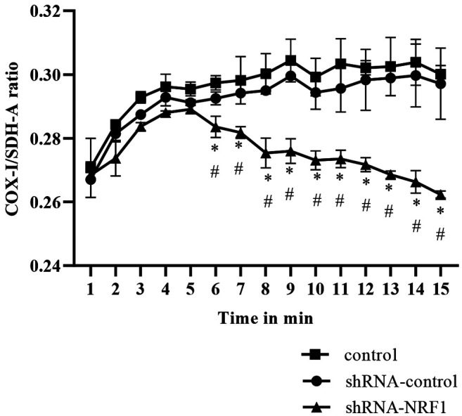
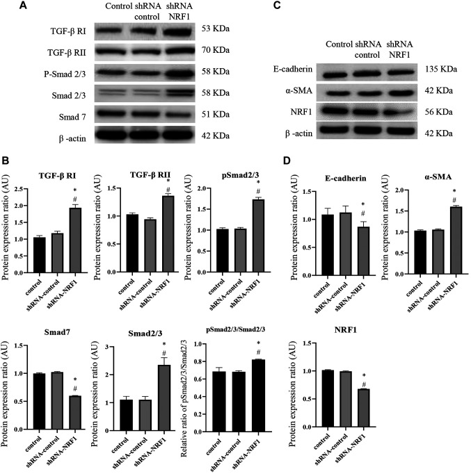
Materials and methods: Cell survival, migration capacity, mitochondrial biogenesis, the expression of TGF-β signaling and EMT markers were examined after overexpression and silencing of NRF1 in PC3 cells.

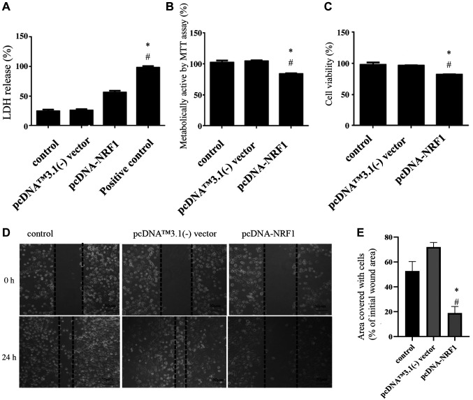
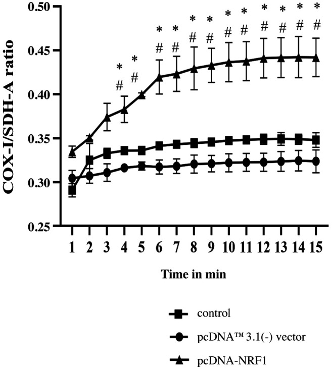
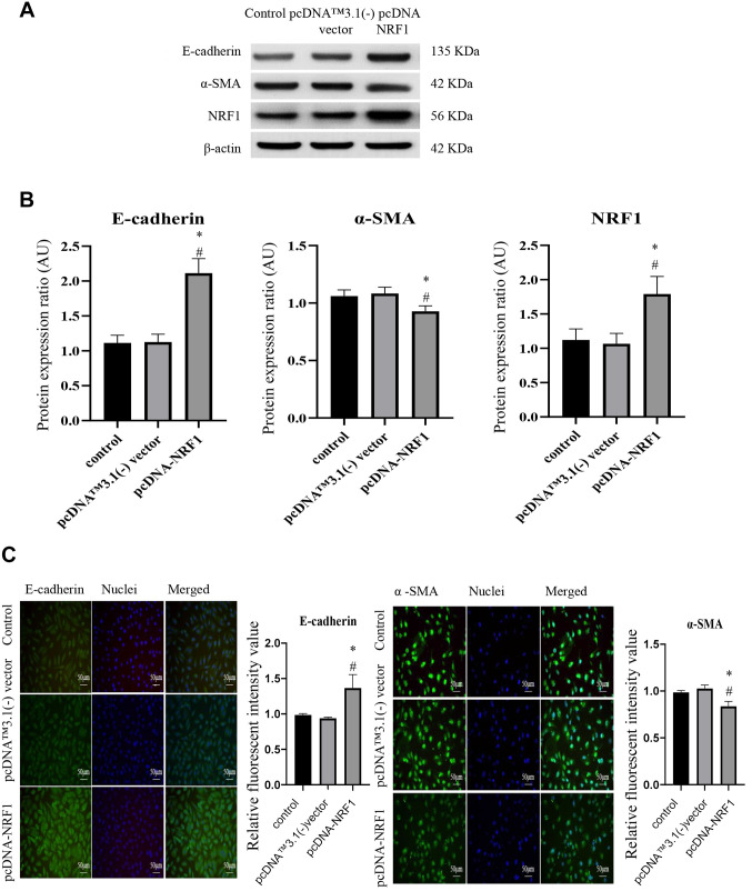
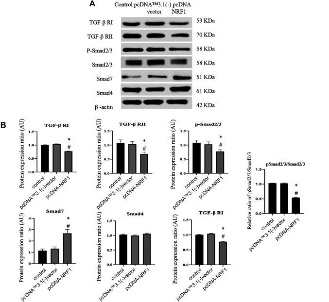
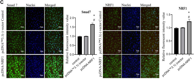
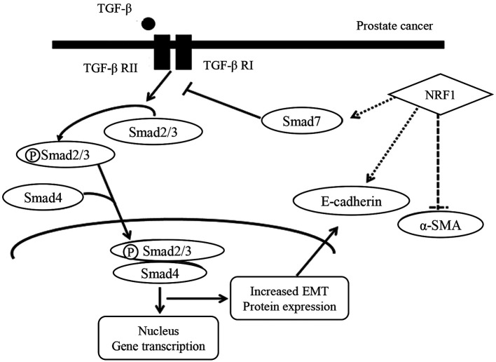
Results: We found that NRF1-overexpressing cells exhibited a decreased cell viability and proliferation ability as well as a reduced migration capacity compared to control cells. Moreover, ectopic expression of NRF1 increased the mitochondrial biogenesis and inhibited the EMT characteristics, including a decrease in the mesenchymal marker, α-SMA and an increase in the epithelial cell marker, E-cadherin. We also demonstrated that overexpression of NRF1 suppressed the expression of TGF-β signaling in PC3 cells. As expected, silencing of NRF1 reversed the abovementioned effects.

Conclusion: This study demonstrated that upregulation of NRF1 holds the potential to inhibit the metastasis of prostate cancer, possibly through an elevation of mitochondrial biogenesis and the subsequent repression of TGF-β-associated EMT. Therapeutic avenues that increase NRF1 expression may serve as an adjunct to conventional treatments of prostate cancer.

Keywords: Nuclear respiratory factor 1; TGF-β signaling; epithelial mesenchymal transition; mitochondrial biogenesis; prostate cancer.


Conflict of interest statement

The Authors declare no conflicts of interest.

Figures

Figure 1
Figure 1. Overexpression of NRF1 reduces the cell survival and migration capacity of PC3 prostate cancer cells. (A) LDH release assay was carried out to show that overexpression of NRF1 increased the cytotoxicity of PC3 cells. Triton X-100 served as a positive control; (B) MTT and (C) trypan blue exclusion assays were conducted to examine the cell proliferation and viability of PC3 cells after transfection of pcDNA-NRF1. (D) Representative migration capacity image and (E) quantitative analysis of wound healing assay in PC3 cells transfected with pcDNA NRF1 or pcDNA™3.1(-) vector. Magnification scale was 200×. The data is presented as mean±SD from three independent experiments. *p<0.05 compared to the control group; #p<0.05 compared to the pcDNA™3.1(-) vector group.
Figure 2
Figure 2. Mitochondrial biogenesis is enhanced after overexpression of NRF1. The COX1/SDH-A ratio was used to show mitochondrial biogenesis over the course of 15 min using the MitoBiogenesis™ InCell ELISA Kit. The data is presented as mean±SD from three independent experiments. *p<0.05 compared to the control group. #p<0.05 compared to the pcDNA™3.1(-) vector group.
Figure 3
Figure 3. Overexpression of NRF1 downregulates the expression of EMT markers. (A) Representative western blots and (B) densitometric analysis of E-cadherin, α-SMA and NRF1; β-actin was used as an internal control; (C) Immunofluorescence staining of E-cadherin and α-SMA in cells transfected with pcDNA NRF1 or pcDNA control (pcDNA™3.1(-) vector). Magnification scale was 200×. 4’,6-diamidino-2-phenylindole (DAPI) was used to detect nuclei. The data is presented as mean±SD from three independent experiments. *p<0.05 compared to the control group; #p<0.05 compared to the pcDNA™3.1(-) vector group.
Figure 4
Figure 4. Ectopic expression of NRF1 suppresses the TGF-β/Smads pathway. (A) Representative western blots and (B) densitometric analysis of TGF-β RI, RII, Smad2/3, p-Smad2/3, Smad4, and Smad7; β-actin served as an internal control; (C) Immunofluorescence staining of Smad7 and NRF1 in cells 24 h after transfection of pcDNA NRF1 or pcDNA control [pcDNA™3.1(-) vector]. Magnification scale was 200X. DAPI was used to detect nuclei. The data is presented as mean±SD from three independent experiments. *p<0.05 compared to the control group; #p<0.05 compared to the pcDNA™3.1(-) vector group.
Figure 5
Figure 5. Silencing NRF1 downregulated mitochondrial biogenesis. The COX1/SDH-A ratio was used to show mitochondrial biogenesis over the course of 15 min using the MitoBiogenesis™ In-Cell ELISA Kit. The data is presented as mean±SD from three independent experiments. *p<0.05 compared to the control group. #p<0.05 compared to the pcDNA™3.1(-) vector group.
Figure 6
Figure 6. Knockdown of NRF1 increased the expression of TGFβ/Smad signaling and EMT markers. (A) Representative western blots of TGF-β RI, RII, Smad2/3, p-Smad2/3, and Smad7; (B) Densitometric analysis of TGF-β RI, RII, Smad2/3, p-Smad2/3 and Smad7. (C) Representative western blots of EMT marker, E-cadherin and α-SMA; (D) Densitometric analysis of E-cadherin and α-SMA in cells 24 h after transfection of shRNAcontrol (scrambled shRNA) or shRNA-NRF1; β-actin served as an internal control. Magnification scale was 200X. DAPI was used to detect nuclei. The data is presented as mean±SD from three independent experiments. *p<0.05 compared to the control group; #p<0.05 compared to the shRNAcontrol group.
Figure 7
Figure 7. Schematic diagram showing the possible mechanism underlying the regulatory effect of NRF1 on TGF-β signaling and EMT markers in PC3 prostate cancer cells.

References

    1. Torre LA, Siegel RL, Ward EM, Jemal A. Global cancer incidence and mortality rates and trends—an update. Cancer Epidemiol Biomarkers Prev. 2016;25(1):16–27. doi: 10.1158/1055-9965.EPI-15-0578. - DOI - PubMed
    1. Siegel RL, Miller KD, Jemal A. Cancer statistics, 2020. CA Cancer J Clin. 2020;70(1):7–30. doi: 10.3322/caac.21590. - DOI - PubMed
    1. Denisenko TV, Gorbunova AS, Zhivotovsky B. Mitochondrial involvement in migration, invasion and metastasis. Front Cell Dev Biol. 2019;7:355. doi: 10.3389/fcell.2019.00355. - DOI - PMC - PubMed
    1. Shiota M, Yokomizo A, Tada Y, Inokuchi J, Tatsugami K, Kuroiwa K, Uchiumi T, Fujimoto N, Seki N, Naito S. Peroxisome proliferator-activated receptor gamma coactivator-1alpha interacts with the androgen receptor (AR) and promotes prostate cancer cell growth by activating the AR. Mol Endocrinol. 2010;24(1):114–127. doi: 10.1210/me.2009-0302. - DOI - PMC - PubMed
    1. Ivanova MM, Luken KH, Zimmer AS, Lenzo FL, Smith RJ, Arteel MW, Kollenberg TJ, Mattingly KA, Klinge CM. Tamoxifen increases nuclear respiratory factor 1 transcription by activating estrogen receptor beta and AP-1 recruitment to adjacent promoter binding sites. FASEB J. 2011;25(4):1402–1416. doi: 10.1096/fj.10-169029. - DOI - PMC - PubMed

Substances