. 2022 Aug 18;14(16):6626-6641.
doi: 10.18632/aging.204235. Epub 2022 Aug 18.

Oxypeucedanin relieves LPS-induced acute lung injury by inhibiting the inflammation and maintaining the integrity of the lung air-blood barrier

Affiliations

Oxypeucedanin relieves LPS-induced acute lung injury by inhibiting the inflammation and maintaining the integrity of the lung air-blood barrier

Li Du et al. Aging (Albany NY). .

Abstract

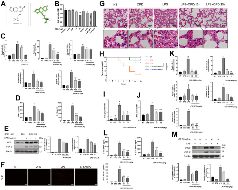
Introduction: Acute lung injury (ALI) is commonly accompanied by a severe inflammatory reaction process, and effectively managing inflammatory reactions is an important therapeutic approach for alleviating ALI. Macrophages play an important role in the inflammatory response, and this role is proinflammatory in the early stages of inflammation and anti-inflammatory in the late stages. Oxypeucedanin is a natural product with a wide range of pharmacological functions. This study aimed to determine the effect of oxypeucedanin on lipopolysaccharide (LPS)-induced ALI.

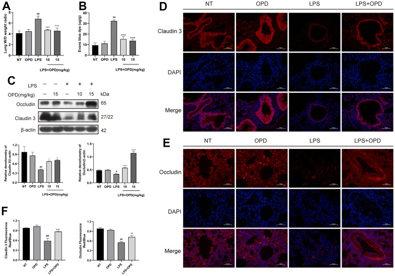
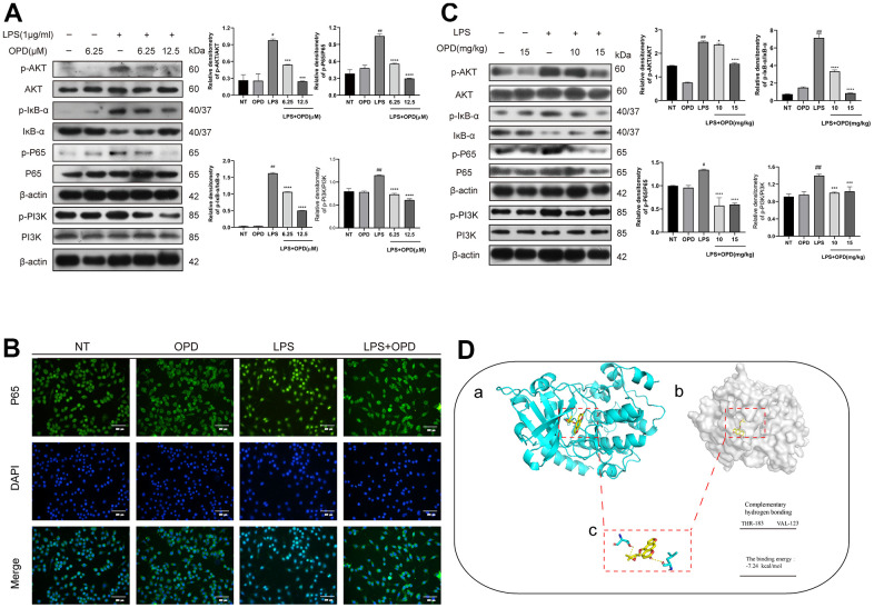
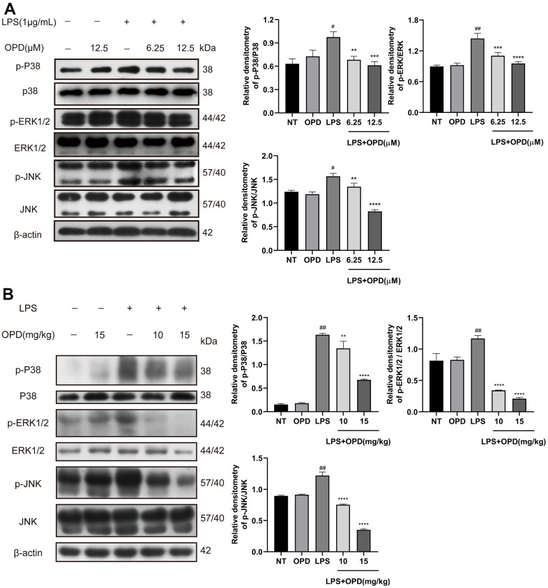
Methods and results: In this study, the following experiments were performed based on LPS-induced models in vivo and in vitro. Using myeloperoxidase activity measurement, ELISA, qRT-PCR, and Western blotting, we found that oxypeucedanin modulated the activity of myeloperoxidase and decreased the expression levels of inflammatory mediators such as TNF-α, IL-6, IL-1β, MPO, COX-2 and iNOS in LPS-induced inflammation models. Meanwhile, oxypeucedanin inhibited the activation of PI3K/AKT and its downstream NF-κB and MAPK signaling pathways. In addition, oxypeucedanin significantly decreased the pulmonary vascular permeability, which was induced by LPSs, and the enhanced expression of tight junction proteins (Occludin and Claudin 3).

Conclusions: In conclusion, this study demonstrated that the anti-inflammatory mechanism of oxypeucedanin is associated with the inhibition of the activation of PI3K/AKT/NF-κB and MAPK signaling pathways and the maintenance of the integrity of the lung air-blood barrier.

Keywords: MAPKs; PI3K/AKT/NF-κB; acute lung injury; lipopolysaccharide; molecular docking simulation.


Conflict of interest statement

CONFLICTS OF INTEREST: The authors have no other relevant affiliations or financial involvement with any organization or entity with a financial interest in or financial conflict with the subject matter or materials discussed in the manuscript apart from those disclosed.

Figures

Figure 1
Figure 1
Oxypeucedanin attenuated changes in inflammation levels in the lung tissue of LPS-induced mice and LPS-induced RAW264.7 cells. (A) The molecular structure of OPD and the three-dimensional structure of the OPD molecule. (B) RAW264.7 cells were cultured in a 37° C cell incubator with 5% CO2. The cells were pretreated with OPD for 1 h, then with or without LPS (1 ug/ml) for 24 h. Different concentrations of OPD were dissolved in DMSO, and the content of DMSO was 0.1%. The effect of OPD on the viability of RAW264.7 cells was determined by the CCK8 assay. (C) After the OPD pretreatment for 1 h, RAW264.7 cells were stimulated with LPS (1 μg/mL) for 6 h, and total RNA was extracted by TRIzol. The mRNA transcription levels of IL-6, IL-1β, TNF-α, iNOS, and COX-2 were determined by qRT–PCR. (D) RAW264.7 cells were stimulated with LPS for 12 h, the culture medium was collected, and the supernatant was collected after centrifugation. IL-6 and TNF-α protein expression levels were determined by ELISA. (E) After 1 h of the OPD (6.25 and 12.5 μM) pretreatment, RAW264.7 cells were stimulated with LPS (1 μg/mL) for 12 h. The protein expression levels of iNOS and COX-2 were determined by Western blotting. The protein bands of iNOS and COX-2 in RAW264.7 cells are shown. The quantitative analysis of iNOS and COX-2 protein was visualized by ImageJ. (F) The fluorescence intensity of ROS levels was read by a fluorescence microscope; scale bar = 50 μm. (G) H&E staining of the lung tissue. The collected lung tissue was processed into paraffin tissue cuts and then soaked in hematoxylin and eosin in sequence, and finally, H&E staining results were obtained, scale bar = 100 μm (H&E: magnification: 200×). (H) After the LPS induction or equivalent volumes of saline and OPD pretreatment, the survival of mice was observed within 12 h (n=8). (I) The lung pathological injury score was determined as follows. Four pathological sections of H&E staining were randomly selected from each group for lung injury scoring with a 200× microscope field of view. (J) MPO of the lung tissue. The supernatant of fresh lung tissue was used as an MPO sample after homogenization (n=5). (K) The mRNA levels of inflammatory factors (IL-6, IL-1β, TNF-α, iNOS, and COX-2) in lung tissue (n=5). (L) The levels of IL-6, IL-1β, and TNF-α in the BALF of LPS-treated mice (n=5). (M) Protein levels of iNOS and COX-2 in LPS-induced mice (n=3). The quantitative analysis of iNOS and COX-2 proteins was performed by ImageJ. The concentrations of OPD in cell and animal experiments were 12.5μM and 15mg/kg, respectively. SEM was used as the error standard for data analysis, and the experiment was repeated three times independently. #p < 0.01, ##p < 0.0001 compared with No-treatment group; **p < 0.01, ***p < 0.001 and ****p < 0.0001 compared with the LPS group. LPS: Lipopolysaccharide; OPD: Oxypeucedanin; NT: No-treatment group. MPO: Myeloperoxidase LPS: Lipopolysaccharide; BALF: Bronchoalveolar Fluid.
Figure 2
Figure 2
Oxypeucedanin maintains the integrity of the lung air-blood barrier in LPS-induced mice. LPS solution was dripped into one nostril of each mouse to establish the ALI model. Twelve hours later, a sample of mouse lung tissue was collected. OPD (10 and 15 mg/kg) was administered by intraperitoneal injection 1 h before the model was constructed. (A) The ratio of wet weight to dry weight of lung tissue. The lung wet/dry weight ratio was determined by the aforementioned method (n=5). (B) The mice were administered a tail vein injection of 1% Evans Blue (40 mg/kg, Sigma–Aldrich, MO, USA) 2 h before euthanasia. The measurement was then performed according to the method mentioned earlier. Pulmonary vascular permeability was observed by the increase in Evans blue dye in the lung tissue (n=5). (C) Protein levels of Occludin and Claudin 3 in LPS-treated mice (n=3). Quantitative analysis of Occludin and Claudin 3 proteins by ImageJ. (DF) Immunofluorescence of Occludin and Claudin 3 proteins in lung tissue. Red indicates Occludin and Claudin 3 proteins, and blue indicates DAPI. Scale bar = 50 μm (n=3). Quantitative analysis of fluorescent pictures of Occludin and Claudin 3 by ImageJ. The concentration of the OPD animal experiment was 15mg/kg. SEM was used as the error standard for data analysis, and the experiment was repeated three times independently. #p < 0.01 and ##p < 0.0001 compared with No-treatment group; *p < 0.05, **p < 0.01 and ****p < 0.0001 compared with the LPS group. LPS: Lipopolysaccharide; OPD: Oxypeucedanin; W/D: Wet/Dry weight.
Figure 3
Figure 3
Oxypeucedanin inhibited the activation of the PI3K/AKT/NF-κB signaling pathways in LPS-treated mice and LPS-treated RAW264.7 cells. (A) The cells were pretreated with oxypeucedanin (6.25 and 12.5 μM) for 1 h and then stimulated with LPS for 1 h before the proteins were harvested. Western blotting was used to analyze the protein bands of the PI3K/AKT and NF-κB signaling pathways in RAW264.7 cells (n=5). The quantitative analysis of the protein levels in the AKT and NF-κB signaling pathways is shown. (B) The fluorescence intensity of the translocation of P65 into the nucleus is in RAW264.7 cells. Green indicates P65 proteins, and blue indicates DAPI. Scale bar = 200 μm (n=5). (C) Western blotting was used to analyze the PI3K/AKT and NF-κB signaling pathway proteins in LPS-induced mice. Quantitative analysis of the proteins in the PI3K/AKT and NF-κB signaling pathways (n=3) is shown. (D) The Pdb experiment database was used to download the pdb format of the AKT protein. Pubchem was used to download the sdf structure of oxypeucedanin molecules. AutoDockTools-1.5.6 and PyMOL software were used for molecular docking and data analysis. Yellow represents oxypeucedanin, and the sum can be <0 with statistical significance. (a, b) The surface binding mode of oxypeucedanin and AKT (30w3); (c) OPD binds to AKT (30w3) amino acid residues through hydrogen bonding. The concentrations of OPD in cell and animal experiments were 12.5μM and 15mg/kg, respectively. SEM was used as the error standard for data analysis, and the experiment was repeated three times independently. #p < 0.01 and ##p < 0.0001 compared with No-treatment group; ***p< 0.001 and ****p < 0.0001 compared with the LPS group. LPS: Lipopolysaccharide; OPD: Oxypeucedanin; NT: No-treatment group.
Figure 4
Figure 4
Oxypeucedanin inhibited the activation of the MAPK signaling pathway in LPS-induced mice and LPS-induced RAW264.7 cells. (A) Western blotting was used to analyze the protein of the MAPK signaling pathway in RAW264.7 cells (n=3). Quantitative analysis of the protein levels in the MAPK signaling pathway by Image J. (B) Western blotting was used to analyze the protein of the MAPK signaling pathway in LPS-induced mice(n=3). Quantitative analysis of protein in MAPK signaling pathway by Image J. The concentrations of OPD in cell and animal experiments were 12.5μM and 15mg/kg, respectively. SEM was used as the error standard for data analysis, and the experiment was repeated three times independently. #p < 0.01 and ##p < 0.0001 compared with No-treatment group; **p < 0.01, ***p < 0.001 and ****p < 0.0001 compared with the LPS group. LPS: Lipopolysaccharide; OPD: Oxypeucedanin; NT: No-treatment group.
Figure 5
Figure 5
Potential mechanism of Oxypeucedanin in alleviating acute lung injury. OPD can significantly reduce LPS-induced acute lung injury inflammatory response and lung air-blood barrier dysfunction, and inhibited the activation of the PI3K/AKT, NF-κB, and MAPK signaling pathways, thus exerting a protective effect on acute lung injury.

Similar articles

Cited by

References

    1. Zhong WT, Wu YC, Xie XX, Zhou X, Wei MM, Soromou LW, Ci XX, Wang DC. Phillyrin attenuates LPS-induced pulmonary inflammation via suppression of MAPK and NF-κB activation in acute lung injury mice. Fitoterapia. 2013; 90:132–9. 10.1016/j.fitote.2013.06.003 - DOI - PubMed
    1. Butt Y, Kurdowska A, Allen TC. Acute Lung Injury: A Clinical and Molecular Review. Arch Pathol Lab Med. 2016; 140:345–50. 10.5858/arpa.2015-0519-RA - DOI - PubMed
    1. Vlaar AP, Juffermans NP. Transfusion-related acute lung injury: a clinical review. Lancet. 2013; 382:984–94. 10.1016/S0140-6736(12)62197-7 - DOI - PubMed
    1. Matthay MA, Zemans RL, Zimmerman GA, Arabi YM, Beitler JR, Mercat A, Herridge M, Randolph AG, Calfee CS. Acute respiratory distress syndrome. Nat Rev Dis Primers. 2019; 5:18. 10.1038/s41572-019-0069-0 - DOI - PMC - PubMed
    1. Zemans RL, Matthay MA. Bench-to-bedside review: the role of the alveolar epithelium in the resolution of pulmonary edema in acute lung injury. Crit Care. 2004; 8:469–77. 10.1186/cc2906 - DOI - PMC - PubMed

Publication types

MeSH terms