. 2022 Aug 19;13(8):723.
doi: 10.1038/s41419-022-05162-4.

METTL3 promotes prostatic hyperplasia by regulating PTEN expression in an m6A-YTHDF2-dependent manner

Affiliations

METTL3 promotes prostatic hyperplasia by regulating PTEN expression in an m6A-YTHDF2-dependent manner

Jiaren Li et al. Cell Death Dis. .

Abstract

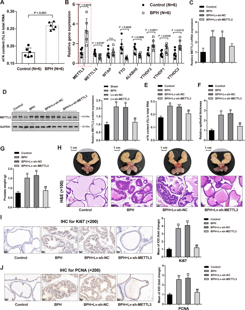
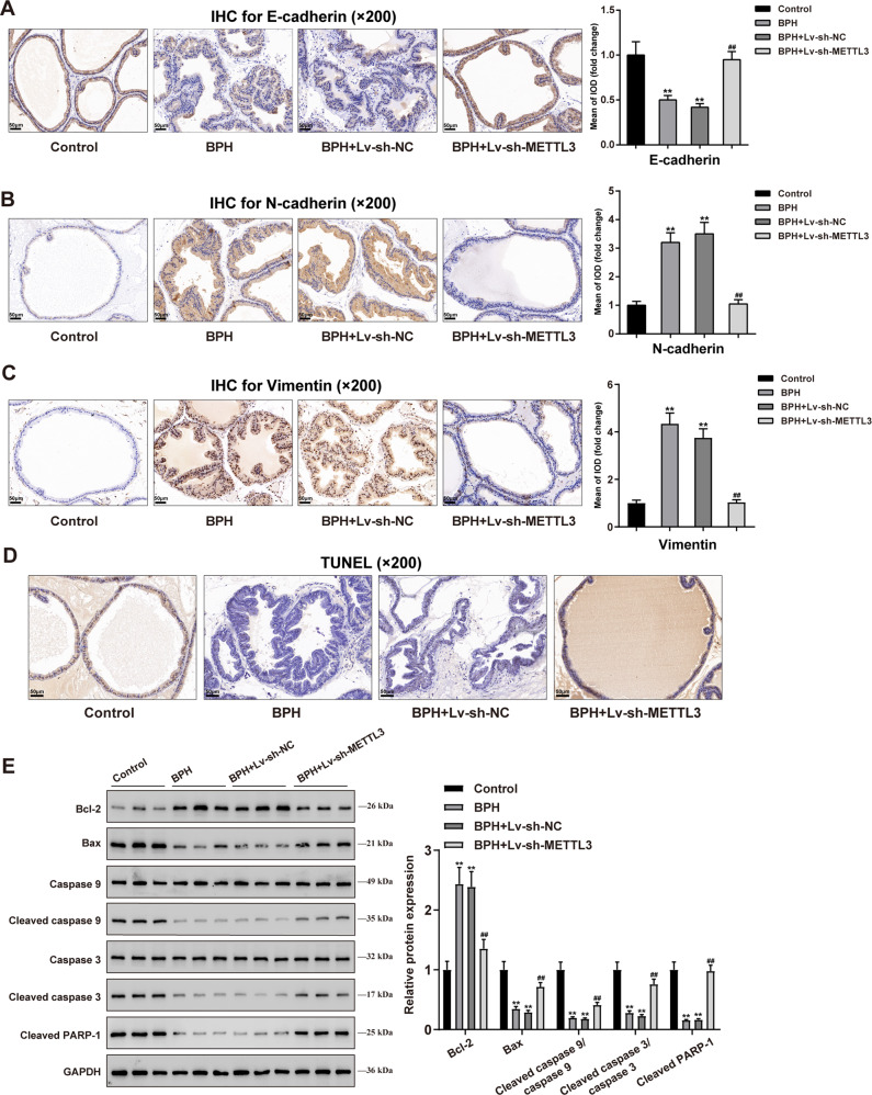
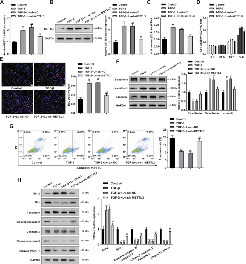
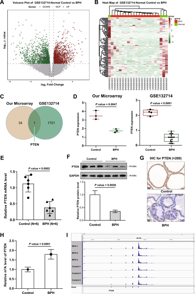
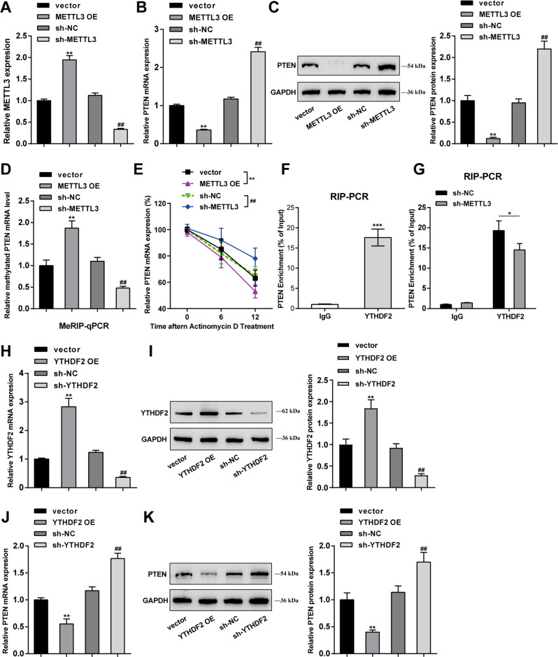
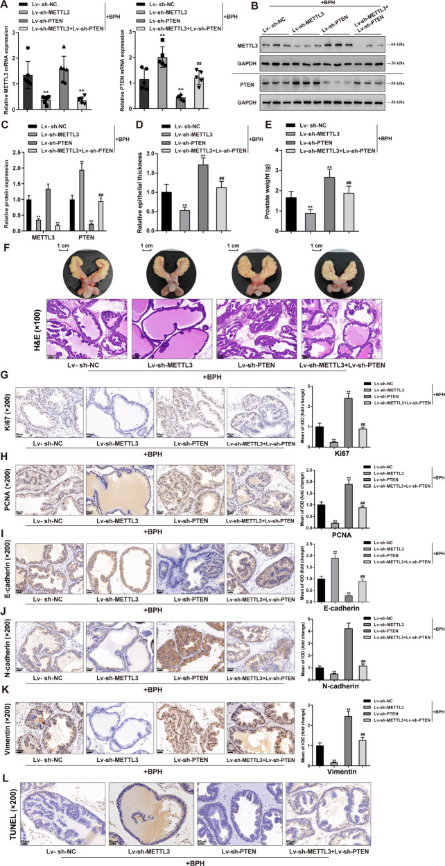
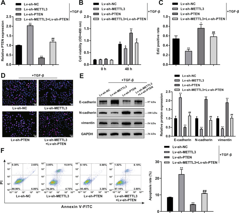
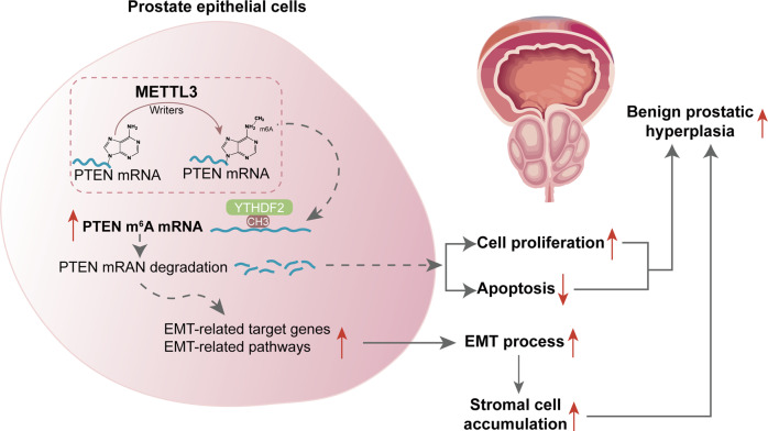
Uncontrolled epithelial cell proliferation in the prostate transition zone and the hyper-accumulation of mesenchymal-like cells derived from the epithelial-mesenchymal transition (EMT) of prostatic epithelium are two key processes in benign prostatic hyperplasia (BPH). m6A RNA modification affects multiple cellular processes, including cell proliferation, apoptosis, and differentiation. In this study, the aberrant up-regulation of methylase METTL3 in BPH samples suggests its potential role in BPH development. Elevated m6A modification in the prostate of the BPH rat was partially reduced by METTL3 knockdown. METTL3 knockdown also partially reduced the prostatic epithelial thickness and prostate weight, significantly improved the histological features of the prostate, inhibited epithelial proliferation and EMT, and promoted apoptosis. In vitro, METTL3 knockdown decreased TGF-β-stimulated BPH-1 cell proliferation, m6A modification, and EMT, whereas promoted cell apoptosis. METTL3 increased the m6A modification of PTEN and inhibited its expression through the reading protein YTHDF2. PTEN knockdown aggravated the molecular, cellular, and pathological alterations in the prostate of BPH rats and amplified TGF-β-induced changes in BPH-1 cells. More importantly, PTEN knockdown partially abolished the improving effects of METTL3 knockdown both in vivo and in vitro. In conclusion, the level of m6A modification is elevated in BPH; the METTL3/YTHDF2/PTEN axis disturbs the balance between epithelial proliferation and apoptosis, promotes EMT, and accelerates BPH development in an m6A modification-related manner.


Conflict of interest statement

The authors declare no competing interests.

Figures

Fig. 1
Fig. 1. Methylases altered in BPH and methylase METTL3 regulates prostatic hyperplasia in model rats.
Prostatic hyperplasia model was induced in rats by subcutaneous injection of testosterone as described in the M&M section. A m6A status of RNA from normal or hyperplasic rat prostate were determined using an m6A RNA Methylation Assay Kit. B The expression of methylation-related enzymes was examined in normal and hyperplasic prostate tissues using qRT-PCR, including methylation enzymes: METTL3, METTL14, WTAP; demethylation enzymes: FTO, ALKBH5; reading proteins: YTHDF2, YTHDF1, YTHDC2. C, D METTL3 knockdown was achieved in rat models by transducing lentivirus containing short hairpin targeting METTL3; METTL3 expression was confirmed in rats in each group using qRT-PCR (C) and Immunoblotting (D). E Methylated N6-methyladenosine (m6A) in RNA was evaluated using an m6A RNA Methylation Assay Kit. F, G Prostatic epithelial thickness (F) and prostate weight (G) were evaluated at the end of the modeling and treatment. H A representative prostate with the urinary bladder from each group were shown (upper) and the histopathological characteristics of rats’ prostate from each group were evaluated using H&E staining (down). Scale bar = 1 cm or 100 μm. I, J The contents of Ki67 and PCNA in rats’ prostate tissues were determined using Immunohistochemical (IHC) staining. Scale bar = 50 μm. **P < 0.01, ***P < 0.001, compared with the control group; #P < 0.05, ##P < 0.01, compared with the BPH + Lv-sh-NC group.
Fig. 2
Fig. 2. METTL3 knockdown inhibits epithelial-mesenchymal transition (EMT) protein expression and promotes apoptosis in rats’ hyperplasic prostate.
AC The contents of E-cadherin (A), N-cadherin (B), and Vimentin (C) in rats’ prostate tissues were determined using IHC staining. D Cell apoptosis in rats’ prostate tissues was determined using a TUNEL assay. E The apoptosis-related proteins (Bcl-2, Bax, Caspase 9, cleaved caspase 9, Caspase 3, cleaved caspase 3, and cleaved PARP-1) levels in prostate tissues in each group were detected by Immunoblotting. All scale bar = 100 μm. **P < 0.01, compared with the control group; ##P < 0.01, compared with the BPH + Lv-sh-NC group.
Fig. 3
Fig. 3. Effects of METTL3 knockdown on proliferation, apoptosis, and EMT of TGF-β-treated BPH-1 cells.
METTL3 knockdown was achieved in TGF-β-treated BPH-1 cells by transducing Lv-sh-METTL3 and then examined for A, B METTL3 expression using qRT-PCR (A) and Immunoblotting (B); C m6A status in RNA using an m6A RNA Methylation Assay Kit; D cell viability using MTT assay; E DNA synthesis using EdU assay; Scale bar = 20 μm; F the protein levels of E-cadherin, N-cadherin, vimentin using Immunoblotting; G cell apoptosis using flow cytometry; H the protein levels of Bcl-2, Bax, Caspase 9, cleaved caspase 9, Caspase 3, cleaved caspase 3, and cleaved PARP-1 using Immunoblotting. **P < 0.01, compared with the control group; ##P < 0.01, compared with the TGF-β + Lv-sh-NC group.
Fig. 4
Fig. 4. PTEN is highly m6A-modified and downregulated in BPH.
A, B The expression profile between control and BPH samples was analyzed using the R language LIMMA package based on GSE132714. C Genes with altered m6A modification status and altered expression levels were compared, and PTEN was selected. D PTEN expression in normal control and BPH samples according to our microarray and GSE132714. E the mRNA level of PTEN in normal control and BPH samples were determined using RT-PCR. F, G PTEN protein levels in normal control and BPH samples were determined using Immunoblotting and IHC staining. H The m6A modification status of PTEN in normal control and BPH samples was determined using an m6A RNA Methylation Assay Kit. I The m6A modification status in PTEN 3′UTR was analyzed using IGV software.
Fig. 5
Fig. 5. METTL3 mediates m6A modification of PTEN and regulates its expression through reading protein YTHDF2.
A METTL3 overexpression or knockdown was achieved in BPH-1 cells by transfecting OE-METTL3 or sh-METTL3 and confirmed using qRT-PCR. BPH-1 cells were transduced with OE-METTL3 or sh-METTL3 and examined for PTEN mRNA expression using qRT-PCR (B); PTEN protein levels using Immunoblotting (C). The m6A modification status of PTEN using Me-RIP assay (D); PTEN mRNA synthesis using actinomycin D assay (E). **P < 0.01, compared with the vector group; ##P < 0.01, compared with the sh-NC group. F The binding between YTHDF2 and PTEN was validated in BPH-1 cells using RIP assay with anti-IgG and anti-YTHDF2. ***P < 0.001, compared with the anti-IgG group. G The binding between YTHDF2 and PTEN was validated in sh-NC or sh-METTL3 transduced BPH-1 cells using RIP assay with anti-IgG and anti-YTHDF2. *P < 0.05, compared with the sh-NC group. (H, I) YTHDF2 overexpression or knockdown was achieved in BPH-1 cells by transfecting OE-YTHDF2 or sh-YTHDF2 and confirmed using qRT-PCR and Immunoblotting, respectively. (J, K) BPH-1 cells were transduced with OE-YTHDF2 or sh-YTHDF2 and examined for PTEN mRNA expression and protein levels using qRT-PCR and Immunoblotting, respectively. **P < 0.01, compared with the vector group; ##P < 0.01, compared with the sh-NC group.
Fig. 6
Fig. 6. METTL3 regulates PTEN and affects cell proliferation and EMT in prostatic hyperplasia model rats.
BPH model was established in rats as described above; rats were injected with Lv-sh-METTL3 and/or Lv-sh-PTEN and examined for AC the mRNA and protein expressions of METTL3 and PTEN in rats’ prostate using qRT-PCR and Immunoblotting; DE prostatic epithelial thickness and prostate weight at the end of the modeling and treatment; F a representative prostate with the urinary bladder from each group (upper) and the histopathological characteristics of rats’ prostate from each group using H&E staining (down); Scale bar = 1 cm or 100 μm; GK the contents of Ki67, PCNA, E-cadherin, N-cadherin, and vimentin in rats’ prostate tissues using Immunohistochemical (IHC) staining; L cell apoptosis in rats’ prostate using TUNEL assay; scale bar = 50 μm. **P < 0.01, compared with the Lv-sh-NC group. ##P < 0.01, compared with the Lv-sh-METTL3 group.
Fig. 7
Fig. 7. METTL3 affects the cell phenotypes of TGF-β-stimulated.
BPH-1 cells through PTEN BPH-1 cells were co-transduced with Lv-sh-METTL3 and Lv-sh-PTEN and examined for PTEN mRNA expression using qRT-PCR (A); cell viability using MTT assay (B); DNA synthesis using EdU assay (C, D); Scale bar = 20 μm; the protein levels of E-cadherin, N-cadherin, and vimentin using Immunoblotting (E); cell apoptosis by Flow cytometry (F). **P < 0.01, compared with the Lv-sh-NC group. ##P < 0.01, compared with the Lv-sh-METTL3 group.
Fig. 8
Fig. 8. A schematic diagram indicates the mechanisms of how METTL3 promotes prostatic hyperplasia of BPH.
The METTL3/YTHDF2/PTEN axis disturbs the balance between epithelial proliferation and apoptosis, promotes EMT, and accelerates BPH development in an m6A modification-related manner.

Similar articles

Cited by

References

    1. Bianchi-Frias D, Vakar-Lopez F, Coleman IM, Plymate SR, Reed MJ, Nelson PS. The effects of aging on the molecular and cellular composition of the prostate microenvironment. PLoS One. 2010;5:e12501. - PMC - PubMed
    1. Welliver C, Feinstein L, Ward JB, Fwu CW, Kirkali Z, Bavendam T, et al. Trends in lower urinary tract symptoms associated with benign prostatic hyperplasia, 2004 to 2013: the Urologic Diseases in America Project. J Urol. 2020;203:171–8. doi: 10.1097/JU.0000000000000499. - DOI - PubMed
    1. Wendell-Smith C. Terminology of the prostate and related structures. Clin Anat. 2000;13:207–13. doi: 10.1002/(SICI)1098-2353(2000)13:3<207::AID-CA9>3.0.CO;2-9. - DOI - PubMed
    1. McNeal JE. Regional morphology and pathology of the prostate. Am J Clin Pathol. 1968;49:347–57. doi: 10.1093/ajcp/49.3.347. - DOI - PubMed
    1. Alonso-Magdalena P, Brossner C, Reiner A, Cheng G, Sugiyama N, Warner M, et al. A role for epithelial-mesenchymal transition in the etiology of benign prostatic hyperplasia. Proc Natl Acad Sci USA. 2009;106:2859–63. doi: 10.1073/pnas.0812666106. - DOI - PMC - PubMed

Publication types