

A lightweight neural network with multiscale feature enhancement for liver CT segmentation
- PMID: 35986015
- PMCID: PMC9391485
- DOI: 10.1038/s41598-022-16828-6
A lightweight neural network with multiscale feature enhancement for liver CT segmentation
Erratum in
-
Publisher Correction: A lightweight neural network with multiscale feature enhancement for liver CT segmentation.Sci Rep. 2022 Sep 21;12(1):15741. doi: 10.1038/s41598-022-20472-5. Sci Rep. 2022. PMID: 36131121 Free PMC article. No abstract available.
Abstract
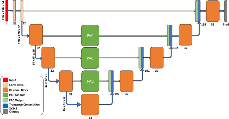
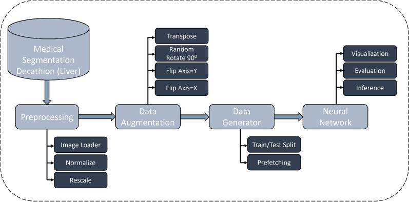
Segmentation of abdominal Computed Tomography (CT) scan is essential for analyzing, diagnosing, and treating visceral organ diseases (e.g., hepatocellular carcinoma). This paper proposes a novel neural network (Res-PAC-UNet) that employs a fixed-width residual UNet backbone and Pyramid Atrous Convolutions, providing a low disk utilization method for precise liver CT segmentation. The proposed network is trained on medical segmentation decathlon dataset using a modified surface loss function. Additionally, we evaluate its quantitative and qualitative performance; the Res16-PAC-UNet achieves a Dice coefficient of 0.950 ± 0.019 with less than half a million parameters. Alternatively, the Res32-PAC-UNet obtains a Dice coefficient of 0.958 ± 0.015 with an acceptable parameter count of approximately 1.2 million.
© 2022. The Author(s).
Conflict of interest statement
The authors declare no competing interests.
Figures


References
-
- Dakua SP. Performance divergence with data discrepancy: A review. Artif. Intell. Rev. 2013;40:429–455. doi: 10.1007/s10462-011-9289-8. - DOI
-
- Dakua SP. Towards left ventricle segmentation from magnetic resonance images. IEEE Sens. J. 2017;17:5971–5981. doi: 10.1109/JSEN.2017.2736641. - DOI
-
- Akhtar Y, et al. Risk assessment of computer-aided diagnostic software for hepatic resection. IEEE Trans. Radiat. Plasma Med. Sci. 2021;6:667. doi: 10.1109/TRPMS.2021.3071148. - DOI
-
- Baâzaoui A, Barhoumi W, Ahmed A, Zagrouba E. Semi-automated segmentation of single and multiple tumors in liver ct images using entropy-based fuzzy region growing. IRBM. 2017;38:98–108. doi: 10.1016/j.irbm.2017.02.003. - DOI
-
- Zhou Z, Xue-chang Z, Si-ming Z, Hua-fei X, Yue-ding S. Semi-automatic liver segmentation in CT images through intensity separation and region growing. Procedia Comput. Sci. 2018;131:220–225. doi: 10.1016/j.procs.2018.04.206. - DOI
Publication types
MeSH terms
LinkOut - more resources
Full Text Sources
Medical
