. 2022 Aug 19;12(1):14182.
doi: 10.1038/s41598-022-17987-2.

Cannabinoid CB1 receptors regulate salivation

Affiliations

Cannabinoid CB1 receptors regulate salivation

Kelsey Andreis et al. Sci Rep. .

Abstract

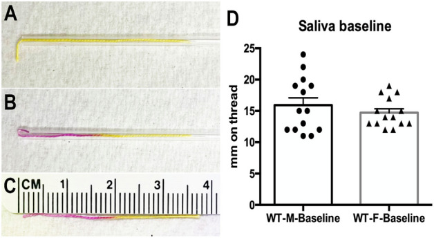
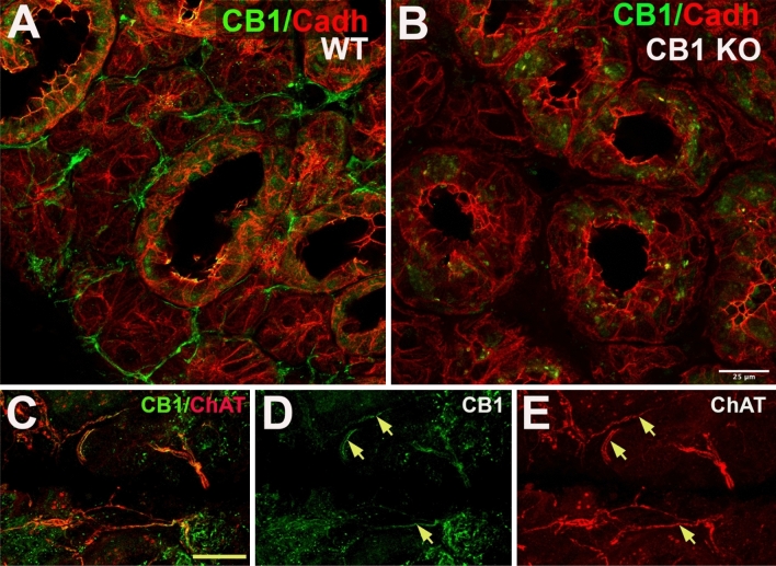
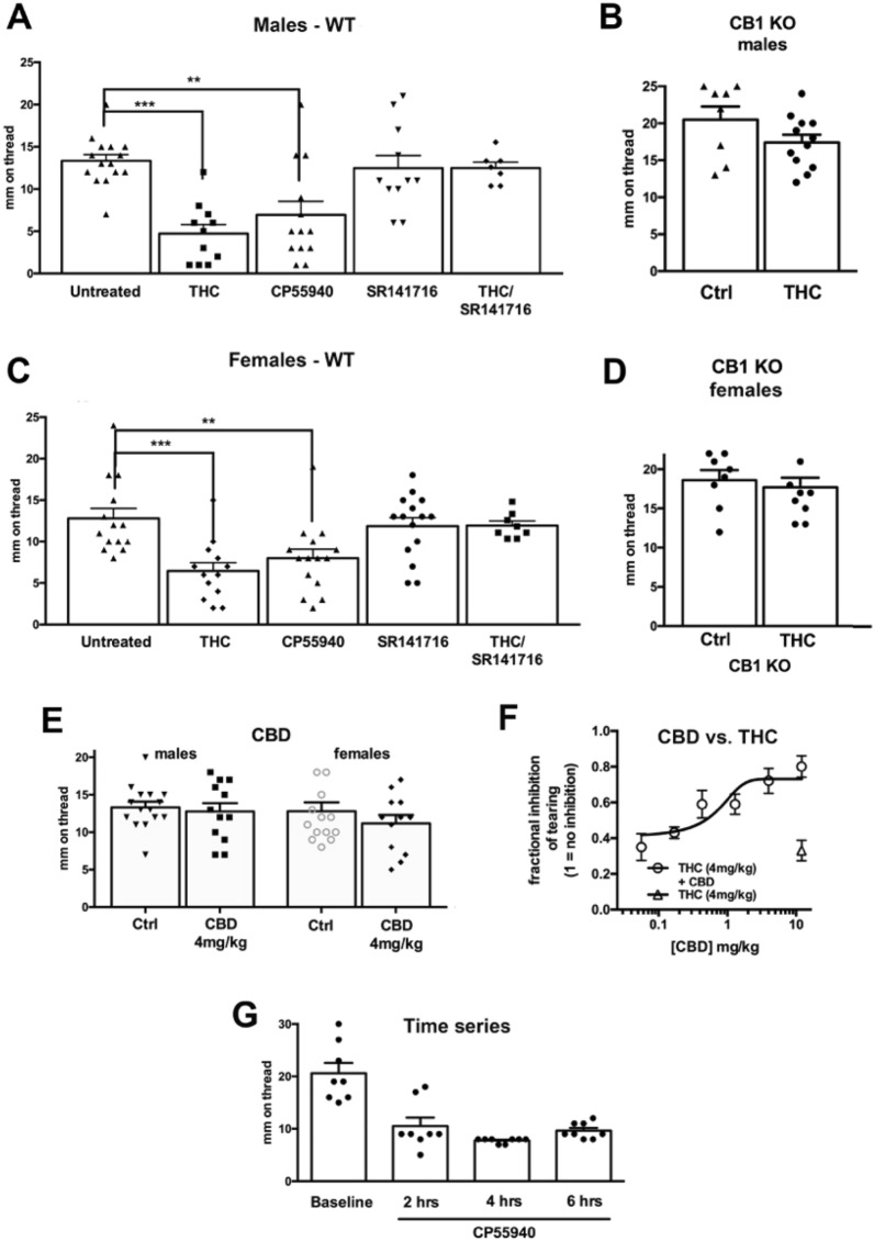
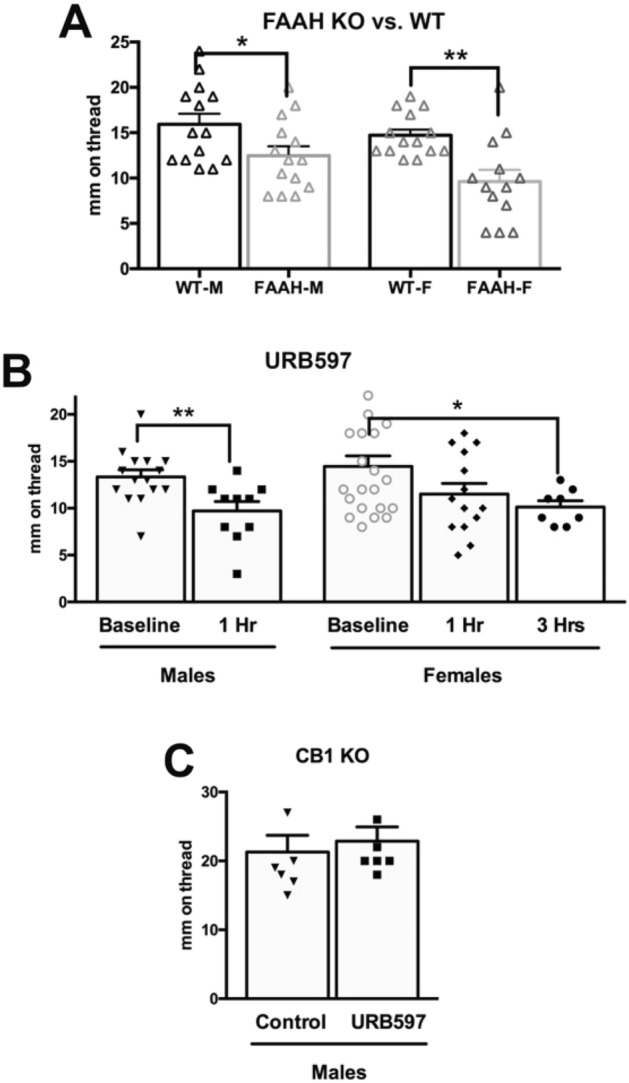
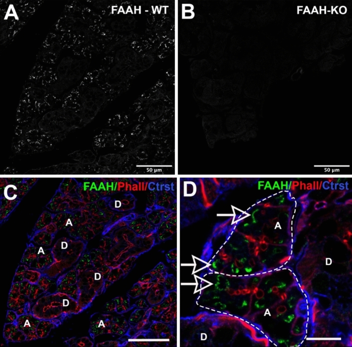
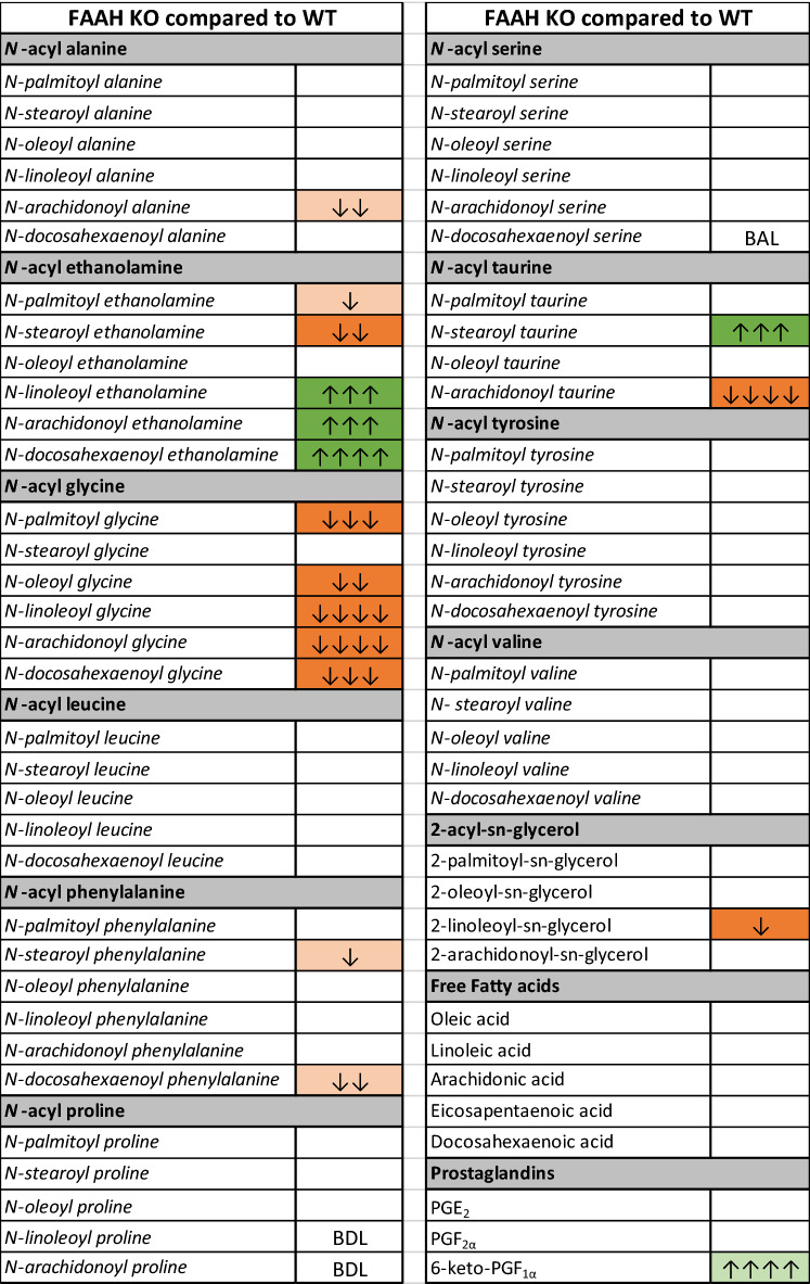
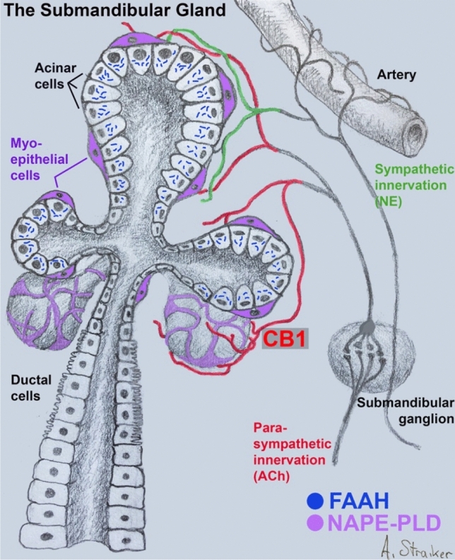
Saliva serves multiple important functions within the body that we typically take for granted, such as helping prepare food for swallowing and defense against oral pathogens. Dry mouth is a primary symptom of Sjӧgren's syndrome and is a side effect of many drug treatments. Cannabis users frequently report dry mouth, but the basis for this is still unknown. If the effects occur via the endogenous cannabinoid signaling system, then this may represent a novel mechanism for the regulation of salivation. We examined expression of cannabinoid CB1 receptors in submandibular salivary gland using immunohistochemistry and tested regulation of salivation by THC and cannabinoid-related ligands. We now report that CB1 receptors are expressed in the axons of cholinergic neurons innervating the submandibular gland. No staining is seen in submandibular gland epithelial cells (acinar and ductal), or myoepithelial cells (MECs). Treatment with THC (4 mg/kg, IP) or the cannabinoid receptor agonist CP55940 (0.5 mg/kg) reduced salivation in both male and female mice 1 h after treatment. CBD had no effect on its own but reversed the effect of THC in a concentration-dependent manner. Neither the CB1 receptor antagonist SR141716 (4 mg/kg) nor the CB2-selective agonist JWH133 (4 mg/kg) had an effect on salivation. We also found that fatty acid amide hydrolase (FAAH), the enzyme that metabolizes the endocannabinoid anandamide and related lipids, regulates salivation. Salivation was reduced in FAAH knockout mice as well as mice treated with the FAAH blocker URB597 (4 mg/kg). URB597 had no effect in CB1 knockout mice. FAAH protein is detected intracellularly in acinar but not ductal epithelial cells. In lipidomics experiments, we found that FAAH knockout mice chiefly had elevated levels of acylethanolamines, including anandamide, and reduced levels of acyglycines. Our results are consistent with a model wherein endocannabinoids activate CB1 receptors on cholinergic axons innervating the submandibular gland. THC likely acts by plugging into this system, activating CB1 receptors to reduce salivation, thus offering a mechanism underlying the dry mouth reported by cannabis users.


Conflict of interest statement

The authors declare no competing interests.

Figures

Figure 1
Figure 1
Capillary based method for measuring basal salivation in mice. (A) Phenol red-coated thread is inserted into a fire-polished glass capillary with 3 mm extending outside the opening. This is then placed under the tongue of an isoflurane anesthetized mouse for 10 s. (B) Saliva travels up the thread, discoloring it. (C) The distance traveled in mm is taken as a measure of basal saliva. (D) Sample baseline data for WT males and females shows that basal levels are similar (n = 14 per condition).
Figure 2
Figure 2
CB1 protein is expressed in a subset of cholinergic axons in submandibular gland. (A,B) Left panel shows CB1 (green) and cadherin (red) in wild type (WT) mouse submandibular gland. Right panel shows that CB1 staining in axon-like processes is absent submandibular gland from CB1 knockout mouse. (C) Double staining in submandibular gland of the mouse shows expression of CB1 (green) and choline acetyl transferase (ChAT, red), a marker for cholinergic axonal inputs. (D,E) CB1 (green) staining is seen in processes of ChAT-positive axons (arrows). Scale bar: (A,B) 25 µm; (CE) 40 µm. Images processed using Adobe Photoshop vsn. 21.2 and FIJI (vsn 2.3.0/1.53q, available at https://imagej.net/Fiji/downloads).
Figure 3
Figure 3
CB1 receptors regulate basal salivation. In male (A) and female (C) mice, THC (mg/kg, IP) or CP55940 (0.5 mg/kg) reduced basal salivation 1 h after treatment. The CB1 antagonist SR141716 (4 mg/kg) had no effect. (B,D) THC had no effect in CB1 knockout males (B) or females (D). (E) CBD (4 mg/kg) had no effect on salivation in males or females. (F) CBD reversed the effects of THC in a concentration-dependent manner. (G) Effect of CP55940 persists for at least six hours. **p < 0.01; ***p < 0.005, one-way ANOVA with Dunnett’s post-hoc test vs. control.
Figure 4
Figure 4
FAAH deletion and blockade reduces salivation. (A) Male and female knockout mice saw lower baseline salivation. (B) Treatment with the FAAH blocker URB597 (4 mg/kg) reduced salivation at 1 h in males and at 3 h in females. (C) URB597 did not alter salivation in male CB1 knockout mice. *p < 0.05, *p < 0.01 unpaired t-test.
Figure 5
Figure 5
FAAH protein expression in acini of submandibular gland. (A,B) FAAH expression in (A) WT and (B) FAAH knockout submandibular gland. (C) Triple-stain from (A) shows FAAH (green) relative to phalloidin (red) and a counterstain that outlines acini. FAAH is seen in acini “A” but not ducts “D”. (D) Higher magnification image shows FAAH staining in acini “A”, outlined but not ducts “D”. Scale bars: (AC) 50 µm; (D) 15 µm. Images processed using Adobe Photoshop vsn. 21.2 and FIJI (vsn 2.3.0/1.53q, available at https://imagej.net/Fiji/downloads).
Figure 6
Figure 6
FAAH protein in male vs. female submandibular gland. (A) FAAH protein levels in males and females (p = 0.058, student’s t test, n = 4). (B) A protein band is detected at the expected molecular weight (~ 63 kDa). Full blot in Supplementary Fig. S1.
Figure 7
Figure 7
Differences in lipid levels in FAAH KO SMG compared to WT. Fold change denoted by arrows 1 = 1–1.49; 2 = 1.5–1.99; 3 = 2–2.99; 4 = 3–9.99; 5 = 10 or more. Green significant increases, orange significant decreases, light green and orange trending changes, white detected but no significant differences, BDL below detection limit, BAL below analytical limits though present in some samples.
Figure 8
Figure 8
CB1 signaling system in submandibular gland. Schematic of submandibular gland showing acinar and ductal epithelial cells as well as enveloping myoepithelial cells (violet). CB1 receptors (red) are seen on the axons of parasympathetic neurons originating in the submandibular ganglion. FAAH, restricted to acinar cells, metabolizes acylethanolamines, including anandamide. NAPE-PLD likely resides in myoepithelial cells.

References

    1. Proctor GB, Carpenter GH. Salivary secretion: Mechanism and neural regulation. Monogr. Oral. Sci. 2014;24:14–29. doi: 10.1159/000358781. - DOI - PubMed
    1. Agostini BA, et al. How common is dry mouth? Systematic review and meta-regression analysis of prevalence estimates. Braz. Dent. J. 2018;29:606–618. doi: 10.1590/0103-6440201802302. - DOI - PubMed
    1. Donaldson M, Goodchild JH. A systematic approach to xerostomia diagnosis and management. Compend. Contin. Educ. Dent. 2018;39:1–9. - PubMed
    1. Khanagar SB, et al. Age-related oral changes and their impact on oral health-related quality of life among frail elderly population: A review. J. Contemp. Dent. Pract. 2020;21:1298–1303. - PubMed
    1. Patel R, Shahane A. The epidemiology of Sjogren's syndrome. Clin. Epidemiol. 2014;6:247–255. doi: 10.2147/CLEP.S47399. - DOI - PMC - PubMed