. 2022 Aug;6(8):979-991.
doi: 10.1038/s41551-022-00922-1. Epub 2022 Aug 19.

Diagnosis of paediatric tuberculosis by optically detecting two virulence factors on extracellular vesicles in blood samples

Affiliations

Diagnosis of paediatric tuberculosis by optically detecting two virulence factors on extracellular vesicles in blood samples

Wenshu Zheng et al. Nat Biomed Eng. 2022 Aug.

Abstract

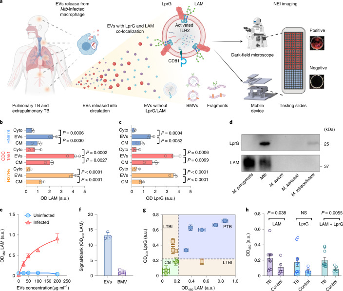
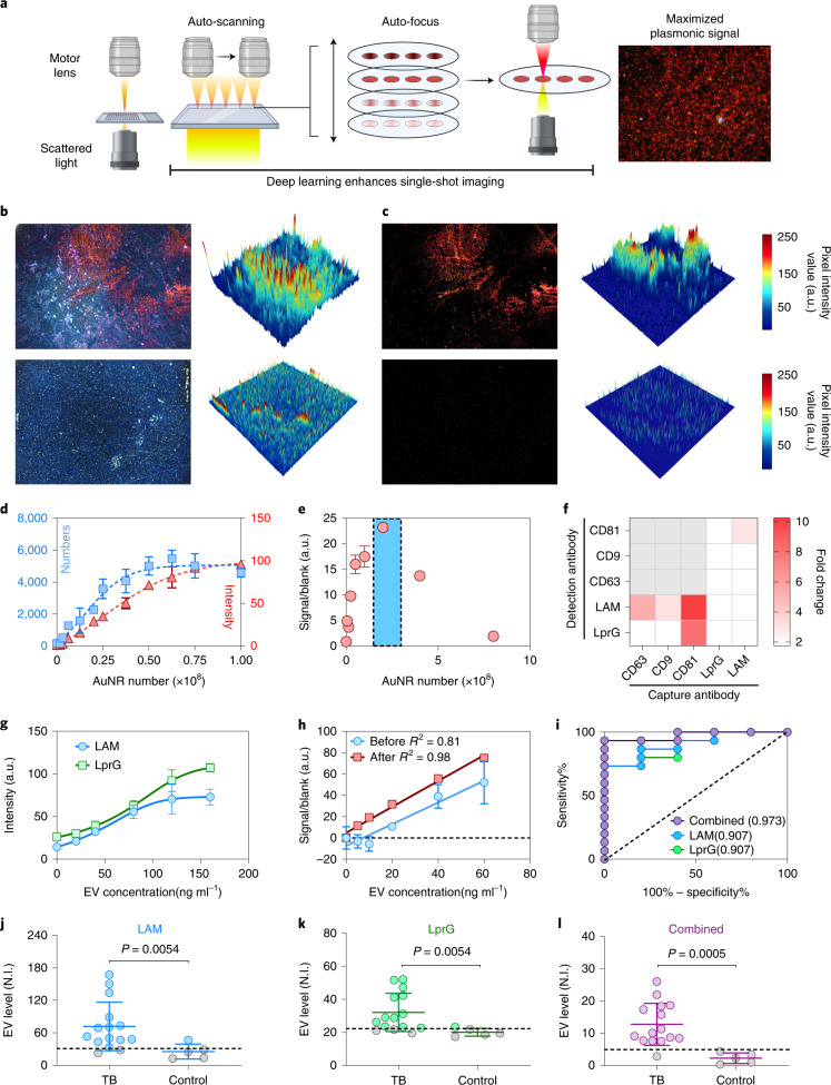
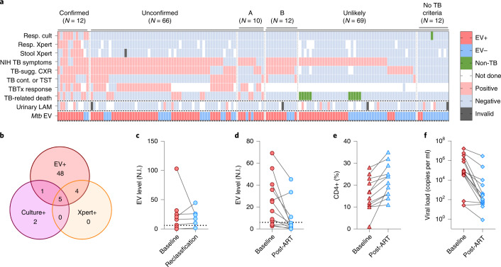
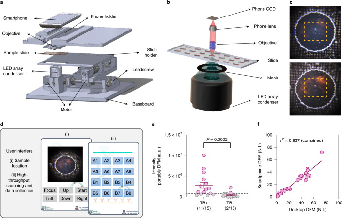
Sensitive and specific blood-based assays for the detection of pulmonary and extrapulmonary tuberculosis would reduce mortality associated with missed diagnoses, particularly in children. Here we report a nanoparticle-enhanced immunoassay read by dark-field microscopy that detects two Mycobacterium tuberculosis virulence factors (the glycolipid lipoarabinomannan and its carrier protein) on the surface of circulating extracellular vesicles. In a cohort study of 147 hospitalized and severely immunosuppressed children living with HIV, the assay detected 58 of the 78 (74%) cases of paediatric tuberculosis, 48 of the 66 (73%) cases that were missed by microbiological assays, and 8 out of 10 (80%) cases undiagnosed during the study. It also distinguished tuberculosis from latent-tuberculosis infections in non-human primates. We adapted the assay to make it portable and operable by a smartphone. With further development, the assay may facilitate the detection of tuberculosis at the point of care, particularly in resource-limited settings.


Conflict of interest statement

T.Y.H. and W.Z. have a provisional patent (‘Method of detecting TB in blood’) submitted through Tulane University. The rest of the authors declare no competing interests.

Figures

Fig. 1
Fig. 1. EV LAM and LprG expression as a biomarker for TB diagnosis.
a, Rationale and assay schematic. Created with BioRender.com. b,c, Western blot densitometry of LAM (b) and LprG (c) expression in cytosolic (Cyto), EV and cell membrane (CM) factions of macrophage cultures infected with the indicated Mtb strains. Data indicate mean ± s.d., n = 3 biologically independent repeats; P values obtained by one-way ANOVA with Dunnett’s post-test. a.u., arbitrary units. d, Western blot analysis of LAM and LprG from CFP samples of patients with sequence-confirmed M. smegmatis, Mtb, M. avium, M. intracellulare and M. kansasii infections. e,f, EV-ELISA for LAM on EVs from Mtb-infected macrophages (e) and BMVs isolated from Mtb culture medium (f) by ultracentrifugation when captured by host EV-specific antibody. g, EV-ELISA for LAM and LprG on serum EVs of non-human primates with pulmonary TB (PTB), LTBI or their healthy controls (Ctrl) (mean ± s.d. of three technical replicates). h, EV-ELISA for LAM, LprG and integrated LAM and LprG (LAM+LprG) expression on isolated EVs from serum of children with TB (N = 10) and without evidence of TB (Ctrl; N = 5). Mean ± s.e.m., P values obtained by two-sided Mann-Whitney U test.
Fig. 2
Fig. 2. NEI optimization for sensitive detection of EV-associated TB biomarkers.
a, Schematic of the NEI image capture workflow and signal. Created with BioRender.com. b,c, NEI images (left) and 3D heat maps (right) before (b) and after (c) image processing to remove artefacts (blue and white signal) to improve detection of AuNRs (red signal) bound to Mtb EVs (top) and negative control EVs (bottom). d, Standard curves for AuNR dilutions quantified by counting positive pixels versus mean pixel intensity. e, AuNR titration to maximize signal-to-noise (highlighted in blue shadow) in samples near the EV-ELISA limit of detection (150 ng EV protein per ml). f, NEI signal increase versus background (signal-to-noise) when using the indicated capture and detection antibody pairs to detect NEI signal from a low-concentration EV sample (150 ng protein per ml) and a negative control (blank) sample. g, EV LAM and LprG NEI signal linearity with an Mtb EV concentration curve generated using EVs from Mtb-infected macrophages. h, Linear regression line and correlation coefficient of NEI EV LAM signal without and after noise reduction in background-normalized serum samples spiked with EVs isolated from Mtb-infected macrophages. For dh: mean ± s.d.; N = 3. i, Receiver operating characteristic (ROC) analysis for the ability of single and integrated (combined) EV LAM and LprG NEI signals to distinguish children with and without TB, indicating areas under the ROC curve. jl, NEI signal for LAM (j), LprG (k) and integrated LAM and LprG (l) expression on serum EVs of children with TB (N = 15) and with no evidence of TB (N = 5), N.I., NEI signal intensity (arbitrary units; a.u.). Solid lines indicate mean ± s.d.; dashed lines indicate the threshold for positive signal determined in corresponding ROC analysis in i. P values were determined by two-sided Mann-Whitney U test.
Fig. 3
Fig. 3. Mtb EV NEI diagnostic performance in children living with HIV at high risk of TB.
a, NEI signal in children with confirmed, unconfirmed and unlikely TB as determined by positive respiratory culture/Xpert or stool Xpert results, TB-related symptoms meeting NIH criteria for the duration, chest X-ray (CXR) findings, close TB contact or positive TST, positive TBTx response, and/or TB-related death. Urine LAM results and serum Mtb EV results were not used for classification. Subgroup A: children reclassified from unlikely to unconfirmed TB on the basis of TBTx (TB treatment) response or TB-related death as determined by an expert review panel (see Supplementary Table 7 for criteria). Subgroup B: children reclassified from unconfirmed to unlikely TB on the basis of symptom improvement following ART initiation without TBTx initiation (alternate data for anti-TBTx response). b, TB cases classified by NEI, Mtb culture and/or Xpert test results. c, Positive baseline NEI signal predicts subsequent TB reclassification in children with unlikely TB assignments at enrolment (Subgroup A in a) who were reclassified to unconfirmed TB by investigators blinded to EV results. df, NEI signal decreases (d) among children diagnosed as unconfirmed TB cases at baseline but reclassified as unlikely TB cases due to symptom improvement without TBTx following ART initiation alone (Subgroup B in a), which corresponded with CD4 cell % increases (e) and HIV viral load reductions (f) following ART initiation.
Fig. 4
Fig. 4. Design and performance of a smartphone-based NEI point-of-care TB diagnosis approach.
ac, Schematic of a portable smartphone-based DFM device for NEI assay readout (a) and its dark-field condenser mask (b), and the effect of this mask on NEI AuNR signal (red) intensity of the targeted Mtb EVs from images (c) collected without (top) and with (bottom) the condenser mask. d, Schematic of smartphone app menu workflow. e, EV NEI signal from serum samples of children with TB (N = 15) and with no evidence of TB (N = 15). Data indicate mean ± s.e.m.; dashed line indicates the TB detection threshold for TB positive (red) or TB negative (grey) sample assignment; P values were determined by two-sided Mann-Whitney U test. f, Comparison of integrated EV LAM and LprG NEI signals obtained for the samples in e using a desktop DFM and the portable smartphone DFM device, indicating the linear regression line and squared Pearson correlation coefficient.

References

    1. Lu LL, et al. IFN-γ-independent immune markers of Mycobacterium tuberculosis exposure. Nat. Med. 2019;25:977–987. - PMC - PubMed
    1. Floyd K, Glaziou P, Zumla A, Raviglione M. The global tuberculosis epidemic and progress in care, prevention, and research: an overview in year 3 of the End TB era. Lancet Respir. Med. 2018;6:299–314. - PubMed
    1. Garrido-Cardenas J, de Lamo-Sevilla C, Cabezas-Fernández M, Manzano-Agugliaro F, Martínez-Lirola M. Global tuberculosis research and its future prospects. Tuberculosis. 2020;121:101917. - PubMed
    1. Harding E. WHO global progress report on tuberculosis elimination. Lancet Respir. Med. 2020;8:19. - PubMed
    1. Dodd PJ, Yuen CM, Sismanidis C, Seddon JA, Jenkins HE. The global burden of tuberculosis mortality in children: a mathematical modelling study. Lancet Glob. Health. 2017;5:e898–e906. - PMC - PubMed

Publication types

Substances