. 2022 Aug 19;13(1):425.
doi: 10.1186/s13287-022-03128-z.

Bone marrow mesenchymal stromal cells in a 3D system produce higher concentration of extracellular vesicles (EVs) with increased complexity and enhanced neuronal growth properties

Affiliations

Bone marrow mesenchymal stromal cells in a 3D system produce higher concentration of extracellular vesicles (EVs) with increased complexity and enhanced neuronal growth properties

Elmira Jalilian et al. Stem Cell Res Ther. .

Abstract

Purpose: Extracellular vesicles (EVs) derived from mesenchymal stromal cells (MSCs) have been demonstrated to possess great potential in preclinical models. An efficient biomanufacturing platform is necessary for scale up production for clinical therapeutic applications. The aim of this study is to investigate the potential differences in neuro-regenerative properties of MSC-derived EVs generated in 2D versus 3D culture systems.

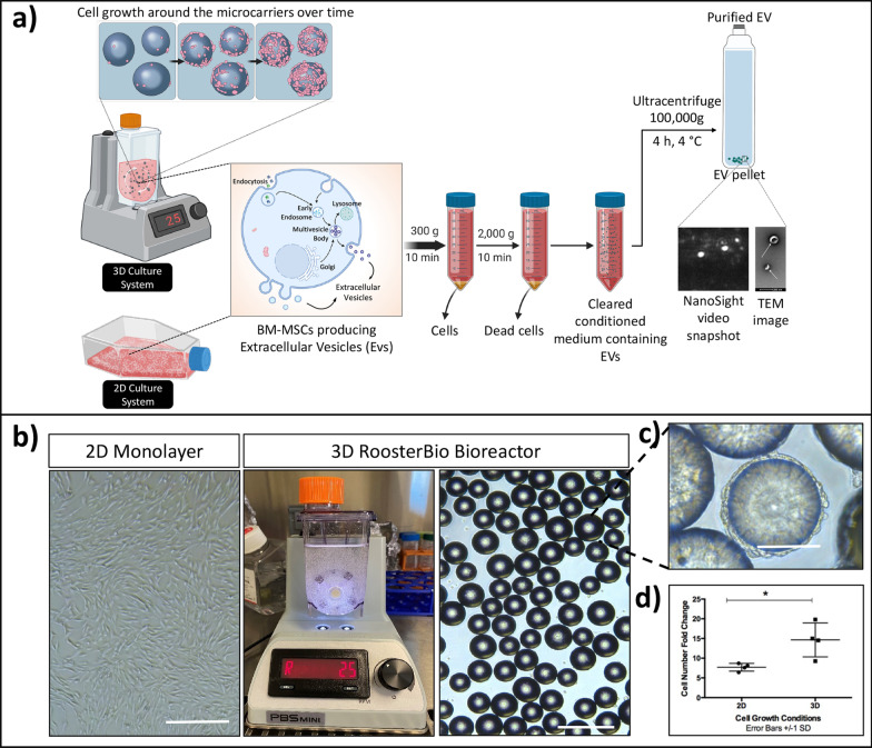
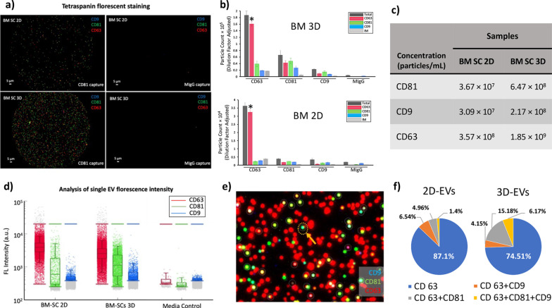
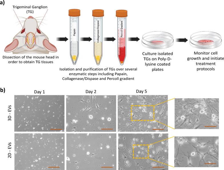
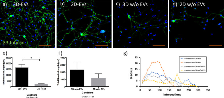
Method: Human bone marrow MSCs (BM-MSCs) were cultured in 2D monolayer and 3D bioreactor systems. EVs were isolated using ultracentrifugation followed by size and concentration measurements utilizing dynamic light scattering (NanoSight) and by fluorescence staining (ExoView). Mouse trigeminal ganglia (TG) neurons were isolated from BALB/c mice and cultured in the presence or absence of EVs derived from 2D or 3D culture systems. Neuronal growth and morphology were monitored over 5 days followed by immunostaining for β3 tubulin. Confocal images were analyzed by Neurolucida software to obtain the density and length of the neurites.

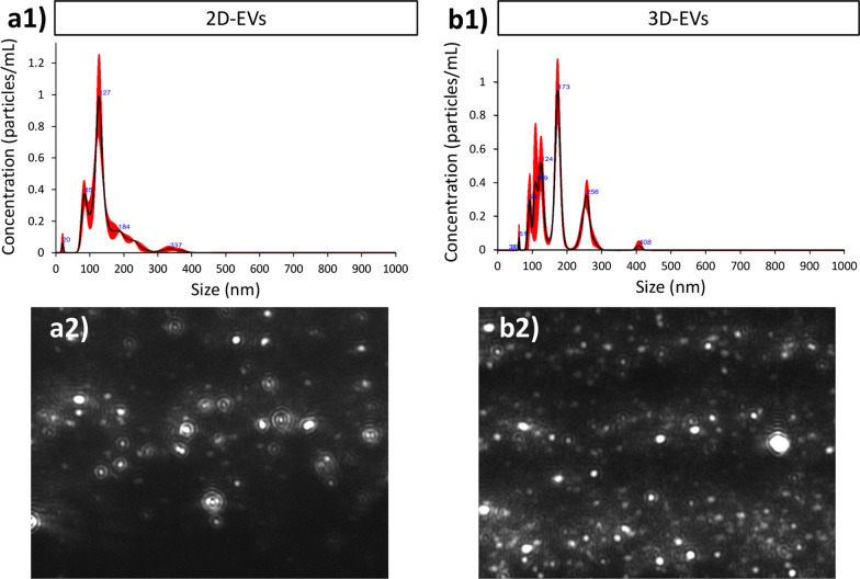
Results: The NanoSight tracking analysis revealed a remarkable increase (24-fold change) in the concentration of EVs obtained from the 3D versus 2D culture condition. ExoView analysis showed a significantly higher concentration of CD63, CD81, and CD9 markers in the EVs derived from 3D versus 2D conditions. Furthermore, a notable shift toward a more heterogeneous phenotype was observed in the 3D-derived EVs compared to those from 2D culture systems. EVs derived from both culture conditions remarkably induced neurite growth and elongation after 5 days in culture compared to untreated control. Neurolucida analysis of the immunostaining images (β3 tubulin) showed a significant increase in neurite length in TG neurons treated with 3D- versus 2D-derived EVs (3301.5 μm vs. 1860.5 μm, P < 0.05). Finally, Sholl analysis demonstrated a significant increase in complexity of the neuronal growth in neurons treated with 3D- versus 2D-derived EVs (P < 0.05).

Conclusion: This study highlights considerable differences in EVs obtained from different culture microenvironments, which could have implications for their therapeutic effects and potency. The 3D culture system seems to provide a preferred environment that modulates the paracrine function of the cells and the release of a higher number of EVs with enhanced biophysical properties and functions in the context of neurite elongation and growth.

Keywords: 2D culture; 3D culture; Bone marrow mesenchymal stromal cell; Exosomes; Extracellular vesicles; Neuronal growth.


Conflict of interest statement

The authors declare that they have no competing interests.

Figures

Fig. 1
Fig. 1
2D versus 3D culture system for BM-MSCs. a Schematic diagram of BM-MSCs grown in a 2D and 3D culture conditions. Cells were either grown in 2D culture flask or 3D bioreactor on microcarriers for expansion. Cells were transferred to collection medium for 3 days and then collected and processed for EV isolation using low and high-speed centrifugation steps. b Morphology of BM-MSCs after 3 days in collection medium and c bioreactor, microcarriers and cell attachment and expansion around microcarriers. d The statistical analysis illustrated significant expansion in cell number in 3D versus 2D culture conditions (P < 0.05). Scale bar 200 px
Fig. 2
Fig. 2
Characterization of particles from 2 and 3D EVs using NTA analysis. Size distribution of particles using NTA in a1 2D and b1 3D condition. Representative image frame of EVs extracted form a a2 2D culture system and a b2 3D culture system
Fig. 3
Fig. 3
Characterization of 2D and 3D EVs using ExoView. a Tetraspanin antibodies-capture allows for the detection and identification of subpopulations according to the different tetraspanin compounds expressed on each particle (right positive stained, left IgG control). b Copy number of each type of particle from 2 and 3D cell culture counted and presented in a bar-graph with a tenfold increase on the Y-axis in BM 3D graph versus that of BM 2D (P < 0.05). c Concentration of each marker per milliliter of the extraction medium. d Fluorescence intensity per EV was measured for each channel. e Representative image of capture spot of colocalization. f Representative pie charts from a single chip that exhibits the percentage of total particles detected with either CD63 (blue), CD63 + CD9 (orange), CD 63 + CD81 (gray), or CD 63 + CD81 + CD9 (yellow) on different capture spots
Fig. 4
Fig. 4
Trigeminal ganglion (TGs) cells isolation and elongation in the presence of EV treatment. a Schematic diagram of experimental procedure for TG cells isolation from BALB/c mice by utilizing different enzymatic procedures and plating the cells in PDL-coated dishes. b Representative images taken using bright-field microscopy from dishes treated with either 3D- or 2D-EVs over 5 days showed considerable neurite growth and elongation in both conditions with a remarkable increase in the number of neurite sprouts in 3D-EVs condition versus 2D-EVs. Scale bars in D1 to D5 images are 200px, and in inlets, they are 100px
Fig. 5
Fig. 5
3D-EVs induce greater neurite elongation and branching than 2D-EVs. a, b Representative confocal microscopy images of TG neurite elongation cultured under 3D-EVs and 2D-EVs. c, d Neurite growth in EV-depleted media derived from 3D or 2D cultures. e Quantification of total neurite growth demonstrated that 3D-EVs induce significant growth than 2D-EVs. f No length difference was observed in cultures treated with EV-depleted media. Statistical significance was tested with an unpaired student t test (two-tailed) for evaluating differences between the two groups, and statistical significance was determined (P < 0.05) (n = 4 biological replicate & n = 16 technical replicates for each condition). g Sholl analysis performed in the dendritic tree demonstrated that 3D-EVs significantly increased the number of dendritic intersections between 50 and 120 radii compared to 2D-EVs and control conditions. Data represent the mean ± 1 SD of 3 independent experiments. P < 0.05. Scale bar 100 µm

References

    1. Yang AY, Chow J, Liu J. Corneal innervation and sensation: the eye and beyond. Yale J Biol Med. 2018;91(1):13–21. - PMC - PubMed
    1. Shaheen BS, Bakir M, Jain S. Corneal nerves in health and disease. Surv Ophthalmol. 2014;59(3):263–285. doi: 10.1016/j.survophthal.2013.09.002. - DOI - PMC - PubMed
    1. Dua HS, Said DG, Messmer EM, Rolando M, Benitez-Del-Castillo JM, Hossain PN, Shortt AJ, Geerling G, Nubile M, Figueiredo FC, Rauz S, Mastropasqua L, Rama P, Baudouin C. Neurotrophic keratopathy. Prog Retin Eye Res. 2018;66:107–131. doi: 10.1016/j.preteyeres.2018.04.003. - DOI - PubMed
    1. Sebbag L, Thomasy SM, Leland A, Mukai M, Kim S, Maggs DJ. Altered corneal innervation and ocular surface homeostasis in FHV-1-exposed cats: a preliminary study suggesting metaherpetic disease. Front Vet Sci. 2020;7:580414. doi: 10.3389/fvets.2020.580414. - DOI - PMC - PubMed
    1. Sebbag L, Pesavento PA, Carrasco SE, Reilly CM, Maggs DJ. Feline dry eye syndrome of presumed neurogenic origin: a case report. JFMS Open Rep. 2018;4(1):2055116917746786. - PMC - PubMed

Publication types

LinkOut - more resources